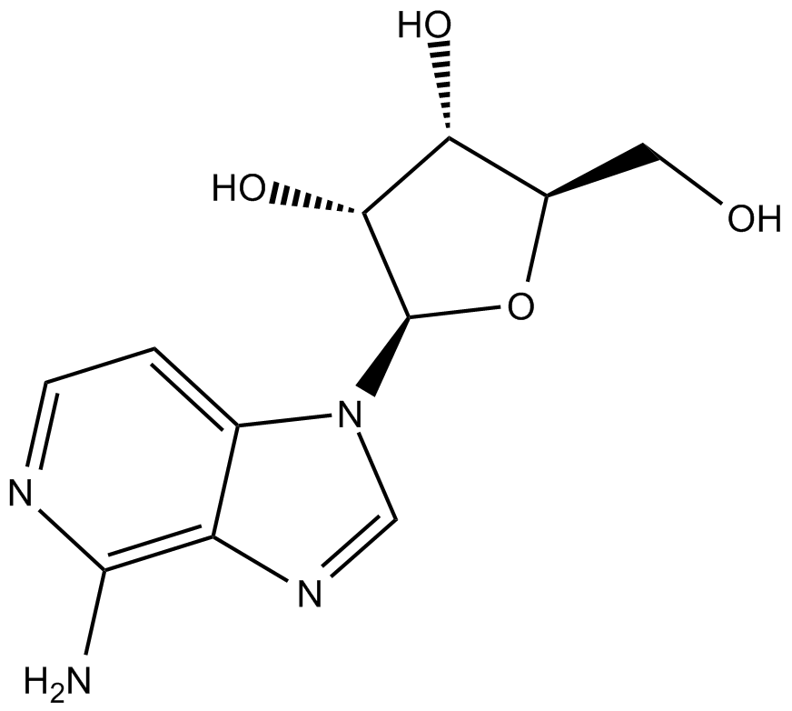
3-Deazaadenosine |
Catalog No.: GC14713 |
3-Deazaadenosine (hydrochloride) is an inhibitor of S-adenosylhomocysteine hydrolase, with a Ki of 3.
Sample solution is provided at 25 µL, 10mM.
Quality Control & SDS
- View current batch:
-
Purity: >98.00%
- COA (Certificate Of Analysis)
- SDS (Safety Data Sheet)
- Datasheet
Kinase experiment [1]: | |
Preparation Method |
Prior to use, the [8-14C] adenosine was checked for purity using isocratic HPLC elution. The assay incubation mixture contained 0.4 IU of enzyme in 50 L. The metabolites were separated by thin-layer chromatography. The radioactivity was quantitated by cutting the plastic backed TLC plates and placing them in scintillation vials, and counting in a Packard 2000 CA scintillation counter |
Applications |
IC50: 0.15 (HIV-1, A012 isolate), 0.20 µM (HIV-1, A018 isolate). 3-Deazaadenosine (hydrochloride) is an inhibitor of S-adenosylhomocysteine hydrolase, with a Ki of 3.9 ?M. |
Cell experiment [2]: | |
Cell lines |
Mouse macrophage RAW 264.7 |
Preparation Method |
RAW 264.7 cells were pretreated with or without 3-Deazaadenosine (100 μMM) for 1 h, and stimulated by the addition of LPS (1 μg/ml). After incubation for 1 h, the cells were washed, and p65 was recovered by immunoprecipitation with anti-p65 and protein A-Sepharose. |
Reaction Conditions |
0-100 μM 3-Deazaadenosine for 1 hour |
Applications |
3-Deazaadenosine (1-100 µM) inhibits LPS-induced expression of TNF-α mRNA, increases DNA binding activity of NF-κB, and causes proteolytic degradation of IκBα, but Not IκBβ in RAW 264.7 cells. 3-Deazaadenosine (100 ?M) enhances nuclear translocation of NF-κB, but blocks LPS-induced NF-κB transcriptional activity, and such inhibition is augmented by the addition of homocysteine. |
Animal experiment [3]: | |
Animal models |
Male, healthy Sprague–Dawley rats (300-350 g) |
Preparation Method |
Animals and balloon injury: rats were fed for 5 days prior and 14 days after the balloon injury with standard chow containing c3Ado at a concentration of 10 mg/kg 3-Deazaadenosine body weight. |
Dosage form |
10 mg/kg 3-Deazaadenosine for 5 days prior and 14 days after the balloon injury |
Applications |
3-deazaadenosine (c3Ado) inhibits atherogenesis in mice. Sprague Dawley rats underwent balloon angioplasty. C3Ado was administered orally, starting 5 days prior to the balloon injury and continued for 2 weeks. Fourteen days after balloon injury the intima/media ratio in the c3Ado-treated group was reduced by 67% and luminal stenosis by 50%. Neointimal cellular density was decreased by 25% and the induction of c-Jun and ki67 was markedly lower. |
References: [1]. Gordon RK, Ginalski K, et,al. Anti-HIV-1 activity of 3-deaza-adenosine analogs. Inhibition of S-adenosylhomocysteine hydrolase and nucleotide congeners. Eur J Biochem. 2003 Sep;270(17):3507-17. doi: 10.1046/j.1432-1033.2003.03726.x. PMID: 12919315. [2]. Sedding DG, Trobs M, et,al.3-Deazaadenosine prevents smooth muscle cell proliferation and neointima formation by interfering with Ras signaling. Circ Res. 2009 May 22;104(10):1192-200. doi: 10.1161/CIRCRESAHA.109.194357. Epub 2009 Apr 16. PMID: 19372464. [3]. Seeger FH, Hess W, et,al.The nucleotide analogue 3-deazaadenosine prevents neointima-formation after balloon injury. Biochem Biophys Res Commun. 2009 Jan 23;378(4):826-31. doi: 10.1016/j.bbrc.2008.11.151. Epub 2008 Dec 12. PMID: 19070587. |
3-Deazaadenosine (hydrochloride) is an inhibitor of S-adenosylhomocysteine hydrolase, with a Ki of 3.9 µM[1].
3-Deazaadenosine dose-dependently prevented the proliferation and migration of human coronary VSMCs in vitro. This was accompanied by an increased expression of the cyclin-dependent kinase inhibitors p21(WAF1/Cip1), p27(Kip1), a decreased expression of G(1)/S phase cyclins, and a lack of retinoblastoma protein hyperphosphorylation. [3]. In the mouse macrophage cell line, RAW264. S-Adenosylhomocysteine accumulated in cells incubated with 3-deazaaristeromycin while S-3-deazaadenosylhomocysteine was the major product in cells incubated with 3-Deazaadenosine and homocysteine thiolactone[4].200 microM 3-Deazaadenosine (c3Ado) prevented this TNF-induced increase in HEC adhesiveness. This effect resulted from interactions of 3-Deazaadenosine with HEC and not with polymorphonuclear neutrophils[5]. 3-Deazaadenosine (DZA), an adenosine analogue, prevented high methionine-induced ICAM-1 and VCAM-1 expression and collagen type-1 synthesis.in vitro 3-Deazaadenosine and CBS gene therapy successfully treated the HHcy-induced inflammatory reaction in the methionine metabolism pathway[6].
3-Deazaadenosine (c3Ado) inhibits atherogenesis in mice. Sprague Dawley rats underwent balloon angioplasty. 3-Deazaadenosine was administered orally, starting 5 days prior to the balloon injury and continued for 2 weeks. Fourteen days after balloon injury the intima/media ratio in the c3Ado-treated group was reduced by 67% and luminal stenosis by 50%. Neointimal cellular density was decreased by 25% and the induction of c-Jun and ki67 was markedly lower. Short-term administration of C3Ado inhibits neointima-formation in rats for at least 3 months after injury[7].
References:
[1]: Gordon RK, Ginalski K, et,al. Anti-HIV-1 activity of 3-deaza-adenosine analogs. Inhibition of S-adenosylhomocysteine hydrolase and nucleotide congeners. Eur J Biochem. 2003 Sep;270(17):3507-17. doi: 10.1046/j.1432-1033.2003.03726.x. PMID: 12919315.
[2]: Jeong SY, Ahn SG, et,al. 3-deazaadenosine, a S-adenosylhomocysteine hydrolase inhibitor, has dual effects on NF-kappaB regulation. Inhibition of NF-kappaB transcriptional activity and promotion of IkappaBalpha degradation. J Biol Chem. 1999 Jul 2;274(27):18981-8. doi: 10.1074/jbc.274.27.18981. PMID: 10383397.
[3]: Sedding DG, TrÖbs M, et,al. 3-Deazaadenosine prevents smooth muscle cell proliferation and neointima formation by interfering with Ras signaling. Circ Res. 2009 May 22;104(10):1192-200. doi: 10.1161/CIRCRESAHA.109.194357. Epub 2009 Apr 16. PMID: 19372464.
[4]: Backlund PS Jr, Carotti D, et,al. Effects of the S-adenosylhomocysteine hydrolase inhibitors 3-deazaadenosine and 3-deazaaristeromycin on RNA methylation and synthesis. Eur J Biochem. 1986 Oct 15;160(2):245-51. doi: 10.1111/j.1432-1033.1986.tb09963.x. PMID: 3769925.
[5]: Jurgensen CH, Huber BE, et,al. 3-deazaadenosine inhibits leukocyte adhesion and ICAM-1 biosynthesis in tumor necrosis factor-stimulated human endothelial cells. J Immunol. 1990 Jan 15;144(2):653-61. PMID: 1967270.
[6]: Sen U, Tyagi N, et,al. Cystathionine-beta-synthase gene transfer and 3-deazaadenosine ameliorate inflammatory response in endothelial cells. Am J Physiol Cell Physiol. 2007 Dec;293(6):C1779-87. doi: 10.1152/ajpcell.00207.2007. Epub 2007 Sep 13. PMID: 17855772.
[7]: Seeger FH, Hess W, et,al. The nucleotide analogue 3-deazaadenosine prevents neointima-formation after balloon injury. Biochem Biophys Res Commun. 2009 Jan 23;378(4):826-31. doi: 10.1016/j.bbrc.2008.11.151. Epub 2008 Dec 12. PMID: 19070587.
Cas No. | 6736-58-9 | SDF | |
Chemical Name | (2R,3R,4S,5R)-2-(4-amino-1H-imidazo[4,5-c]pyridin-1-yl)-5-(hydroxymethyl)tetrahydrofuran-3,4-diol | ||
Canonical SMILES | NC1=C2C(N(C=N2)[C@H]3[C@@H]([C@@H]([C@H](O3)CO)O)O)=CC=N1 | ||
Formula | C11H14N4O4 | M.Wt | 266.25 |
Solubility | ≥ 26.6mg/mL in DMSO | Storage | Store at -20°C |
General tips | For obtaining a higher solubility , please warm the tube at 37 ℃ and shake it in the ultrasonic bath for a while.Stock solution can be stored below -20℃ for several months. | ||
Shipping Condition | Evaluation sample solution : ship with blue ice All other available size: ship with RT , or blue ice upon request |
Step 1: Enter information below (Recommended: An additional animal making an allowance for loss during the experiment)
Step 2: Enter the in vivo formulation (This is only the calculator, not formulation. Please contact us first if there is no in vivo formulation at the solubility Section.)
Calculation results:
Working concentration: mg/ml;
Method for preparing DMSO master liquid: mg drug pre-dissolved in μL DMSO ( Master liquid concentration mg/mL, Please contact us first if the concentration exceeds the DMSO solubility of the batch of drug. )
Method for preparing in vivo formulation: Take μL DMSO master liquid, next addμL PEG300, mix and clarify, next addμL Tween 80, mix and clarify, next add μL ddH2O, mix and clarify.
Method for preparing in vivo formulation: Take μL DMSO master liquid, next add μL Corn oil, mix and clarify.
Note: 1. Please make sure the liquid is clear before adding the next solvent.
2. Be sure to add the solvent(s) in order. You must ensure that the solution obtained, in the previous addition, is a clear solution before proceeding to add the next solvent. Physical methods such as vortex, ultrasound or hot water bath can be used to aid dissolving.
3. All of the above co-solvents are available for purchase on the GlpBio website.
3-deazaadenosine (3DA) alleviates senescence to promote cellular fitness and cell therapy efficiency in mice
Cellular senescence is a stable type of cell cycle arrest triggered by different stresses. As such, senescence drives age-related diseases and curbs cellular replicative potential. Here, we show that 3-deazaadenosine (3DA), an S-adenosyl homocysteinase (AHCY) inhibitor, alleviates replicative and oncogene-induced senescence. 3DA-treated senescent cells showed reduced global Histone H3 Lysine 36 trimethylation (H3K36me3), an epigenetic modification that marks the bodies of actively transcribed genes. By integrating transcriptome and epigenome data, we demonstrate that 3DA treatment affects key factors of the senescence transcriptional program. Remarkably, 3DA treatment alleviated senescence and increased the proliferative and regenerative potential of muscle stem cells from very old mice in vitro and in vivo. Moreover, ex vivo 3DA treatment was sufficient to enhance the engraftment of human umbilical cord blood (UCB) cells in immunocompromised mice. Together, our results identify 3DA as a promising drug enhancing the efficiency of cellular therapies by restraining senescence.
Convenient Synthesis of 3-Deazapurine Nucleosides (3-Deazainosine, 3-Deazaadenosine and 3-Deazaguanosine) Using Inosine as a Starting Material
A convenient synthetic method for preparing 3-deazapurine nucleosides (3-deazainosine, 3-deazaadenosine, and 3-deazaguanosine) from inosine via a 5-ethynyl-1-β-D-ribofuranosylimidazole-4-carboxamide (EICAR) derivative, which is a key intermediate, is described. A large-scale synthesis of an EICAR derivative starting from inosine was achieved in six steps via dinitrophenylation at the N1 position followed by ring opening, iodination of the resulting 5-amino group, and a palladium-catalyzed cross-coupling reaction. The resulting EICAR derivative was then converted into 3-deazainosine, 3-deazaadenosine, and 3-deazaguanosine. This route enabled us to synthesize 3-deazapurine nucleosides conveniently in good yields. ? 2021 Wiley Periodicals LLC. Basic Protocol 1: Preparation of 5-ethynyl-1-β-D-ribofuranosylimidazole-4-carboxamide (EICAR) derivative 6 Basic Protocol 2: Preparation of 3-deazapurine nucleosides 8, 11, and 14.
3-Deazaadenosine, an S-adenosylhomocysteine hydrolase inhibitor, attenuates lipopolysaccharide-induced inflammatory responses via inhibition of AP-1 and NF-κB signaling
3-Deazadenosine (3-DA) is a general methylation inhibitor that depletes S-adenosylmethionine, a methyl donor, by blocking S-adenosylhomocysteine hydrolase (SAHH). In this study, we investigated the inhibitory activity and molecular mechanisms of 3-DA in inflammatory responses. 3-DA suppressed the secretion of inflammatory mediators such as nitric oxide (NO) and prostaglandin E2 (PGE2) in lipopolysaccharide-treated RAW264.7 cells and phorbol 12-myristate 13-acetate (PMA)-differentiated U937 cells. It also reduced mRNA expression of inducible nitric oxide synthase, cyclooxygenase-2, tumor necrosis factor-α, interleukin-1β (IL-1 β), and IL-6, indicating that 3-DA has anti-inflammatory properties in murine and human macrophages. Moreover, 3-DA strongly blocked AP-1 and NF-κB luciferase activity under PMA-, MyD88-, and TRIF-stimulated conditions and decreased the translocation of c-Jun, c-Fos, p65, and p50 into the nucleus. In addition, the p-ERK level in AP-1 signaling and the p-IκBα level in NF-kB signaling were diminished by 3-DA treatment. Interestingly, 3-DA did not alter the phosphorylation of MEK1/2, an ERK modulator, or IKKα/β, an IκBα regulator. Instead, 3-DA prevented MEK1/2 and IKKα/β from combining with ERK and IκBα, respectively, and directly suppressed MEK1/2 and IKKα/β kinase activity. These results indicate that MEK1/2 and IKKα/β are direct targets of 3-DA. In addition, suppression of SAHH by siRNA or treatment with adenosine dialdehyde, another SAHH inhibitor, showed inhibitory patterns against p-ERK and IκBα similar to those of 3-DA. Taken together, this study demonstrates that 3-DA inhibits AP-1 and NF-κB signaling by directly blocking MEK1/2 and IKKα/β or indirectly mediating SAHH, resulting in anti-inflammatory activity.
Groove modification of siRNA duplexes to elucidate siRNA-protein interactions using 7-bromo-7-deazaadenosine and 3-bromo-3-deazaadenosine as chemical probes
Elucidation of dynamic interactions between RNA and proteins is essential for understanding the biological processes regulated by RNA, such as RNA interference (RNAi). In this study, the logical chemical probes, comprising 7-bromo-7-deazaadenosine (Br7C7A) and 3-bromo-3-deazaadenosine (Br3C3A), to investigate small interfering RNA (siRNA)-RNAi related protein interactions, were developed. The bromo substituents of Br7C7A and Br3C3A are expected to be located in the major and the minor grooves, respectively, and to act as a steric hindrance in each groove when these chemical probes are incorporated into siRNAs. A comprehensive investigation using siRNAs containing these chemical probes revealed that (i) Br3C3A(s) at the 5'-end of the passenger strand enhanced their RNAi activity, and (ii) the direction of RISC assembly is determined by the interaction between Argonaute2, which is the main component of RISC, and siRNA in the minor groove near the 5'-end of the passenger strand. Utilization of these chemical probes enables the investigation of the dynamic interactions between RNA and proteins.
3-Deazaadenosine-induced disorganization of macrophage microfilaments
3-Deazaadenosine (c3Ado) has been reported to inhibit a number of cellular functions. These biological effects of c3Ado have generally been attributed to its ability to act as inhibitor and substrate of S-adenosylhomocysteine hydrolase. In this report, it is revealed by fluorescence microscopy that c3Ado caused disorganization of the microfilament system of mouse macrophages at concentrations (greater than or equal to 5 microM) similar to those that inhibited antibody-dependent phagocytosis and zymosan-stimulated H2O2 production by these cells. Inhibition of phagocytosis and perturbation of microfilaments by c3Ado were completely abrogated by washing the macrophages free of this agent and allowing the cells a 30-min recovery period. Furthermore, these effects of c3Ado on phagocytosis and microfilaments appeared to be independent of the increase in S-adenosylhomocysteine and S-3-deazaadenosylhomocysteine that occurred in these macrophages. First, periodate-oxidized adenosine and 3-deaza(+/-)aristeromycin, two other inhibitors of S-adenosylhomocysteine hydrolase that caused greater increases in macrophage S-adenosylhomocysteine than did c3Ado, had no effect on either phagocytosis or microfilaments. Second, pretreatment of macrophages with periodate-oxidized adenosine (to inhibit S-adenosylhomocysteine hydrolase) prevented the subsequent metabolism of c3Ado to S-3-deazaadenosylhomocysteine but did not diminish the effects of c3Ado on phagocytosis or microfilaments. These results demonstrate that c3Ado can perturb the microfilament system of cells and provide an alternative mechanism for the biological effects of c3Ado.
Average Rating: 5
(Based on Reviews and 2 reference(s) in Google Scholar.)GLPBIO products are for RESEARCH USE ONLY. Please make sure your review or question is research based.
Required fields are marked with *