Angiotensin 1/2 (1-6) (Synonyms: H2N-Asp-Arg-Val-Tyr-Ile-His-OH ) |
Catalog No.GP10067 |
Products are for research use only. Not for human use. We do not sell to patients.
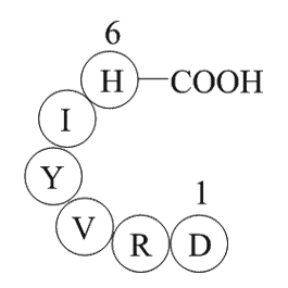
Cas No.:47896-63-9
Sample solution is provided at 25 µL, 10mM.
Quality Control & SDS
- View current batch:
- Purity: >98.00%
- COA (Certificate Of Analysis)
- SDS (Safety Data Sheet)
- Datasheet
Cas No. | 47896-63-9 | SDF | |
Synonyms | H2N-Asp-Arg-Val-Tyr-Ile-His-OH | ||
Chemical Name | 2-amino-4,6-dimethyl-3-oxo-1-N,9-N-bis[(3R,6S,7S,10S,16S)-7,11,14-trimethyl-2,5,9,12,15-pentaoxo-3,10-di(propan-2-yl)-8-oxa-1,4,11,14-tetrazabicyclo[14.3.0]nonadecan-6-yl]phenoxazine-1,9-dicarboxamide | ||
Canonical SMILES | CC1C(C(=O)NC(C(=O)N2CCCC2C(=O)N(CC(=O)N(C(C(=O)O1)C(C)C)C)C)C(C)C)NC(=O)C3=C4C(=C(C=C3)C)OC5=C(C(=O)C(=C(C5=N4)C(=O)NC6C(OC(=O)C(N(C(=O)CN(C(=O)C7CCCN7C(=O)C(NC6=O)C(C)C)C)C)C(C)C)C)N)C | ||
Formula | C36H55N11O10 | M.Wt | 801.89 |
Solubility | ≥80.2mg/mL in DMSO | Storage | Store at -20°C |
General tips | Please select the appropriate solvent to prepare the stock solution according to the solubility of the product in different solvents; once the solution is prepared, please store it in separate packages to avoid product failure caused by repeated freezing and thawing.Storage method and period of the stock solution: When stored at -80°C, please use it within 6 months; when stored at -20°C, please use it within 1 month. To increase solubility, heat the tube to 37°C and then oscillate in an ultrasonic bath for some time. |
||
Shipping Condition | Evaluation sample solution: shipped with blue ice. All other sizes available: with RT, or with Blue Ice upon request. |
Step 1: Enter information below (Recommended: An additional animal making an allowance for loss during the experiment)
Step 2: Enter the in vivo formulation (This is only the calculator, not formulation. Please contact us first if there is no in vivo formulation at the solubility Section.)
Calculation results:
Working concentration: mg/ml;
Method for preparing DMSO master liquid: mg drug pre-dissolved in μL DMSO ( Master liquid concentration mg/mL, Please contact us first if the concentration exceeds the DMSO solubility of the batch of drug. )
Method for preparing in vivo formulation: Take μL DMSO master liquid, next addμL PEG300, mix and clarify, next addμL Tween 80, mix and clarify, next add μL ddH2O, mix and clarify.
Method for preparing in vivo formulation: Take μL DMSO master liquid, next add μL Corn oil, mix and clarify.
Note: 1. Please make sure the liquid is clear before adding the next solvent.
2. Be sure to add the solvent(s) in order. You must ensure that the solution obtained, in the previous addition, is a clear solution before proceeding to add the next solvent. Physical methods such as vortex, ultrasound or hot water bath can be used to aid dissolving.
3. All of the above co-solvents are available for purchase on the GlpBio website.
A Comparative Effectiveness Meta-Analysis of Drugs for the Prophylaxis of Migraine Headache
Objective: To compare the effectiveness and side effects of migraine prophylactic medications. Design: We performed a network meta-analysis. Data were extracted independently in duplicate and quality was assessed using both the JADAD and Cochrane Risk of Bias instruments. Data were pooled and network meta-analysis performed using random effects models. Data sources: PUBMED, EMBASE, Cochrane Trial Registry, bibliography of retrieved articles through 18 May 2014. Eligibility criteria for selecting studies: We included randomized controlled trials of adults with migraine headaches of at least 4 weeks in duration. Results: Placebo controlled trials included alpha blockers (n = 9), angiotensin converting enzyme inhibitors (n = 3), angiotensin receptor blockers (n = 3), anticonvulsants (n = 32), beta-blockers (n = 39), calcium channel blockers (n = 12), flunarizine (n = 7), serotonin reuptake inhibitors (n = 6), serotonin norepinephrine reuptake inhibitors (n = 1) serotonin agonists (n = 9) and tricyclic antidepressants (n = 11). In addition there were 53 trials comparing different drugs. Drugs with at least 3 trials that were more effective than placebo for episodic migraines included amitriptyline (SMD: -1.2, 95% CI: -1.7 to -0.82), -flunarizine (-1.1 headaches/month (ha/month), 95% CI: -1.6 to -0.67), fluoxetine (SMD: -0.57, 95% CI: -0.97 to -0.17), metoprolol (-0.94 ha/month, 95% CI: -1.4 to -0.46), pizotifen (-0.43 ha/month, 95% CI: -0.6 to -0.21), propranolol (-1.3 ha/month, 95% CI: -2.0 to -0.62), topiramate (-1.1 ha/month, 95% CI: -1.9 to -0.73) and valproate (-1.5 ha/month, 95% CI: -2.1 to -0.8). Several effective drugs with less than 3 trials included: 3 ace inhibitors (enalapril, lisinopril, captopril), two angiotensin receptor blockers (candesartan, telmisartan), two anticonvulsants (lamotrigine, levetiracetam), and several beta-blockers (atenolol, bisoprolol, timolol). Network meta-analysis found amitriptyline to be better than several other medications including candesartan, fluoxetine, propranolol, topiramate and valproate and no different than atenolol, flunarizine, clomipramine or metoprolol. Conclusion: Several drugs good evidence supporting efficacy. There is weak evidence supporting amitriptyline's superiority over some drugs. Selection of prophylactic medication should be tailored according to patient preferences, characteristics and side effect profiles.
Adeno-Associated Virus Overexpression of Angiotensin-Converting Enzyme-2 Reverses Diabetic Retinopathy in Type 1 Diabetes in Mice
Angiotensin-converting enzyme (ACE)-2 is the primary enzyme of the vasoprotective axis of the renin angiotensin system that regulates the classic renin angiotensin system axis. We aimed to determine whether local retinal overexpression of adenoassociated virus (AAV)-ACE2 prevents or reverses diabetic retinopathy. Green fluorescent protein (GFP)-chimeric mice were generated to distinguish resident (retinal) from infiltrating bone marrow-derived inflammatory cells and were made diabetic using streptozotocin injections. Retinal digestion using trypsin was performed and acellular capillaries enumerated. Capillary occlusion by GFP(+) cells was used to measure leukostasis. Overexpression of ACE2 prevented (prevention cohort: untreated diabetic, 11.3 ± 1.4; ACE2 diabetic, 6.4 ± 0.9 per mm(2)) and partially reversed (reversal cohort: untreated diabetic, 15.7 ± 1.9; ACE2 diabetic, 6.5 ± 1.2 per mm(2)) the diabetes-associated increase of acellular capillaries and the increase of infiltrating inflammatory cells into the retina (F4/80(+)) (prevention cohort: untreated diabetic, 24.2 ± 6.7; ACE2 diabetic, 2.5 ± 1.6 per mm(2); reversal cohort: untreated diabetic, 56.8 ± 5.2; ACE2 diabetic, 5.6 ± 2.3 per mm(2)). In both study cohorts, intracapillary bone marrow-derived cells, indicative of leukostasis, were only observed in diabetic animals receiving control AAV injections. These results indicate that diabetic retinopathy, and possibly other diabetic microvascular complications, can be prevented and reversed by locally restoring the balance between the classic and vasoprotective renin angiotensin system.
Genetic Polymorphism of Angiotensin II Type 1 Receptors and Their Effect on the Clinical Outcome of Captopril Treatment in Arab Iraqi Patients with Acute Coronary Syndrome (Mid Euphrates)
Genetic variation in the angiotensin II type 1 receptor (AT1R) has an important effect on the outcome of acute coronary syndrome (ACS) initiated treatment with captopril. This study aims to investigate the impact of genetic polymorphism of AT1R (rs5186 and rs275651) on the ACS outcome in Iraqi patients treated with captopril. A total of 250 Iraqi individuals with ACS were included in this case-control study and they were divided into two study groups; Study group 1 included 125 participants who were prescribed captopril, 25 mg twice daily and study group 2 included 125 participants who received no captopril as part of their ACS treatment (control study). The AT1R gene (rs5186) CC genotype was found to be associated with ST-elevation myocardial infarction (STEMI) (Odd's ratio (O.R) = 1.2, P = 0.7), while AC was associated with Non-ST-elevation myocardial infarction (NSTEMI) and unstable angina (UA) (O.R = 1.2, P = 0.8). AC genotype is more prone to have Percutaneous coronary intervention (PCI) after ACS attack (O.R = 1.2, P = 0.6). CC genotype had a risk to get less improvement (O.R = 1.6, P = 0.5), so might require higher doses of captopril during acute coronary insult. The AT1R gene (rs275651) AA genotype was associated with UA (O.R = 1.3, P = 0.9). AA and AT genotypes were more prone to have PCI after ACS attack (O.R = 3.9 P = 0.2, O.R = 3.5, P = 0.3 respectively) and thus requiring higher doses of captopril. We conclude that the AT1R rs5186, rs275651 genetic polymorphisms might partially affect the clinical outcome of ACS patients treated with captopril and might have captopril resistance which requires higher doses.
Angiotensin converting enzyme 2 polymorphisms and postexercise hypotension in hypertensive medicated individuals
The renin-angiotensin aldosterone system (RAAS) is associated with diverse physiological responses and adaptations to exercise. The angiotensin converting enzyme (ACE) 2 has vasodilatory effects, which might be associated with the blood pressure (BP) responses to acute exercise. The aim of this study was to investigate the role of ACE2 polymorphisms in postexercise hypotension (PEH). Thirty-four medicated hypertensive (61·3 ± 1·7 years, 76·1 ± 2·7 kg, 160 ± 1·6 cm) men (n = 12) and women (n = 22), participated in a control and a moderate intensity exercise session in a randomized order. After both experimental sessions, they left the laboratory wearing an ambulatory BP device for 24-h monitoring. ACE2 polymorphisms (Int-1 and Int-3) were assessed by polymerase chain reaction. Over the course of 5-h monitoring, we observed a significant reduction in SBP and DBP following exercise in the AA/AG of the Int-1 polymorphism (p-interaction = 0·02 and 0·001, respectively), whereas this could not be found in the individuals homozygous G (p-interaction = 0·76 and 0·51, respectively). With regard to Int-3 polymorphism, individuals AA/AG showed a significant reduction in SBP following exercise (p-interaction <0·0001) but not for DBP (p-interaction = 0·06) whereas GG individuals showed only a significant reduction in DBP following exercise (p-interaction = 0·02). Our results suggest that ACE2 polymorphism could affect PEH; however, larger trials are needed to confirm our findings.
Angiotensin II and angiotensin-(1-7) effects on free cytosolic sodium, intracellular pH, and the Na(+)-H+ antiporter in vascular smooth muscle
The aim of the present study was to define the effects of angiotensin II (Ang II) and Ang-(1-7) on free cytosolic Na+ (Na+i), intracellular pH (pHi), and the Na(+)-H+ antiporter in cultured vascular smooth muscle cells from rat aorta. Cells were loaded with either BCECF-AM or SBFI-AM for measurement of pHi and Na+i, respectively. Ang II (10(-6) mol/L) caused a rapid rise in Na+i followed by a progressive increase that peaked at about 10 minutes (from 11 +/- 1.5 to 16 +/- 1.5 mmol/L, P < .001), whereas Ang-(1-7) (10(-6) mol/L) did not affect Na+i significantly (from 11.5 +/- 1.1 to 11.8 +/- 0.07 mmol/L). The effect of Ang II on Na+i was concentration dependent (delta Na+i, 5.1 +/- 0.9, 3.8 +/- 0.6, 1.6 +/- 0.6, and 0.14 +/- 0.18 mmol/L with decreasing concentrations of 10(-6), 10(-7), 10(-8), and 10(-9) mol/L, respectively). Ang II caused a brief acidification followed by an increase in pHi (from 7.34 +/- 0.03 to 7.43 +/- 0.03 after 10 minutes, P < .005), and Ang-(1-7) had no significant effect on pHi (from 7.23 +/- 0.03 to 7.23 +/- 0.03). To investigate whether pHi and Na+i changes induced by Ang II were due to cell Na+ entry via stimulation of the Na(+)-H+ antiporter, we pretreated cells with EIPA (25 mumol/L) or ouabain (2.0 mmol/L). Ang II in the presence of ouabain caused a greater increase than that seen with ouabain alone (delta Na+i, 13 +/- 1.5 versus 6.3 +/- 1.2 mmol/L, P < .0025). EIPA by itself decreased Na+i and pHi. After EIPA, Ang II failed to increase both Na+i and pHi, demonstrating that the Na(+)-H+ antiporter is responsible for the rises in Na+i and pHi during stimulation with Ang II. To further characterize the mechanism of Ang II action, we exposed cells to an Ang II type I receptor antagonist (L-158,809, 10(-6) mol/L) or two different type 2 receptor antagonists (PD 123177 and CGP 421112A, 10(-6) mol/L). L-158,809 completely blocked the rise in pHi caused by Ang II, whereas PD 123177 and CGP 421112A did not. We conclude that Ang II increases both Na+i and pHi, and both effects are mediated by stimulation of the Na(+)-H+ antiporter. Ang-(1-7), by contrast, has no significant effect on Na+i, pHi, or the Na(+)-H+ antiporter. Stimulation of this antiporter by Ang II is exerted through the type 1 receptor.
Average Rating: 5
(Based on Reviews and 30 reference(s) in Google Scholar.)GLPBIO products are for RESEARCH USE ONLY. Please make sure your review or question is research based.
Required fields are marked with *