


Ultra-high Sensitivity ECL kit
The ultra-high sensitivity ECL kit is also defined as the electrochemiluminescence kit which is designed to identify a wide variety of antigens. It works on the principle of luminance in which it gives luminance upon the oxidative reaction of luminol in the presence of peroxide and HRP. HRP is called horseradish peroxidase, which in the presence of peroxide, converts luminol into a product that is in oxidized form due to oxidative reaction, giving fluorescence. Then the results of these samples are optimized with the help of any imaging device or any X films. This ultra-high sensitivity ECL kit produces a more uniform and stable color upon a reaction that in return is beneficial to identify low abundant proteins within the sample. Due to its high sensitivity, it can be able to detect the femtogram range of proteins within the samples. The light is produced in the form of photons due to the oxidative reaction of the luminal substrate. These photons are then captured by any imaging device and X film to give desired results. In different studies, scientists also use high ultra sensitivity ECL western bloating (Immunoblotting). Immune blotting is the widely used process to detect the desired proteins with the samples for protein expression. In this process, proteins within the sample are separated with the help of Electrophoresis. In this process, these proteins are separated with the help of sodium dodecyl sulfate on the polyacrylamide gel. Then these separated proteins are transferred to the membrane along with the block’s regions. These blocks are required for the prevention of non-specific protein binding onto the membrane. The primary antibodies are combined with the proteins. After that HRP conjugated secondary antibodies are combined with primary antibodies. These HRP conjugated secondary antibodies proceed with the reaction with the help of substrate to give fluorescence. This is how the Electro chemiluminescence kit works(Mruk and Cheng, 2011).
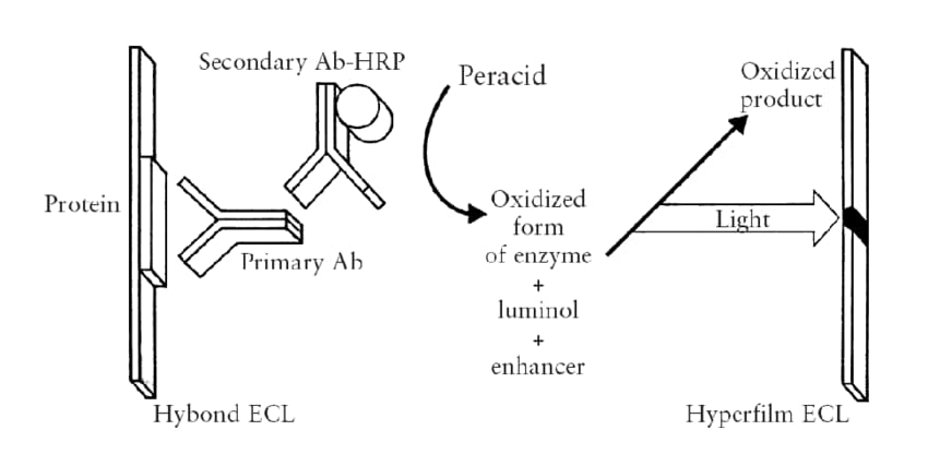
Figure 1:Diagrammatic explanation of working principle of ECL kit western blotting
Similarly, this ECL kit can be beneficial for the electrochemiluminescence of rat insulin. Studies suggest that the rats are of great concern. In diabetes research, scientists use them for in-vivo studies to determine the level of insulin in rats and rodents after treatment or in different stages related to pathophysiology. Various techniques are available for quantification of the level of Insulin in in- vivo studies. These include ELISA and radioimmunoassay etc. But the reliability of these techniques is of major concern in the research. This study suggests that the electrochemiluminescence assay is of the greatest importance for determining the level of insulin in rats. When the level of Insulin is determined via ECL, it is observed that the value of Z′ is about 0.82 ± 0.03, and the (S/B) is 133, which reflects its reliability. So, using the ECL assay for insulin quantification is a robust and reliable way. It is because when the insulin is determined via radioimmunoassay, there is non-specific interference from the matrix of serum includes, including proteases, serum proteins, cations, etc. Similarly, the ELISA also has a limited ability to determine the level of insulin in different pathological conditions of the sample. This requires the ELISA kit of high sensitivity, medium sensitivity, etc. for different conditions. On the other hand, ECL is a robust way for insulin detection, it is because, in this process, the non-specific interference with the serum matrix is diminished with the help of immunoaffinity chromatography. This ECL assay consists of the use of two monoclonal antibodies. These include Ruthenium and biotin. These two antibodies are used in the sandwich assay and are specific for the different epitopes of insulin in rats. Upon reaction, due to the different epitope combining competition, these antibodies bind to the insulin within the sample to make complex. After that, the formed complex combines with the streptavidin-coated magnetic bead. This complex formed due to magnetic strength. After that upon flow cell, the antibodies which are not bound to the bead, are washed out. Then upon cyclic conversion, the ORI-tag label oxidized into the red-ox form, which in return gives electrochemiluminescence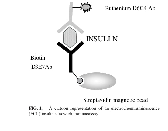
Figure 2:Diagrammatic explanation of electroluminescence immunoassay.
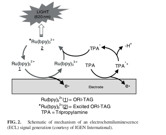
Figure 3:Representation of mechanism for fluorescence in Electrochemiluminescence
We also know that the world is full of Various varieties of plant and animal viruses. But to determine the plant virus is a difficult task to do. So, it is a need for scientists to develop a technique that can be able to develop an accurate method for virus detection. So, researchers combine the reverse transcriptase PCR with electrochemiluminescence to detect the plant RNA virus for the first time. This study suggests that the amplified product of the PCR can be combined with the biotin probe to select the target for identification. This, in turn, reduces the chances of results that are pseudo-positive for the proper detection, a specific nucleotide sequence is attached to the 5’ end of the primer. This will allow the hybridization between the TBR and PCR product. At the same time, the PCR chain attached to 2 Ru-probes, in return intensifies the ECL. The study also explained that via the use of ECL, plant viruses (Sorghum mosaic virus, sugar cane mosaic virus, etc ) can simply be identified from the diseased plant samples more accurately with less cost.
Studies also suggest the use of an ECL assay kit to measure enzyme activity, so an ECL kit can also be used to detect the outcomes of reactions that are enzyme-linked. For example, for the detection of the phosphorylation of amino acids which is induced by tyrosine- kinase, labeled antibodies are used which are specific for the phosphorylation reaction, in the kit which has substrate placed on the magnetic bead or electrodes of carbon. So, the results can be explained based on the activity of the enzyme which is related to the accumulation of the label antibodies from the solid phase. For example, to measure the activity of the protease, protein linkers are used which measure the release of labels (combine to solid phase). In the same way, ligation activity for the HIV integrase enzyme can be measured by measuring the extent of ligation of the oligonucleotide which is labeled to the immobilized oligonucleotide.


Comments