3-Methyladenine (Synonyms: 3-MA) |
Katalog-Nr.GC10710 |
3-Methyladenin ist ein klassischer Autophagie-Inhibitor.
Products are for research use only. Not for human use. We do not sell to patients.
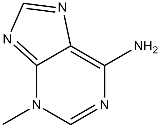
Cas No.: 5142-23-4
Sample solution is provided at 25 µL, 10mM.
- Phytother Res 37.1 (2023):211-230.PMID:36086852
- Acta Pharmacol Sin (2024):1-14.PMID:38992120
- Front Immunol 15 (2024):1521080.
- Stem Cell Transl (2024):szae028.PMID:38736295
- J Ethnopharmacol (2024):118758.PMID:39222762
- Mol Nutr Food Res (2024):2300123.PMID:38196088
- Am J Cancer Res 14.3 (2024):1121.PMID:38590396
- J Funct Foods 126 (2025):106695.
- Langmuir (2024).PMID:39589485
- Neurochem Res 50.1 (2025):81.PMID:39831923
- Exp Eye Res(2024):110021.PMID:39117136
- Front Med-Lausanne 11 (2025):1534120.PMID:39882536
- J Reprod Develop (2023).PMID:37779094
- Redox Biol (2024):103062.
- Bba Mol Basis Dis (2023):166887.
- Inflammation (2024):1-15.PMID:38977539
3 Methyladenine is a classic autophagy inhibitor. It inhibits phosphatidylinositol 3-kinase (PI3K), which is located upstream of the IGF/PI3K/mTOR/ULK pathway.[1] 3-Methyladenine is capable to induce a consistent and abrupt decrease in cell viability across a series of ontologically unrelated human cell lines. In addition, 3 Methyladenine-induced cytotoxicity was not driven by the inhibition of the AKT/mTOR axis.[2]
In vitro study indicated that the inhibition of autophagy by 3 Ma abolished uric acid-induced differentiation of renal fibroblasts to myofibroblasts and activation of transforming growth factor-β1 (TGF-β1), epidermal growth factor receptor (EGFR), and Wnt signaling pathways in cultured renal interstitial fibroblasts. Moreover, 3-Methyladenine was effective in attenuating renal deposition of extracellular matrix (ECM) proteins and expression of α-smooth muscle actin (α-SMA) and reducing renal epithelial cells arrested at the G2/M phase of cell cycle.[3]
In vivo study demonstrated that the administration of 3 Ma inhibited Wnt/β-catenin and Notch/Jagged-1 signaling pathways as well as suppresses EGFR/ERK1/2 signaling pathway. Furthermore, 3-MA treatment remarkably inhibited the infiltration of macrophages and lymphocytes as well as release of multiple profibrogenic cytokines/chemokines in the injured kidney.[3]
References:
[1]. Yang F, et al. Rapamycin and 3-Methyladenine Influence the Apoptosis, Senescence, and Adipogenesis of Human Adipose-Derived Stem Cells by Promoting and Inhibiting Autophagy: An In Vitro and In Vivo Study. Aesthetic Plast Surg. 2021 Jun;45(3):1294-1309.
[2].Chicote J, et al. Cell Death Triggered by the Autophagy Inhibitory Drug 3-Methyladenine in Growing Conditions Proceeds With DNA Damage. Front Pharmacol. 2020 Oct 15;11:580343.
[3].Bao J, et al. Pharmacological inhibition of autophagy by 3-MA attenuates hyperuricemic nephropathy. Clin Sci (Lond). 2018 Nov 2;132(21):2299-2322.
Cell experiment [1]: | |
Cell lines |
Rat renal interstitial fibroblasts (NRK-49F) cells |
Preparation Method |
Rat renal interstitial fibroblasts (NRK-49F) were cultured in DMEM with F12 containing 10% FBS, 1%penicillin and streptomycin in an atmosphere of 5%CO2, and 95% air at 37◦C. Cells were starved for 24h with DMEM containing 0.5% FBS. |
Reaction Conditions |
Cells were exposed to uric acid (800 μM) for 36h in the presence or absence of 3-Methyladenine (0–10 mM). |
Applications |
3-Methyladenine could abolish uric acid-induced α-SMA and collagen I expression. Uric acid also triggered a significant up-regulation of LC3II/I and Beclin-1. 3-Methyladenine dose-dependently suppressed these responses. In addition, treatment with 3-MA decreased TGF-βRI expression levels and the ratio of p-Smad3/Smad3 in a dose-dependent manner. |
Animal experiment [2]: | |
Animal models |
male Sprague-Dawley (SD) rats (2-months old; weight 200±20 g) |
Preparation Method |
Mice were maintained on a 12h light/dark cycle and provided access to food and water ad libitum. All rats were pretrained to swim in the absence of a load for one week. The saline-treatment group was intravenously injected with 500 μL sterile saline by using 261/2 gauge needle via tail vein;3-MA-treatment group was intravenously injected with 500 μL 3-Methyladenine by using a 261/2 gauge needle via the tail vein. |
Dosage form |
15 mg/kg |
Applications |
3-Methyladenine could decrease the distortion of myocardium fibers as well as significantly decrease the width of cardiomyocytes of mice. In addition, 3-Methyladenine treatment obviously reduced autophagosome formation in the myocardium. 3-Methyladenine could also prevent the reduction of Bcl-2/Bax in the left ventricle of OE rats. |
References: [1]. Bao J, et al. Pharmacological inhibition of autophagy by 3-MA attenuates hyperuricemic nephropathy. Clin Sci (Lond). 2018 Nov 2;132(21):2299-2322. [2]. Liu H, et al. Autophagy inhibitor 3-methyladenine alleviates overload-exercise- induced cardiac injury in rats. Acta Pharmacol Sin. 2017 Jul;38(7):990-997. |
Cas No. | 5142-23-4 | SDF | |
Überlieferungen | 3-MA | ||
Chemical Name | 3-methylpurin-6-amine | ||
Canonical SMILES | CN1C=NC(=C2C1=NC=N2)N | ||
Formula | C6H7N5 | M.Wt | 149.15 |
Löslichkeit | ≥ 7.45mg/mL in DMSO, ≥ 5mg/mL in Water | Storage | Store at 2-8°C,Solutions of 3-MA are best fresh-prepared |
General tips | Please select the appropriate solvent to prepare the stock solution according to the
solubility of the product in different solvents; once the solution is prepared, please store it in
separate packages to avoid product failure caused by repeated freezing and thawing.Storage method
and period of the stock solution: When stored at -80°C, please use it within 6 months; when stored
at -20°C, please use it within 1 month. To increase solubility, heat the tube to 37°C and then oscillate in an ultrasonic bath for some time. |
||
Shipping Condition | Evaluation sample solution: shipped with blue ice. All other sizes available: with RT, or with Blue Ice upon request. |
Prepare stock solution | |||
![]() |
1 mg | 5 mg | 10 mg |
1 mM | 6.7047 mL | 33.5233 mL | 67.0466 mL |
5 mM | 1.3409 mL | 6.7047 mL | 13.4093 mL |
10 mM | 0.6705 mL | 3.3523 mL | 6.7047 mL |
Step 1: Enter information below (Recommended: An additional animal making an allowance for loss during the experiment)
Step 2: Enter the in vivo formulation (This is only the calculator, not formulation. Please contact us first if there is no in vivo formulation at the solubility Section.)
Calculation results:
Working concentration: mg/ml;
Method for preparing DMSO master liquid: mg drug pre-dissolved in μL DMSO ( Master liquid concentration mg/mL, Please contact us first if the concentration exceeds the DMSO solubility of the batch of drug. )
Method for preparing in vivo formulation: Take μL DMSO master liquid, next addμL PEG300, mix and clarify, next addμL Tween 80, mix and clarify, next add μL ddH2O, mix and clarify.
Method for preparing in vivo formulation: Take μL DMSO master liquid, next add μL Corn oil, mix and clarify.
Note: 1. Please make sure the liquid is clear before adding the next solvent.
2. Be sure to add the solvent(s) in order. You must ensure that the solution obtained, in the previous addition, is a clear solution before proceeding to add the next solvent. Physical methods such as vortex, ultrasound or hot water bath can be used to aid dissolving.
3. All of the above co-solvents are available for purchase on the GlpBio website.
Quality Control & SDS
- View current batch:
- Purity: >96.00%
- COA (Certificate Of Analysis)
- SDS (Safety Data Sheet)
- Datasheet
-
Related Biological Data
Autophagy inhibition augments the SAHA-mediated enhancement of NK cell cytotoxicity to BCSCs. D) The autophagy level of ALDH+ cells modulated by SAHA or 3MA, Scale bar = 2 μm.
NK cell infusion with intratumor injection of hydrogel encapsulating SAHA and 3MA (GlpBio, USA), or NK cell infusion with intravenous injection of SAHA (15mg kg−1) and 3MA (10mg kg−1).
Advanced Science 9.23 (2022): 2201271. PMID: 35712750 IF: 15.1004 -
Related Biological Data
Autophagosome accumulation is critical for efficient RABV VLP budding ACCEPTED MANUSCRIPT in cells and viral virulence in mice. (C) N2a cells were infected with RABV at an MOI of 0.01 for indicated time points, and treated with 3-MA (1mM) for 24h. Cell culture supernatants were collected for viral titration.
RABV-infected (MOI = 0.01, 36h) N2a cells were mock-treated or treated with 3-MA (1mM) (GlpBio, USA) for 24h.
Autophagy just-accepted (2024). PMID: 38566321 IF: 13.3003 -
Related Biological Data
VDR activated mitophagy was beneficial for ameliorating fibrosis and mitochondrial function. L. MitoSOX Red and JC-1 staining (magnification ×630, scale bar: 5μm).
HK-2 cells were randomly divided into several groups: control group: D-glucose 5mM, high glucose (30mM) and TGF-β(50ng/ml) intervention group, high glucose and TGF-β with or without other interventions: paricalcitol (200nM), 3-MA (10mM)(GlpBio, USA) for indicated time (72h) based on previous studies.
Redox Biology (2024): 103062. PMID: 38320454 IF: 11.3997 -
Related Biological Data
Autophagy inhibitors exacerbated TGF-β1-induced fibrosis in primary ConFB. (C) Invasive ability of primary ConFB stimulated with 1ng/ml TGF-β1 for 48h, with cotreatment of 2mM 3-MA or 2μM CQ for 18h, determined by Transwell assay (left).
Cells were treated with 1ng/ml TGF-β1 (100-21) for 48h, with or without co-treatment of 2μM rapamycin, serum starvation (0.5% FBS), 2mM 3-MA (GC10710, GLPBIO), or 2μM CQ (GC19549, GLPBIO) for 18h.
J Mol Cell Biol(2023): mjad009. PMID: 36792067 IF: 8.185 -
Related Biological Data
Melatonin enhances autophagy in HSFs. (F) Western blot indicated that treatment with 3-MA blocked the melatonin-enhanced autophagy in HSFs.
HSFs were treated with vehicle (DMSO) or 200µM melatonin in the presence or absence of 5mM 3-MA(GlpBio, USA) for 24h.
Bba-Mol Basis Dis(2023): 166887. PMID: 37739092 IF: 6.2001 -
Related Biological Data
Inhibition of autophagy increased upregulated S100P-enhanced chemosensitivity. D. Inhibition of autophagy reversed upregulated S100P-enhanced cells death. HL-60 and Jurkat cells were transfected with S100P shRNA or control shRNA and then pre-treated with 3-MA. Cells were subsequently treated for 24h with ADM, then active caspase-3 levels were assayed by western blot.
HL-60 and Jurkat cells were transfected with S100P vector or empty vector and then pre-treated with 3-MA (10mM)(GlpBio, USA) for 1h.
Am J Cancer Res 14.3 (2024): 1121. PMID: 38590396 IF: 5.2999
Average Rating: 5
(Based on Reviews and 17 reference(s) in Google Scholar.)GLPBIO products are for RESEARCH USE ONLY. Please make sure your review or question is research based.
Required fields are marked with *