


D-luciferin
D-Luciferin is basically a substrate for many metabolic enzymes. It is used in different techniques like assays in which it is used to measure luciferase enzymes in the presence of adenosine triphosphate (ATP). Almost all the bioluminescence insects have specific types of luciferase enzymes but all they share a common substrate called D luciferin which is present in almost 40 different species of beetles or firefly insects(White et al., 1963, De Wet et al., 1985). It has a wide variety of applications, but more commonly it is a bioluminescent agent which is used in different in-vivo and in-vitro research related to stem cells, gene expression, growth of cancer, etc.
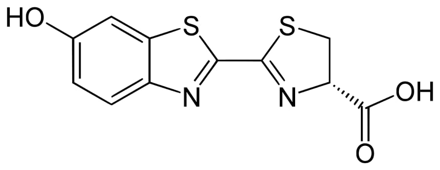
Figure 1: D luciferin
It is reported as a bioluminescent agent for in-vivo imaging for luciferase expression. As we have already discussed that it acts as a substrate for luciferase enzyme, so in the presence of Mg2+ (which acts as a co-factor) and oxygen, it utilizes ATP and emits yellow-green light, which in-vivo imaging, shifts to red color at 37°C. So, this reaction can be helpful to indicate the availability of ATP (energy) and indirectly is an indication of life because here it acts as a stain for life and death(McCapra, 1994).
D-luciferin is a 4,5-Dihydro-2-(6-hydroxy-2-benzothiazole)-4-thiazole carboxylic acid which is a common reagent used in different biotechnology techniques more commonly in in-vivo imaging. This D-Luciferin is labeled with either stem cells, infectious diseases, or cancerous cells. After that, they are inoculated in experimental animals like rats or mice for investigation. This allows us to investigate the pattern and progression of the disease or the efficacy of different drugs via non-invasive or real-time techniques in the experimental animals through bioluminescent imaging. Along with the in-vivo study, it is also used in in-vitro studies, for example, in different assays like ATP assays, a gene reporter assay, contamination assays, and luciferase assays.
As we discussed above that D-Luciferin is used in many assays, let us take a relevant example in which researchers work on the luciferase assays to determine the infectivity of SARS coronavirus within the host cells. During the interaction between the SARS Coronavirus and the human cell, a key step for the entry of the virus into the human host cell is the attachment of the spikes of the virus onto the Angiotensin-Converting Enzyme 2. During an assay, a plasmid is labeled with luciferase and the coronavirus, both transfected into the human host cell and stored for 72 hours in an incubator (incubation). After 72 hours of incubation, pseudoviruses are formed, which contain the spikes as well as the genes for the luciferase enzyme. After the collection of these pseudoviruses, they are incubated with human cells which have ACE 2 receptors for 60 hours. If the successful infection of the human cell is occurred due to the pseudoviruses which have spikes similar to coronavirus and luciferase coding along with plasmid, there will be the production of viruses due to replication of these pseudoviruses and Luciferase plasmid within the cells. After that, when D-Luciferin is applied to these viruses, they will emit light which would be an indication of infection. So, D-Luciferin could be used in such of these assays to measure the infectivity, and progression of the disease. Also, with the help of D-Luciferin, we can measure the drug efficacy as well(Shang et al., 2020).
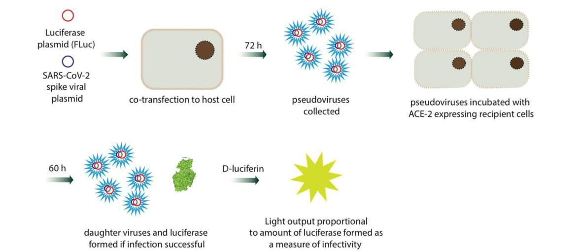
Figure 2:A Fluc/D-luciferin based assay used to determine the infectivity of coronaviruses in different host cells – image shows process used for
D-Luciferin plays a major role in bioluminescence imaging (BLI). With the help of these biotechnology techniques, we can measure the progression of various types of bacterial infections including klebsiella pneumonia and antibiotic resistance for different bacteria. Along with these bacterial infection monitoring, with in-vivo imaging with D-Luciferin, we can also monitor parasite infections along with their progression like infection caused by parasites Trypanosoma cruzi, Trypanosoma gondiii & Leishmania, etc.
D-Luciferin mainly consists of three forms which are given as follows:
- D-Luciferin in Free Acid form.
- D-Luciferin in Sodium salt form.
- D-Luciferin in Potassium salt form.
D-Luciferin in Free Acid form
It has better stability and is available in the form of dried powder in freeze Conditions. It is not soluble in water but can be soluble in K/Na bicarbonates and then dilute with buffers like PBS to get desired concentration. If the 10mg/ml of D-Luciferin is added to a base of equi molar concentration, then we have to shake it for 30 min to completely dissolve it. On the other hand, while opening the vial, it is necessary to let out the carbonate, because this step makes the process faster. The reason behind this is that when a high amount of carbonate is present in the vial, it makes it more acidic.
D-Luciferin in Sodium salt form
D-Luciferin in the form of Na-salt has greater solubility as compared to the D-Luciferin in K-salt form. Due to its greater solubility, it can be dissolved in water as well as in any buffer solution like PBS. It usually takes a few swirls while dissolving.
D-Luciferin in Potassium salt form
D-Luciferin in the form of K- salt has great stability as compared to the D-Luciferin in Na- salt form. It is also soluble in water as well as any buffer solution (PBS). Dissolution also requires very few swirls. For experimental purposes, Na and K salts of D-Luciferin are easier and more convenient to use in imaging (preferably in-vivo) because of their greater solubility in H2O and buffer.
Solubility
D-Luciferin in the form of free acid can be dissolved at a rate of 10mg/ml in the solution of the base of equi molar concentration like TRIS, and bicarbonate of Na and K. When it is dissolved, it could take almost 30 min to completely solubilize in base solution. The one factor which can increase the solubility of D-Luciferin is while opening the vial, there must be the removal of carbonates that are performed. If it retains, it may decrease the solubility by a decrease in the pH of the solution (making the solution more acidic, which interferes with solubility). If it is available in a higher concentration, it requires washing with Argon gas.
Stability
In general, D-Luciferin is sensitive toward oxygen and light before and after its dissolution in solution. During the continuous opening and closing of the lid of the vial, it is recommended that you have to fill the vial with any inert gas. The reason behind using inert gas as compared to air is the lesser density of inert gas. Argon gas is generally recommended for filling before closing the container.
Storage conditions
In the form of solid material, D-Luciferin can be stored at -20°C, while in dissolving form, it must be stored at -80°C.


 Chemical Structure")
 Chemical Structure")


Kommentare