3-Methyladenine (Synonyms: 3-MA) |
Catalog No.GC10710 |
3-Methyladenine est un inhibiteur classique de l'autophagie.
Products are for research use only. Not for human use. We do not sell to patients.
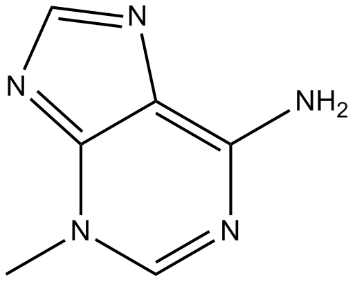
Cas No.: 5142-23-4
Sample solution is provided at 25 µL, 10mM.
- Phytother Res 37.1 (2023):211-230.PMID:36086852
- Acta Pharmacol Sin (2024):1-14.PMID:38992120
- Front Immunol 15 (2024):1521080.
- Stem Cell Transl (2024):szae028.PMID:38736295
- J Ethnopharmacol (2024):118758.PMID:39222762
- Mol Nutr Food Res (2024):2300123.PMID:38196088
- Am J Cancer Res 14.3 (2024):1121.PMID:38590396
- J Funct Foods 126 (2025):106695.
- Langmuir (2024).PMID:39589485
- Neurochem Res 50.1 (2025):81.PMID:39831923
- Exp Eye Res(2024):110021.PMID:39117136
- Front Med-Lausanne 11 (2025):1534120.PMID:39882536
- J Reprod Develop (2023).PMID:37779094
- Redox Biol (2024):103062.
- Bba Mol Basis Dis (2023):166887.
- Inflammation (2024):1-15.PMID:38977539
3-Methyladenine est un inhibiteur classique de l'autophagie. Il inhibe la phosphatidylinositol 3-kinase (PI3K), qui se trouve en amont de la voie IGF/PI3K/mTOR/ULK. 3-Méthyladénine peut induire une diminution cohérente et brutale de la viabilité cellulaire dans une série de lignées cellulaires humaines ontologiquement non liées. De plus, la cytotoxicité induite par 3-Méthyladénine n'était pas liée à l'inhibition de l'axe AKT/mTOR.
Une étude in vitro a indiqué que l'inhibition de l'autophagie par 3-Méthyladénine abolissait la différenciation induite par l'acide urique des fibroblastes rénaux en myofibroblastes et l'activation des voies de signalisation TGF-β1, EGFR et Wnt dans des fibroblastes interstitiels rénaux cultivés. De plus, 3-Méthyladénine était efficace pour atténuer la déposition de protéines de matrice extracellulaire (ECM) rénale et l'expression de l'α-actine musculaire lisse (α-SMA), tout en réduisant les cellules épithéliales rénales arrêtées au stade G2/M du cycle cellulaire.
Une étude in vivo a démontré que l'administration de 3-Méthyladénine inhibait les voies de signalisation Wnt/β-caténine et Notch/Jagged-1, ainsi que supprime la voie de signalisation EGFR/ERK1/2. De plus, le traitement par 3-MA inhibait de manière remarquable l'infiltration des macrophages et des lymphocytes, ainsi que la libération de multiples cytokines/chemokines profibrogéniques dans le rein blessé.
References:
[1]. Yang F, et al. Rapamycin and 3-Methyladenine Influence the Apoptosis, Senescence, and Adipogenesis of Human Adipose-Derived Stem Cells by Promoting and Inhibiting Autophagy: An In Vitro and In Vivo Study. Aesthetic Plast Surg. 2021 Jun;45(3):1294-1309.
[2].Chicote J, et al. Cell Death Triggered by the Autophagy Inhibitory Drug 3-Methyladenine in Growing Conditions Proceeds With DNA Damage. Front Pharmacol. 2020 Oct 15;11:580343.
[3].Bao J, et al. Pharmacological inhibition of autophagy by 3-MA attenuates hyperuricemic nephropathy. Clin Sci (Lond). 2018 Nov 2;132(21):2299-2322.
Expériences cellulaires [1]: | |
Lignées cellulaires | Fibroblastes interstitiels rénaux de rat (NRK-49F) |
Méthode de préparation | Les fibroblastes interstitiels rénaux de rat (NRK-49F) ont été cultivés dans du DMEM avec F12 contenant 10 % de FBS, 1 % de pénicilline et de streptomycine, dans une atmosphère de 5 % de CO2 et 95 % d'air à 37 °C. Les cellules ont été privées de nutriments pendant 24 heures avec du DMEM contenant 0,5 % de FBS. |
Conditions de réaction | Les cellules ont été exposées à l'acide urique (800 μM) pendant 36 heures en présence ou en absence de 3-Méthyladénine (0–10 mM). |
Domaines d'application | La 3-Méthyladénine pouvait abolir l'expression de l'α-SMA et du collagène I induite par l'acide urique. L'acide urique a également déclenché une régulation significative à la hausse de LC3II/I et de Beclin-1. La 3-Méthyladénine a supprimé ces réponses de manière dose-dépendante. De plus, le traitement par 3-MA a diminué les niveaux d'expression de TGF-βRI et le ratio de p-Smad3/Smad3 de manière dose-dépendante. |
Expériences animales [2]: | |
Modèles animaux | Rats mâles Sprague-Dawley (SD) (2 mois; poids 200 ± 20 g) |
Méthode de préparation | Les rats ont été maintenus dans un cycle lumineux/nocturne de 12 heures et avaient accès à de la nourriture et de l'eau ad libitum. Tous les rats ont été préentraînés à nager sans charge pendant une semaine. Le groupe traité avec du sérum physiologique a reçu une injection intraveineuse de 500 μL de sérum physiologique stérile à l'aide d'une aiguille de calibre 26 1/2 par la veine caudale; le groupe traité avec 3-MA a reçu une injection intraveineuse de 500 μL de 3-Méthyladénine à l'aide d'une aiguille de calibre 26 1/2 par la veine caudale. |
Forme de dosage | 15 mg/kg |
Domaines d'application | La 3-Méthyladénine pouvait diminuer la distorsion des fibres myocardiques ainsi que réduire significativement la largeur des cardiomyocytes des souris. De plus, le traitement par 3-Méthyladénine a réduit de manière évidente la formation d'autophagosomes dans le myocarde. La 3-Méthyladénine pouvait également prévenir la réduction de Bcl-2/Bax dans le ventricule gauche des rats OE. |
Références : [1]. Bao J, et al. Pharmacological inhibition of autophagy by 3-MA attenuates hyperuricemic nephropathy. Clin Sci (Lond). 2018 Nov 2;132(21):2299-2322. [2]. Liu H, et al. Autophagy inhibitor 3-methyladenine alleviates overload-exercise-induced cardiac injury in rats. Acta Pharmacol Sin. 2017 Jul;38(7):990-997. |
Cas No. | 5142-23-4 | SDF | |
Synonymes | 3-MA | ||
Chemical Name | 3-methylpurin-6-amine | ||
Canonical SMILES | CN1C=NC(=C2C1=NC=N2)N | ||
Formula | C6H7N5 | M.Wt | 149.15 |
Solubility | ≥ 7.45mg/mL in DMSO, ≥ 5mg/mL in Water | Storage | Store at 2-8°C,Solutions of 3-MA are best fresh-prepared |
General tips | Please select the appropriate solvent to prepare the stock solution according to the
solubility of the product in different solvents; once the solution is prepared, please store it in
separate packages to avoid product failure caused by repeated freezing and thawing.Storage method
and period of the stock solution: When stored at -80°C, please use it within 6 months; when stored
at -20°C, please use it within 1 month. To increase solubility, heat the tube to 37°C and then oscillate in an ultrasonic bath for some time. |
||
Shipping Condition | Evaluation sample solution: shipped with blue ice. All other sizes available: with RT, or with Blue Ice upon request. |
Prepare stock solution | |||
![]() |
1 mg | 5 mg | 10 mg |
1 mM | 6.7047 mL | 33.5233 mL | 67.0466 mL |
5 mM | 1.3409 mL | 6.7047 mL | 13.4093 mL |
10 mM | 0.6705 mL | 3.3523 mL | 6.7047 mL |
Step 1: Enter information below (Recommended: An additional animal making an allowance for loss during the experiment)
Step 2: Enter the in vivo formulation (This is only the calculator, not formulation. Please contact us first if there is no in vivo formulation at the solubility Section.)
Calculation results:
Working concentration: mg/ml;
Method for preparing DMSO master liquid: mg drug pre-dissolved in μL DMSO ( Master liquid concentration mg/mL, Please contact us first if the concentration exceeds the DMSO solubility of the batch of drug. )
Method for preparing in vivo formulation: Take μL DMSO master liquid, next addμL PEG300, mix and clarify, next addμL Tween 80, mix and clarify, next add μL ddH2O, mix and clarify.
Method for preparing in vivo formulation: Take μL DMSO master liquid, next add μL Corn oil, mix and clarify.
Note: 1. Please make sure the liquid is clear before adding the next solvent.
2. Be sure to add the solvent(s) in order. You must ensure that the solution obtained, in the previous addition, is a clear solution before proceeding to add the next solvent. Physical methods such as vortex, ultrasound or hot water bath can be used to aid dissolving.
3. All of the above co-solvents are available for purchase on the GlpBio website.
Quality Control & SDS
- View current batch:
- Purity: >96.00%
- COA (Certificate Of Analysis)
- SDS (Safety Data Sheet)
- Datasheet
-
Related Biological Data
Autophagy inhibition augments the SAHA-mediated enhancement of NK cell cytotoxicity to BCSCs. D) The autophagy level of ALDH+ cells modulated by SAHA or 3MA, Scale bar = 2 μm.
NK cell infusion with intratumor injection of hydrogel encapsulating SAHA and 3MA (GlpBio, USA), or NK cell infusion with intravenous injection of SAHA (15mg kg−1) and 3MA (10mg kg−1).
Advanced Science 9.23 (2022): 2201271. PMID: 35712750 IF: 15.1004 -
Related Biological Data
Autophagosome accumulation is critical for efficient RABV VLP budding ACCEPTED MANUSCRIPT in cells and viral virulence in mice. (C) N2a cells were infected with RABV at an MOI of 0.01 for indicated time points, and treated with 3-MA (1mM) for 24h. Cell culture supernatants were collected for viral titration.
RABV-infected (MOI = 0.01, 36h) N2a cells were mock-treated or treated with 3-MA (1mM) (GlpBio, USA) for 24h.
Autophagy just-accepted (2024). PMID: 38566321 IF: 13.3003 -
Related Biological Data
VDR activated mitophagy was beneficial for ameliorating fibrosis and mitochondrial function. L. MitoSOX Red and JC-1 staining (magnification ×630, scale bar: 5μm).
HK-2 cells were randomly divided into several groups: control group: D-glucose 5mM, high glucose (30mM) and TGF-β(50ng/ml) intervention group, high glucose and TGF-β with or without other interventions: paricalcitol (200nM), 3-MA (10mM)(GlpBio, USA) for indicated time (72h) based on previous studies.
Redox Biology (2024): 103062. PMID: 38320454 IF: 11.3997 -
Related Biological Data
Autophagy inhibitors exacerbated TGF-β1-induced fibrosis in primary ConFB. (C) Invasive ability of primary ConFB stimulated with 1ng/ml TGF-β1 for 48h, with cotreatment of 2mM 3-MA or 2μM CQ for 18h, determined by Transwell assay (left).
Cells were treated with 1ng/ml TGF-β1 (100-21) for 48h, with or without co-treatment of 2μM rapamycin, serum starvation (0.5% FBS), 2mM 3-MA (GC10710, GLPBIO), or 2μM CQ (GC19549, GLPBIO) for 18h.
J Mol Cell Biol(2023): mjad009. PMID: 36792067 IF: 8.185 -
Related Biological Data
Melatonin enhances autophagy in HSFs. (F) Western blot indicated that treatment with 3-MA blocked the melatonin-enhanced autophagy in HSFs.
HSFs were treated with vehicle (DMSO) or 200µM melatonin in the presence or absence of 5mM 3-MA(GlpBio, USA) for 24h.
Bba-Mol Basis Dis(2023): 166887. PMID: 37739092 IF: 6.2001 -
Related Biological Data
Inhibition of autophagy increased upregulated S100P-enhanced chemosensitivity. D. Inhibition of autophagy reversed upregulated S100P-enhanced cells death. HL-60 and Jurkat cells were transfected with S100P shRNA or control shRNA and then pre-treated with 3-MA. Cells were subsequently treated for 24h with ADM, then active caspase-3 levels were assayed by western blot.
HL-60 and Jurkat cells were transfected with S100P vector or empty vector and then pre-treated with 3-MA (10mM)(GlpBio, USA) for 1h.
Am J Cancer Res 14.3 (2024): 1121. PMID: 38590396 IF: 5.2999
Average Rating: 5
(Based on Reviews and 17 reference(s) in Google Scholar.)GLPBIO products are for RESEARCH USE ONLY. Please make sure your review or question is research based.
Required fields are marked with *