3X FLAG Peptide
|
Catalog No.GP10149 |
3X FLAG Peptide est un polypeptide synthétique contenant trois séquences répétées d'acides aminés DYKXXD, souvent utilisé pour se lier de manière compétitive aux anticorps anti-Flag.
Products are for research use only. Not for human use. We do not sell to patients.
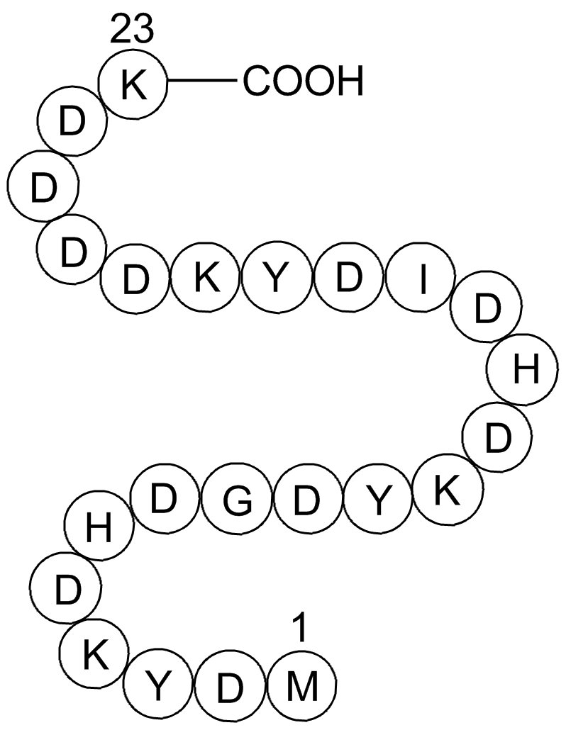
Cas No.: 402750-12-3
Sample solution is provided at 25 µL, 10mM.
- Sci Adv 11.2 (2025):eadr7943.PMID:39792683
- Gene Dev (2024).PMID:38503515
- Gene Dev 33.19-20 (2019):1397-1415.PMID:31467087
- Cell Rep 39.10 (2022).PMID:35675778
- EMBO Rep (2023):e56834.PMID:37306046
- NPJ Precis Oncol 7.1 (2023):33.PMID:36966223
- J Biol Chem 299.2 (2023).PMID:36621624
- Viruses 13.10 (2021):1980.PMID:34696410
- Drug Metab Dispos 51.10 (2023):1342-1349.
- bioRxiv (2022):2022-09.
- Method Enzymol.Vol.673.Academic Press,2022.191-225.
3X FLAG Peptide est un polypeptide synthétique contenant trois séquences répétées d'acides aminés DYKXXD, souvent utilisé pour se lier de manière compétitive aux anticorps anti-Flag. Le peptide 3X FLAG est principalement utilisé pour l'identification et la purification des protéines, comme l'élution des protéines de fusion Flag liées aux anticorps Anti-Flag lors de l'immunoprécipitation[1-5].
References:
[1]. Hopp, T., Prickett, K., et al. A Short Polypeptide Marker Sequence Useful for Recombinant Protein Identification and Purification. Nat Biotechnol 6, 1204-1210 (1988). https://doi.org/10.1038/nbt1088-1204
[2]. Einhauer A, Jungbauer A. Affinity of the monoclonal antibody M1 directed against the FLAG peptide. J Chromatogr A. 2001 Jun 29;921(1):25-30. doi: 10.1016/s0021-9673(01)00831-7. PMID: 11461009.
[3].Gao XK, Rao XS, et al. Phase separation of insulin receptor substrate 1 drives the formation of insulin/IGF-1 signalosomes. Cell Discov. 2022 Jun 28;8(1):60. doi: 10.1038/s41421-022-00426-x. PMID: 35764611; PMCID: PMC9240053.
[4].Gao J, Huang G, et al.PROTEIN S-ACYL TRANSFERASE 13/16 modulate disease resistance by S-acylation of the nucleotide binding, leucine-rich repeat protein R5L1 in Arabidopsis. J Integr Plant Biol. 2022 Sep;64(9):1789-1802. doi: 10.1111/jipb.13324. Epub 2022 Jul 28. PMID: 35778928.
[5]. Lian H, Jiang K, et al.The Salmonella effector protein SopD targets Rab8 to positively and negatively modulate the inflammatory response. Nat Microbiol. 2021 May;6(5):658-671. doi: 10.1038/s41564-021-00866-3. Epub 2021 Feb 18. PMID: 33603205; PMCID: PMC8085087.
Protocole pour l'expression et la purification des protéines avec le peptide 3×FLAG [1] :
1. Les plasmides contenant les gènes étiquetés FLAG ont été transfectés dans des cellules 293T pendant 2 jours.
2. Les cellules ont été récoltées par centrifugation à 14 000 rpm pendant 10 min à 4℃, puis lysées avec un tampon HEPES (150 mM de chlorure de sodium, 50 mM de HEPES, pH 7,4, 5 mM d'EDTA, 1 % (p/v) de désoxycholate de sodium, 1 % (v/v) de Triton X-100, 0,2 % de fluorure de sodium, 0,1 % d'orthovanadate de sodium et un cocktail d'inhibiteurs de protéase).
3. Des billes de gel Anti-FLAG M2 ont été ajoutées et incubées sur un agitateur rotatif à 4℃ pendant 2 heures. Les billes de gel M2 ont été récoltées par centrifugation à 3 000 rpm pendant 1 min et lavées quatre fois dans un tampon HEPES.
4. Les protéines étiquetées FLAG ont été purifiées par compétition avec le peptide 3×FLAG. Les billes M2 ont été resuspendues avec un tampon contenant 1,5 mg/mL de peptide 3×FLAG et incubées à 4℃ pendant 2 heures.
5. Après centrifugation à 5 000 rpm pendant 1 min, le surnageant a été purifié par filtration sur gel en utilisant une colonne SuperdexTM 200 augmentée, équilibrée dans un tampon de stockage (50 mM Tris-HCl, 37 mM NaCl, 1 mM EDTA, 5 mM DTT).
6. Toutes les protéines purifiées ont été concentrées par filtration centrifuge et stockées en aliquotes à -80℃.
7. La protéine purifiée a été quantifiée à l'aide d'un spectrophotomètre ND-2000 NanoDrop avec une OD de 280 nm et vérifiée par coloration au bleu de Coomassie.
Ce protocole fournit uniquement une ligne directrice et doit être modifié en fonction de vos besoins spécifiques.
Protocole pour les analyses Co-IP et Co-IP-MS utilisant le peptide 3×FLAG [2] :
1. Les lysats cellulaires ont été centrifugés à 13 300 rpm pendant 15 min à 4℃ pour éliminer les cellules intactes.
2. Le surnageant a été incubé soit avec de l'immunoglobuline G (IgG) de contrôle, soit avec un anticorps primaire toute la nuit dans un tampon IP (150 mM de NaCl, 20 mM HEPES à pH 7,4, 1 % Triton X-100, 12,5 mM β-glycérophosphate, 1,5 mM MgCl2, 2 mM EGTA avec des inhibiteurs de phosphatase et de protéase), suivi d'une incubation avec 20 μl de billes de protéine A/G resuspendues pendant 2 heures à 4℃ pour extraire les protéines liées.
3. Pour l'IP séquentiel, la protéine liée a été élue des billes par incubation avec 5 volumes de gel compact de solution d'élution de peptide 3×FLAG (concentration finale de 150 ng/mL) à 4℃ pendant 2 heures, puis immunoprécipitée avec un second anticorps toute la nuit à 4℃.
4. Les billes ont été centrifugées à 1 000g pendant 5 minutes à 4℃ pour éliminer le surnageant, lavées 4 fois avec le tampon IP, puis bouillies pendant 20 min à 95℃. Les échantillons ont été analysés sur un gel SDS-PAGE, suivi d'une coloration au bleu brillant de Coomassie R-250.
5. Les bandes de gel ont ensuite été excisées, décolorées, trypsinisées et soumises à une analyse MS pour identifier les protéines individuelles à l'aide de la chromatographie liquide couplée à la spectrométrie de masse (LC-MS).
Ce protocole fournit uniquement une ligne directrice et doit être modifié en fonction de vos besoins spécifiques.
Références :
[1]. Gao XK, Rao XS, et al. Phase separation of insulin receptor substrate 1 drives the formation of insulin/IGF-1 signalosomes. Cell Discov. 2022 Jun 28;8(1):60. doi: 10.1038/s41421-022-00426-x. PMID: 35764611; PMCID: PMC9240053.
[2]. Cong M, Wang Y, et al. MTSS1 suppresses mammary tumor-initiating cells by enhancing RBCK1-mediated p65 ubiquitination. Nat Cancer. 2020 Feb;1(2):222-234. doi: 10.1038/s43018-019-0021-y. Epub 2020 Jan 20. PMID: 35122005.
Cas No. | 402750-12-3 | SDF | |
Synonymes | H-Met-Asp-Tyr-Lys-Asp-His-Asp-Gly-Asp-Tyr-Lys-Asp-His-Asp-Ile-Asp-Tyr-Lys-Asp-Asp-Asp-Asp-Lys-OH | ||
Chemical Name | 3X FLAG Peptide | ||
Canonical SMILES | CCC(C)C(C(=O)NC(CC(=O)O)C(=O)NC(CC1=CC=C(C=C1)O)C(=O)NC(CCCCN)C(=O)NC(CC(=O)O)C(=O)NC(CC(=O)O)C(=O)NC(CC(=O)O)C(=O)NC(CC(=O)O)C(=O)NC(CCCCN)C(=O)O)NC(=O)C(CC(=O)O)NC(=O)C(CC2=CNC=N2)NC(=O)C(CC(=O)O)NC(=O)C(CCCCN)NC(=O)C(CC3=CC=C(C=C3)O)NC(=O)C(CC(=O)O)NC( | ||
Formula | C120H169N31O49S | M.Wt | 2861.87 |
Solubility | ≥143.1mg/mL in DMSO, <14.35mg/mL in EtOH, ≥143.4mg/mL in Water with gentle warming | Storage | Desiccate at -20°C |
General tips | Please select the appropriate solvent to prepare the stock solution according to the
solubility of the product in different solvents; once the solution is prepared, please store it in
separate packages to avoid product failure caused by repeated freezing and thawing.Storage method
and period of the stock solution: When stored at -80°C, please use it within 6 months; when stored
at -20°C, please use it within 1 month. To increase solubility, heat the tube to 37°C and then oscillate in an ultrasonic bath for some time. |
||
Shipping Condition | Evaluation sample solution: shipped with blue ice. All other sizes available: with RT, or with Blue Ice upon request. |
Prepare stock solution | |||
![]() |
1 mg | 5 mg | 10 mg |
1 mM | 0.3494 mL | 1.7471 mL | 3.4942 mL |
5 mM | 0.0699 mL | 0.3494 mL | 0.6988 mL |
10 mM | 0.0349 mL | 0.1747 mL | 0.3494 mL |
Step 1: Enter information below (Recommended: An additional animal making an allowance for loss during the experiment)
Step 2: Enter the in vivo formulation (This is only the calculator, not formulation. Please contact us first if there is no in vivo formulation at the solubility Section.)
Calculation results:
Working concentration: mg/ml;
Method for preparing DMSO master liquid: mg drug pre-dissolved in μL DMSO ( Master liquid concentration mg/mL, Please contact us first if the concentration exceeds the DMSO solubility of the batch of drug. )
Method for preparing in vivo formulation: Take μL DMSO master liquid, next addμL PEG300, mix and clarify, next addμL Tween 80, mix and clarify, next add μL ddH2O, mix and clarify.
Method for preparing in vivo formulation: Take μL DMSO master liquid, next add μL Corn oil, mix and clarify.
Note: 1. Please make sure the liquid is clear before adding the next solvent.
2. Be sure to add the solvent(s) in order. You must ensure that the solution obtained, in the previous addition, is a clear solution before proceeding to add the next solvent. Physical methods such as vortex, ultrasound or hot water bath can be used to aid dissolving.
3. All of the above co-solvents are available for purchase on the GlpBio website.
Quality Control & SDS
- View current batch:
- Purity: >98.00%
- COA (Certificate Of Analysis)
- SDS (Safety Data Sheet)
- Datasheet
Average Rating: 5
(Based on Reviews and 29 reference(s) in Google Scholar.)GLPBIO products are for RESEARCH USE ONLY. Please make sure your review or question is research based.
Required fields are marked with *