ω-Conotoxin GVIA |
Catalog No.GC13886 |
La ω-Conotoxin GVIA est une toxine de l'escargot conique qui bloque sélectivement les canaux de type N dans les neurones.
Products are for research use only. Not for human use. We do not sell to patients.
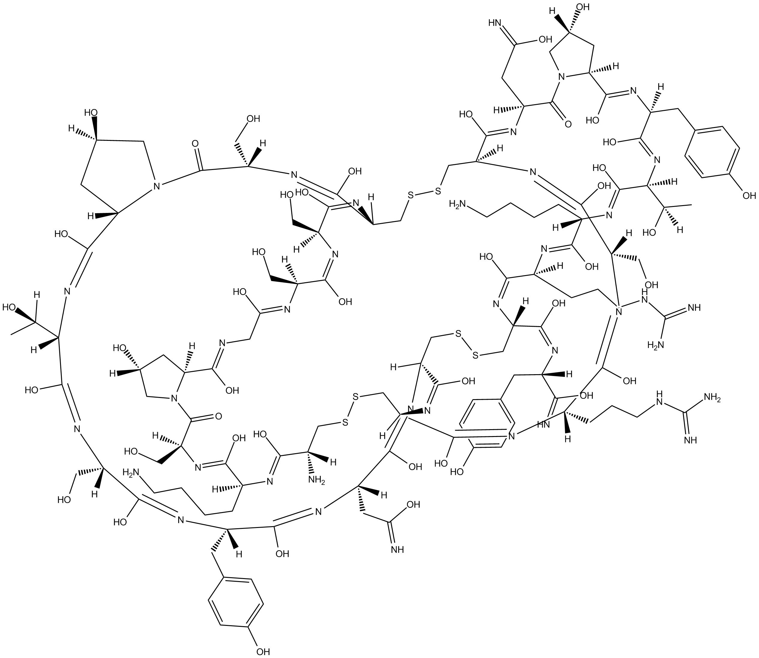
Cas No.: 106375-28-4
Sample solution is provided at 25 µL, 10mM.
La ω-Conotoxin GVIA est une toxine de l'escargot conique qui bloque sélectivement les canaux de type N dans les neurones[1]. La ω-Conotoxin GVIA se lie aux membranes plasmatiques synaptiques du néocortex humain, de l'hippocampe du rat et du cerveau du poussin (IC50 = 4,6, 60 et 150 pM, respectivement, dans les essais de liaison par radioligand) [3,4,5]. La ω-Conotoxin GVIA a considérablement réduit l'amplitude des contractions tétaniques du muscle tibial antérieur chez la souris, mais la facilitation tétanique n'a pas été altérée. Les contractions musculaires provoquées par une stimulation électrique directe n'ont pas été significativement modifiées par la ω-Conotoxin GVIA. La ω-Conotoxin GVIA n'a pas provoqué de changements significatifs de la pression artérielle systémique [5].
References:[1]. H.D. Mansvelder, J.C. Stoof, K.S. Kits. Dihydropyridine block of ω-agatoxin IVA- and ω-conotoxin GVIA-sensitive Ca2+ channels in rat pituitary melanotropic cells.Eur. J. Pharmacol., 311 (1996), pp. 293-304[2]. Feuerstein, T.J., Dooley, D.J., and Seeger, W. Inhibition of norepinephrine and acetylcholine release from human neocortex by ω-conotoxin GVIA. J. Pharmacol. Exp. Ther. 252(2), 778-785 (1990).[3]. Lampe, R.A., Lo, M.M., Keith, R.A., et al. Effects of site-specific acetylation on ω-conotoxin GVIA binding and function. Biochemistry 32(13), 3255-3260 (1993).[4]. Sato, K., Park, N.G., Kohno, T., et al. Role of basic residues for the binding of ω-conotoxin GVIA to N-type calcium channels. Biochem. Biophys. Res. Commun. 194(3), 1292-1296 (1993).[5]: Rossoni G, Berti F, La Maestra L, Clementi F. ω-Conotoxin GVIA binds to and blocks rat neuromuscular junction. Neuroscience Letters. 1994;176:185-188.Expériences cellulaires [1]: | |
Lignées cellulaires | Cellules mélanotropes hypophysaires de rat |
Méthode de préparation | Le ω-Conotoxine GVIA a été administré via un système de tube en Y à gravité ou un jet de pression. La chambre d'enregistrement a été perfusée en continu à un débit d'environ 1,5 ml/min, entraîné par une pression d'air, et le volume du bain a été maintenu constant par une aspiration continue. Des tests électrophysiologiques ont été effectués. |
Conditions de réaction | 1 ou 10 µM |
Domaines d'application | Le ω-Conotoxine GVIA à 1 µM a bloqué le courant activé à haut voltage de 25,5 ± 3,6 %. Le blocage à 10 µM ω-Conotoxine GVIA était de 22,9 ± 2,5 % (pas significativement différent du blocage à 1 µM ; n = 3), indiquant que le ω-Conotoxine GVIA produit un effet saturant. |
Expériences animales [2]: | |
Modèles animaux | Rats mâles COBS CD (SD) |
Méthode de préparation | Après injection intraveineuse de différentes doses de ω-Conotoxine GVIA (1-2 nmol/kg) chez des rats, le nerf sciatique des rats a été stimulé et la pression artérielle des rats a été mesurée via l'artère carotide. |
Forme de dosage | Injection intraveineuse, 1-2 nmol/kg |
Domaines d'application | L'injection intraveineuse a provoqué une perte progressive du développement de la tension du muscle tibial, induite indirectement par la stimulation électrique du nerf sciatique. Le ω-Conotoxine GVIA (1 nmol/kg i.v.) a réduit la force de contraction musculaire de 75 % en environ 27 minutes (de 39,5 ± 1,8 g à 9,9 ± 0,4 g). |
Références : [1] : H.D. Mansvelder, J.C. Stoof, K.S. Kits. Blocage dihydropyridine des canaux Ca2+ sensibles à l'ω-agatoxine IVA et à l'ω-conotoxine GVIA dans les cellules mélanotropes hypophysaires de rat. Eur. J. Pharmacol., 311 (1996), pp. 293-304 [2] : Rossoni G, Berti F, La Maestra L, Clementi F. Le ω-Conotoxine GVIA se lie et bloque la jonction neuromusculaire chez le rat. Neuroscience Letters. 1994;176:185-188. |
Cas No. | 106375-28-4 | SDF | |
Canonical SMILES | C[C@](O)([H])[C@@](/N=C(O)/[C@](/N=C(O)/[C@]1([H])C[C@](O)([H])CN12)([H])CC3=CC=C(O)C=C3)([H])/C(O)=N/[C@@](/C(O)=N/[C@@](/C(O)=N\[C@@](/C(O)=N\[C@@](C(O)=N)([H])CC4=CC=C(O)C=C4)([H])CSSC[C@@](N=C(O)[C@](N=C(O)[C@](N=C(O)[C@](N=C(O)[C@](N=C(O)[C@]5([H])C[ | ||
Formula | C120H182N38O43S6 | M.Wt | 3037.35 |
Solubility | Soluble to 1 mg/ml in Water | Storage | Desiccate at -20°C |
General tips | Please select the appropriate solvent to prepare the stock solution according to the
solubility of the product in different solvents; once the solution is prepared, please store it in
separate packages to avoid product failure caused by repeated freezing and thawing.Storage method
and period of the stock solution: When stored at -80°C, please use it within 6 months; when stored
at -20°C, please use it within 1 month. To increase solubility, heat the tube to 37°C and then oscillate in an ultrasonic bath for some time. |
||
Shipping Condition | Evaluation sample solution: shipped with blue ice. All other sizes available: with RT, or with Blue Ice upon request. |
Prepare stock solution | |||
![]() |
1 mg | 5 mg | 10 mg |
1 mM | 0.3292 mL | 1.6462 mL | 3.2923 mL |
5 mM | 0.0658 mL | 0.3292 mL | 0.6585 mL |
10 mM | 0.0329 mL | 0.1646 mL | 0.3292 mL |
Step 1: Enter information below (Recommended: An additional animal making an allowance for loss during the experiment)
Step 2: Enter the in vivo formulation (This is only the calculator, not formulation. Please contact us first if there is no in vivo formulation at the solubility Section.)
Calculation results:
Working concentration: mg/ml;
Method for preparing DMSO master liquid: mg drug pre-dissolved in μL DMSO ( Master liquid concentration mg/mL, Please contact us first if the concentration exceeds the DMSO solubility of the batch of drug. )
Method for preparing in vivo formulation: Take μL DMSO master liquid, next addμL PEG300, mix and clarify, next addμL Tween 80, mix and clarify, next add μL ddH2O, mix and clarify.
Method for preparing in vivo formulation: Take μL DMSO master liquid, next add μL Corn oil, mix and clarify.
Note: 1. Please make sure the liquid is clear before adding the next solvent.
2. Be sure to add the solvent(s) in order. You must ensure that the solution obtained, in the previous addition, is a clear solution before proceeding to add the next solvent. Physical methods such as vortex, ultrasound or hot water bath can be used to aid dissolving.
3. All of the above co-solvents are available for purchase on the GlpBio website.
Quality Control & SDS
- View current batch:
- Purity: >98.00%
- COA (Certificate Of Analysis)
- SDS (Safety Data Sheet)
- Datasheet
Average Rating: 5
(Based on Reviews and 28 reference(s) in Google Scholar.)GLPBIO products are for RESEARCH USE ONLY. Please make sure your review or question is research based.
Required fields are marked with *