


Calcein AM
Calcein AM is the short form of the calcein-acetoxymethyl ester, which is basically a non-fluorescent and cell permeant, i.e., that essentially means that it does not exhibit fluorescence and also the cell membrane allows its free movement across itself owing to its lipophilic nature. Chemically, it is a acetoxymethyl ester of calcein. It passively enters the cell, without any energy expenditure through a gap junction, where it gets biochemically converted, i.e., via the biochemical hydrolysis that is catalyzed by the activity of the esterase enzymes that reside in the cytoplasm, into Calcein, which is acidic in nature as it holds a negative charge being an anion. Moreover, the latter is cell-impermeant, i.e., which essentially means that it does not have the ability to come out of the cell and thus get trapped inside the cell. Furthermore, the important point is that the latter one is fluorophore. The key difference between how Calcein AM and Calcein interact with the cell is shown in Figure 1.
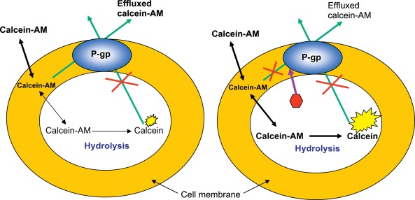
Figure 1 Calcein AM and Calcein: P-gp is the protein that may affect the transportation of Calcein AM but not of a Calcein. Red hexagon in the right image represents the P-gp inhibitor, which are discussed later in the article
It has some characteristics that makes it quite a useful while studying the structural features as well as the functions of the alive cells in the fields of microscopy and fluorometry. These favorable characteristics are low cytotoxicity, real-time signal stability, high bleach resistance and insensitivity to the cytoplasmic pH. A number of the structural features of the cells have been studied using Calcein AM; they include, competence of the cell membrane which is the ability to accept the circular foreign DNA molecule in the form of plasmid. on the other hand, a number of the physiological features of the cells have been studied using Calcein AM; they include, the phenomenon of apoptosis, migration of lymphocytes, and Multi Drug Resistance MDR, it also includes the determination of certain parameter for example, cell viability and cell to cell communication via the gap junctions.
In a research study, Calcein AM has been successfully employed to visualize and quantify the increased oxidation state which is oxidative stress in the alive fibroblast cells using confocal microscopy. Oxidative stress is the indication of the hemostatic imbalance between the production of Reactive Oxygen Species ROS as a part of normal physiologic anerobic metabolism and its subsequent either neutralization or degradation via the activity of the body’s antioxidant system. All the major vital structural as well as the functional macromolecules of the cells under the oxidative stress are prone to irreparable damage. This happens via the peroxidation of lipids, genetic mutations in the DNA and Protein denaturation which is complete loss of the protein functions. Oxidative stress is a major landmark of the some pathological conditions, for example, atherosclerosis, cancers and neurodegenerative diseases.
In that research, first of all, the alive fibroblast cells were loaded with cell permeable molecule Calcein AM, which converted into Calcein, fluorophore having the cyan colored fluorescence. Then, confocal microscopic images were taken; these are shown in Figure 2. in the baseline images, the fluorescence signal is dominant in the nuclei while weak in the cytoplasmic region. In the micrographs taken after 10 minutes, small illuminated dots also appear which are indicated by small arrows in the micrographs; these are actually the cytoplasmic mitochondria. Moreover, in this image, the nuclear fluorescence has also increased. In the micrographs taken after 30 minutes, both the nuclear as well as the cytoplasmic fluorescence have increased a lot. This data is graphically presented in Figure 3.
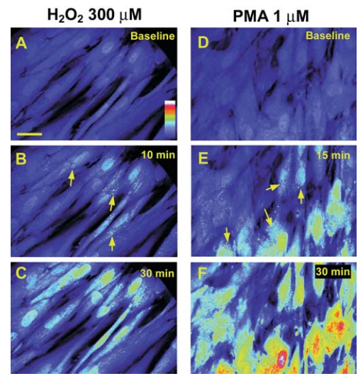
Figure 2 Confocal Micrographs of the Calcein AM treated fibroblasts, which are taken at different time intervals
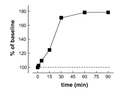
Figure 3 The intensity of the fluorescence is presented as a function of time. After 30 minutes of Calcein AM treatment, maximum fluorescence intensity reached; that becomes constant when further time is given
Multi Drug Resistance MDR is the biggest challenge, that is encountered when it comes to the clinical applicability of the chemotherapy against the broad spectrum of cancers/ neoplasms. And, its accurate detection as well as quantification is utmost important. Calcein AM has been successfully shown as a means to accurately quantify the Multi Drug Resistance MDR in the cancer cell lines via the retention assay. Actually, Multi Drug Resistance MDR is a phenotype that is characterized by the over expression/ over activity of a protein molecule, which is MRP1 and it is actually an efflux pump that means that it moves its ligand/ substrate out of the cell. Interestingly, Calcein AM acts as a ligand/ substrate for this protein. In a research study, Calcein AM is loaded in the cells expressing MRP1 and in the cells not expressing MRP1. For the cells expressing MRP1, it is theoretically expected to exhibit fluorescence out of the cell, while for the cells not expressing MRP1, it is theoretically expected to exhibit fluorescence inside the cell and graphically presented in Figure 4.
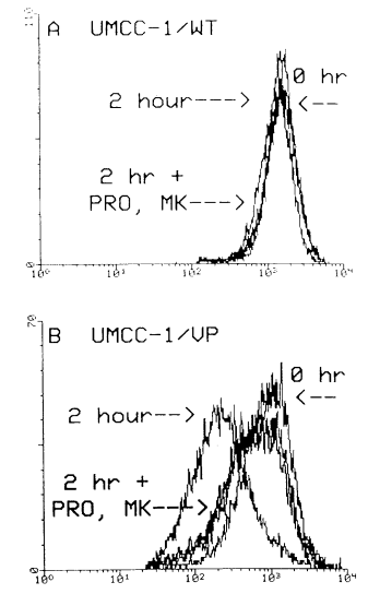
Figure 4 The Fluorescence intensity in the MPR1 expressing cell which are UMCC-1/VP shown in the below graph, and in the cells not expressing MPR1 which are UMCC-1/WT shown in the above graph. MK and PRO indicate MK571 and probenecid, respectively. Both of them are the inhibitors of MPR1
Apoptosis is equally important part of normal physiology as well as of abnormal pathological conditions. At a normal pace, being a part of the normal physiology, it is a key player in the reconstruction of the normal tissues and in the removal of the damaged or injured tissues. At either extreme, at an abnormally high or low pace, it becomes a crucial process in a broad spectrum of pathologies. At an extremely low pace, it becomes a crucial part of diseases, for example, Rheumatoid arthritis and cancers. On the other hand, at an extremely high pace, it becomes a crucial part of diseases, Ischemic heart disease, AIDS, Alzheimer’s disease and Parkinson’s disease; the last two are the neurodegenerative in nature. Therefore, accurate detection and quantification of the apoptosis is clinically significant. Calcein AM along with the ethidium homodimer (EthD-1) is used in the detection and quantification of the apoptosis. Calcein produces green fluorescence. As the structure of the plasma membrane is collapsed, which is the landmark of the apoptosis; the catalytic activity of the esterase stops, while the ethidium homodimer (EthD-1) becomes adherent to the hydrogen bonding of the DNA molecules and exhibits a 40 X stronger red fluorescence. In this protocol, Calcein AM indicates the cell viability via a decreased esterase activity which is indicated by the decreased degree of fluorescence; while the ethidium homodimer (EthD-1) fluorescence indicates all the physical as well as chemical modifications in the architecture of the plasma membrane. The computer-generated experimental results are shown in Figure 5.
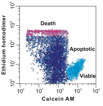
Figure 5 The detection of cell viability and degree of apoptosis via the usage of Calcein AM along with the ethidium homodimer (EthD-1)


 Chemical Structure")
 Chemical Structure")


Commentaires