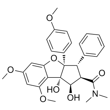
Rocaglamide (Rocaglamide A) |
Numéro de catalogue: GC33426 |
Rocaglamide (Rocaglamide A) (Roc-A) est isolé du genre Aglaia et peut être utilisé pour la toux, les blessures, l'asthme et les maladies inflammatoires de la peau. Le Rocaglamide (Rocaglamide A) est un puissant inhibiteur de l'activation de NF-κB dans les lymphocytes T. Le rocaglamide (Rocaglamide A) est un inhibiteur puissant et sélectif de l'activation du facteur de choc thermique 1 (HSF1) avec une IC50 d'environ 50 nM. Rocaglamide (Rocaglamide A) inhibe la fonction du facteur d'initiation de la traduction eIF4A. Rocaglamide (Rocaglamide A) a également des propriétés anticancéreuses dans la leucémie.
Sample solution is provided at 25 µL, 10mM.
Contrôle de qualité & Fiche de Données de Sécurité
- Afficher le lot actuel :
Cell experiment [1]: | |
Cell lines |
HepG2 and Huh-7 cells |
Preparation Method |
HepG2 and Huh-7 cells were seeded in 96-well plates in complete culture medium and incubated for 24 h. The cells were then exposed to 100 nM Rocaglamide (Rocaglamide A) and TRAIL for 24 h. |
Reaction Conditions |
100 nM;24h |
Applications |
Rocaglamide (Rocaglamide A) significantly enhanced TRAIL induced apoptosis. |
Animal experiment [2]: | |
Animal models |
Female SCID mice (6-week-old) |
Preparation Method |
The Huh-7 cells, suspended in 100 µl mix, were implanted subcutaneously into the right flank of 10 female SCID mice (6-week-old) and then randomly divided into two equal groups, one of which received an intraperitoneal injection of Rocaglamide (Rocaglamide A) (2.5 mg/kg in 80 µl olive oil; n=5) and the other, used as a vehicle contro. These treatments were performed once daily for 32 days and the tumor volumes and body weights of the animals were measured twice a week. |
Dosage form |
2.5 mg/kg; i.p.; 32 days |
Applications |
Rocaglamide (Rocaglamide A) induced tumor cell apoptosis in a SCID mouse model (Huah-7 cells) without causing weight loss in mice, and no significant signs of toxicity were observed during treatment, suggesting that Rocaglamide is generally well tolerated in vivo. |
References: [1]. Luan Z, He Y, et,al. Rocaglamide overcomes tumor necrosis factor-related apoptosis-inducing ligand resistance in hepatocellular carcinoma cells by attenuating the inhibition of caspase-8 through cellular FLICE-like-inhibitory protein downregulation. Mol Med Rep. 2015 Jan;11(1):203-11. doi: 10.3892/mmr.2014.2718. Epub 2014 Oct 21. PMID: 25333816; PMCID: PMC4237083. |
Rocaglamide (Rocaglamide A) is isolated from the genus Aglaia (family Meliaceae). Rocaglamide (Rocaglamide A)(IC50 of 50 nM) inhibits the function of the translation initiation factor eIF4A, a DEAD box RNA helicase [1,2].
Rocaglamide (Rocaglamide A) (100 nM;24h) significantly enhanced TRAIL induced apoptosis[4]. Rocaglamide (Rocaglamide A) (20- 100 nM;48h)can induce 10-30% apoptosis of L1236 and KM-H2 cells [3]. Rocaglamide (Rocaglamide A) are able to suppress the PMA-induced expression of NF-kappaB target genes and sensitize leukemic T cells to apoptosis induced by TNFalpha, cisplatin, and gamma-irradiation [5]. Rocaglamide (Rocaglamide A)( 30/50 nM;21d) prevented TNF-α mediated inhibition of osteoblast differentiation, and promoted osteoblast differentiation directly, in both C2C12 and primary mesenchymal stromal cells [6].
Rocaglamide (Rocaglamide A)( 2.5 mg/kg; i.p.; 32 days) induced tumor cell apoptosis in a SCID mouse model (Huah-7 cells) without causing weight loss in mice, and no significant signs of toxicity were observed during treatment, suggesting that Rocaglamide is generally well tolerated in vivo[4]. Rocaglamide (Rocaglamide A)( 0.5 mg/kg; i.p.; five times per week for two weeks) overcomes CPT resistance in U266 in vitro and significant increases in anti-tumor efficacies of CPT in mice xenografted with U266[7].
References:
[1]. Santagata S, Mendillo ML, et,al. Tight coordination of protein translation and HSF1 activation supports the anabolic malignant state. Science. 2013 Jul 19;341(6143):1238303. doi: 10.1126/science.1238303. PMID: 23869022; PMCID: PMC3959726.
[2]. Kim S, Salim AA, Swanson SM and Kinghorn AD: Potential of cyclopenta[b]benzofurans from Aglaia species in cancer chemotherapy. Anticancer Agents Med Chem. 6:319-345. 2006.
[3]. Giaisi M, KÖhler R, et,al. Rocaglamide and a XIAP inhibitor cooperatively sensitize TRAIL-mediated apoptosis in Hodgkin's lymphomas. Int J Cancer. 2012 Aug 15;131(4):1003-8. doi: 10.1002/ijc.26458. Epub 2011 Nov 8. PMID: 21952919.
[4]. Luan Z, He Y, et,al. Rocaglamide overcomes tumor necrosis factor-related apoptosis-inducing ligand resistance in hepatocellular carcinoma cells by attenuating the inhibition of caspase-8 through cellular FLICE-like-inhibitory protein downregulation. Mol Med Rep. 2015 Jan;11(1):203-11. doi: 10.3892/mmr.2014.2718. Epub 2014 Oct 21. PMID: 25333816; PMCID: PMC4237083.
[5]. Baumann B, Bohnenstengel F, et,al. Rocaglamide derivatives are potent inhibitors of NF-kappa B activation in T-cells. J Biol Chem. 2002 Nov 22;277(47):44791-800. doi: 10.1074/jbc.M208003200. Epub 2002 Sep 16. PMID: 12237314.
[6]. Li A, Yang L, et,al. Rocaglamide-A Potentiates Osteoblast Differentiation by Inhibiting NF-κB Signaling. Mol Cells. 2015 Nov;38(11):941-9. doi: 10.14348/molcells.2015.2353. Epub 2015 Nov 6. PMID: 26549505; PMCID: PMC4673408.
[7]. Wu Y, Giaisi M, et,al. Rocaglamide breaks TRAIL-resistance in human multiple myeloma and acute T-cell leukemia in vivo in a mouse xenogtraft model. Cancer Lett. 2017 Mar 28;389:70-77. doi: 10.1016/j.canlet.2016.12.010. Epub 2016 Dec 18. PMID: 27998762.
Cas No. | 84573-16-0 | SDF | |
Synonymes | Rocaglamide A; Roc-A | ||
Canonical SMILES | O1C2=CC(OC)=CC(OC)=C2[C@]2(O)[C@H](O)[C@H](C(N(C)C)=O)[C@@H](C3=CC=CC=C3)[C@]12C1=CC=C(OC)C=C1 |&1:11,13,15,21,28,r| | ||
Formula | C29H31NO7 | M.Wt | 505.56 |
Solubilité | 150 mg/mL in DMSO | Storage | Store at -20°C |
Conseils généraux | Afin d'obtenir une solubilité plus élevée, veuillez chauffer le tube à 37 °C et le secouer dans le bain à ultrasons pendant un certain temps. La solution mère peut être conservée à une température inférieure à -20 °C pendant plusieurs mois. | ||
Condition d'expédition | Solution d'échantillon d'évaluation : livré avec la glace bleue Toute autre taille disponible : livré avec RT ou la glace bleue sur demande |
Step 1: Enter information below (Recommended: An additional animal making an allowance for loss during the experiment)
Step 2: Enter the in vivo formulation (This is only the calculator, not formulation. Please contact us first if there is no in vivo formulation at the solubility Section.)
Calculation results:
Working concentration: mg/ml;
Method for preparing DMSO master liquid: mg drug pre-dissolved in μL DMSO ( Master liquid concentration mg/mL, Please contact us first if the concentration exceeds the DMSO solubility of the batch of drug. )
Method for preparing in vivo formulation: Take μL DMSO master liquid, next addμL PEG300, mix and clarify, next addμL Tween 80, mix and clarify, next add μL ddH2O, mix and clarify.
Method for preparing in vivo formulation: Take μL DMSO master liquid, next add μL Corn oil, mix and clarify.
Note: 1. Please make sure the liquid is clear before adding the next solvent.
2. Be sure to add the solvent(s) in order. You must ensure that the solution obtained, in the previous addition, is a clear solution before proceeding to add the next solvent. Physical methods such as vortex, ultrasound or hot water bath can be used to aid dissolving.
3. All of the above co-solvents are available for purchase on the GlpBio website.
PHB2 (prohibitin 2) promotes PINK1-PRKN/Parkin-dependent mitophagy by the PARL-PGAM5-PINK1 axis
Mitophagy, which is a conserved cellular process for selectively removing damaged or unwanted mitochondria, is critical for mitochondrial quality control and the maintenance of normal cellular physiology. However, the precise mechanisms underlying mitophagy remain largely unknown. Prior studies on mitophagy focused on the events in the mitochondrial outer membrane. PHB2 (prohibitin 2), which is a highly conserved membrane scaffold protein, was recently identified as a novel inner membrane mitophagy receptor that mediates mitophagy. Here, we report a new signaling pathway for PHB2-mediated mitophagy. Upon mitochondrial membrane depolarization or misfolded protein aggregation, PHB2 depletion destabilizes PINK1 in the mitochondria, which blocks the mitochondrial recruitment of PRKN/Parkin, ubiquitin and OPTN (optineurin), leading to an inhibition of mitophagy. In addition, PHB2 overexpression directly induces PRKN recruitment to the mitochondria. Moreover, PHB2-mediated mitophagy is dependent on the mitochondrial inner membrane protease PARL, which interacts with PHB2 and is activated upon PHB2 depletion. Furthermore, PGAM5, which is processed by PARL, participates in PHB2-mediated PINK1 stabilization. Finally, a ligand of PHB proteins that we synthesized, called FL3, was found to strongly inhibit PHB2-mediated mitophagy and to effectively block cancer cell growth and energy production at nanomolar concentrations. Thus, our findings reveal that the PHB2-PARL-PGAM5-PINK1 axis is a novel pathway of PHB2-mediated mitophagy and that targeting PHB2 with the chemical compound FL3 is a promising strategy for cancer therapy.Abbreviations: AIFM1: apoptosis inducing factor mitochondria associated 1; ATP5F1A/ATP5A1: ATP synthase F1 subunit alpha; BAF: bafilomycin A1; CALCOCO2/NDP52: calcium binding and coiled-coil domain 2; CCCP: chemical reagent carbonyl cyanide m-chlorophenyl hydrazine; FL3: flavaglines compound 3; HSPD1/HSP60: heat shock protein family D (Hsp60) member 1; LC3B/MAP1LC3B: microtubule associated protein 1 light chain 3 beta; MEF: mouse embryo fibroblasts; MPP: mitochondrial-processing peptidase; MT-CO2/COX2: mitochondrially encoded cytochrome c oxidase II; MTS: mitochondrial targeting sequence; OA: oligomycin and antimycin A; OPTN: optineurin; OTC: ornithine carbamoyltransferase; PARL: presenilin associated rhomboid like; PBS: phosphate-buffered saline; PGAM5: PGAM family member 5, mitochondrial serine/threonine protein phosphatase; PHB: prohibitin; PHB2: prohibitin 2; PINK1: PTEN induced kinase 1; PRKN/Parkin: parkin RBR E3 ubiquitin protein ligase; Roc-A: rocaglamide A; TOMM20: translocase of outer mitochondrial membrane 20; TUBB: tubulin beta class I.
Inhibitory effects of Rocaglamide-A on PPARγ-driven adipogenesis through regulation of mitotic clonal expansion involving the JAK2/STAT3 pathway
Inhibition of adipogenesis is an important strategy for obesity treatment. Rocaglamide-A (Roc-A) is a natural herbal medicine isolated from the genus Aglaia (family Meliaceae), which has a cyclopenta[b]benzofuran core structure. Roc-A exhibits various pharmacological effects against diverse human cancer cells. However, the exact role of Roc-A during adipogenesis in adipocytes has not been studied at all. In this study, we demonstrate that Roc-A is crucial for reducing adipogenesis via downregulating PPARγ transcriptional activity. Consistently, Western-blot and RT-PCR analyses clearly showed that Roc-A inhibits the expression of PPARγ target genes and lipogenic markers in a dose-dependent manner along with suppression of lipid accumulation, in both 3T3-L1 cells and mouse adipose-derived stem cells. Mechanistically, Roc-A significantly decreased STAT3 phosphorylation in a dose-dependent manner in 3T3-L1 adipocytes. In particular, we confirmed that Roc-A effectively suppressed the expression of genes involved in cell-cycle regulation, such as cyclin A, B, D1, and E1, early during mitotic clonal expansion in 3T3-L1 adipocytes, and this effect was abolished by the JAK2/STAT3 activator FGF2. Taken together, our results demonstrated that Roc-A reduces adipogenesis by inhibiting PPARγ transactivation and STAT3 phosphorylation and thus may serve as a therapeutic target in obesity.
Rocaglamide and silvestrol: a long story from anti-tumor to anti-coronavirus compounds
Covering: up to the beginning of 2020Many natural substances have been transformed again and again with regard to their pharmaceutical-medical potential, including new members of a growing class of natural products, the flavaglines. Important representatives are rocaglamide and silvestrol, isolated from the Aglaia species, which are highlighted here. These products started as potential anti-tumor agents five decades ago and have recently proved to be very promising antiviral agents, especially against RNA viruses. Today they are discussed as potential starting compounds for developing drug candidates and therapeutics.
Rocaglamide-A Potentiates Osteoblast Differentiation by Inhibiting NF-κB Signaling
Rheumatoid arthritis is a chronic inflammatory disease that leads to bone and cartilage erosion. The inhibition of osteoblast differentiation by the inflammatory factor TNF-α is critical for the pathogenesis of rheumatoid arthritis. To modulate TNF-α mediated inhibition of osteoblast differentiation is required to improve therapeutic efficacy of rheumatoid arthritis. Here, we explored the potential role of rocaglamide-A, a component of Aglaia plant, in osteoblast differentiation. Rocaglamide-A prevented TNF-α mediated inhibition of osteoblast differentiation, and promoted osteoblast differentiation directly, in both C2C12 and primary mesenchymal stromal cells. Mechanistically, Rocaglamide-A inhibited the phosphorylation of NF-κB component p65 protein and the accumulation of p65 in nucleus, which resulted in the diminished NF-κB responsible transcriptional activity. Oppositely, overexpression of p65 reversed rocaglamide-A's protective effects on osteoblast differentiation. Collectively, rocaglamide-A protected and stimulated osteoblast differentiation via blocking NF-κB pathway. It suggests that rocaglamide-A may be a good candidate to develop as therapeutic drug for rheumatoid arthritis associated bone loss diseases.
Rocaglamide promotes the infiltration and antitumor immunity of NK cells by activating cGAS-STING signaling in non-small cell lung cancer
Background: Natural killer (NK) cell-based immunotherapy is clinically limited due to insufficient tumor infiltration in solid tumors. We have previously found that the natural product rocaglamide (RocA) can enhance NK cell-mediated killing of non-small cell lung cancer (NSCLC) cells by inhibiting autophagy, and autophagic inhibition has been shown to increase NK cell tumor infiltration in melanoma. Therefore, we hypothesized that RocA could increase NK cell infiltration in NSCLC by autophagy inhibition. Methods: Flow cytometry, RNA-sequencing, real-time PCR, Western blotting analysis, and xenograft tumor model were utilized to assess the infiltration of NK cells and the underlying mechanism. Results: RocA significantly increased the infiltration of NK cells and the expressions of CCL5 and CXCL10 in NSCLC cells, which could not be reversed by the inhibitions of autophagy/ULK1, JNK and NF-κB. However, such up-regulation could be suppressed by the inhibitions of TKB1 and STING. Furthermore, RocA dramatically activated the cGAS (cyclic GMP-AMP synthase)-STING (stimulator of interferon genes) signaling pathway, and the inhibition/depletion of STING ablated the up-regulation of CCL5 and CXCL10, NK cell infiltration, and tumor regression induced by RocA. Besides, RocA damaged mitochondrial DNA (mtDNA) and promoted the cytoplasmic release of mtDNA. The mPTP inhibitor cyclosporin A could reverse RocA-induced cytoplasmic release of mtDNA. Conclusions: RocA could promote NK cell infiltration by activating cGAS-STING signaling via targeting mtDNA, but not by inhibiting autophagy. Taken together, our current findings suggested that RocA was a potent cGAS-STING agonist and had a promising potential in cancer immunotherapy, especially in NK cell-based immunotherapy.
Average Rating: 5
(Based on Reviews and 39 reference(s) in Google Scholar.)GLPBIO products are for RESEARCH USE ONLY. Please make sure your review or question is research based.
Required fields are marked with *