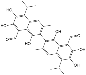
Apoptosis
As one of the cellular death mechanisms, apoptosis, also known as programmed cell death, can be defined as the process of a proper death of any cell under certain or necessary conditions. Apoptosis is controlled by the interactions between several molecules and responsible for the elimination of unwanted cells from the body.
Many biochemical events and a series of morphological changes occur at the early stage and increasingly continue till the end of apoptosis process. Morphological event cascade including cytoplasmic filament aggregation, nuclear condensation, cellular fragmentation, and plasma membrane blebbing finally results in the formation of apoptotic bodies. Several biochemical changes such as protein modifications/degradations, DNA and chromatin deteriorations, and synthesis of cell surface markers form morphological process during apoptosis.
Apoptosis can be stimulated by two different pathways: (1) intrinsic pathway (or mitochondria pathway) that mainly occurs via release of cytochrome c from the mitochondria and (2) extrinsic pathway when Fas death receptor is activated by a signal coming from the outside of the cell.
Different gene families such as caspases, inhibitor of apoptosis proteins, B cell lymphoma (Bcl)-2 family, tumor necrosis factor (TNF) receptor gene superfamily, or p53 gene are involved and/or collaborate in the process of apoptosis.
Caspase family comprises conserved cysteine aspartic-specific proteases, and members of caspase family are considerably crucial in the regulation of apoptosis. There are 14 different caspases in mammals, and they are basically classified as the initiators including caspase-2, -8, -9, and -10; and the effectors including caspase-3, -6, -7, and -14; and also the cytokine activators including caspase-1, -4, -5, -11, -12, and -13. In vertebrates, caspase-dependent apoptosis occurs through two main interconnected pathways which are intrinsic and extrinsic pathways. The intrinsic or mitochondrial apoptosis pathway can be activated through various cellular stresses that lead to cytochrome c release from the mitochondria and the formation of the apoptosome, comprised of APAF1, cytochrome c, ATP, and caspase-9, resulting in the activation of caspase-9. Active caspase-9 then initiates apoptosis by cleaving and thereby activating executioner caspases. The extrinsic apoptosis pathway is activated through the binding of a ligand to a death receptor, which in turn leads, with the help of the adapter proteins (FADD/TRADD), to recruitment, dimerization, and activation of caspase-8 (or 10). Active caspase-8 (or 10) then either initiates apoptosis directly by cleaving and thereby activating executioner caspase (-3, -6, -7), or activates the intrinsic apoptotic pathway through cleavage of BID to induce efficient cell death. In a heat shock-induced death, caspase-2 induces apoptosis via cleavage of Bid.
Bcl-2 family members are divided into three subfamilies including (i) pro-survival subfamily members (Bcl-2, Bcl-xl, Bcl-W, MCL1, and BFL1/A1), (ii) BH3-only subfamily members (Bad, Bim, Noxa, and Puma9), and (iii) pro-apoptotic mediator subfamily members (Bax and Bak). Following activation of the intrinsic pathway by cellular stress, pro‑apoptotic BCL‑2 homology 3 (BH3)‑only proteins inhibit the anti‑apoptotic proteins Bcl‑2, Bcl-xl, Bcl‑W and MCL1. The subsequent activation and oligomerization of the Bak and Bax result in mitochondrial outer membrane permeabilization (MOMP). This results in the release of cytochrome c and SMAC from the mitochondria. Cytochrome c forms a complex with caspase-9 and APAF1, which leads to the activation of caspase-9. Caspase-9 then activates caspase-3 and caspase-7, resulting in cell death. Inhibition of this process by anti‑apoptotic Bcl‑2 proteins occurs via sequestration of pro‑apoptotic proteins through binding to their BH3 motifs.
One of the most important ways of triggering apoptosis is mediated through death receptors (DRs), which are classified in TNF superfamily. There exist six DRs: DR1 (also called TNFR1); DR2 (also called Fas); DR3, to which VEGI binds; DR4 and DR5, to which TRAIL binds; and DR6, no ligand has yet been identified that binds to DR6. The induction of apoptosis by TNF ligands is initiated by binding to their specific DRs, such as TNFα/TNFR1, FasL /Fas (CD95, DR2), TRAIL (Apo2L)/DR4 (TRAIL-R1) or DR5 (TRAIL-R2). When TNF-α binds to TNFR1, it recruits a protein called TNFR-associated death domain (TRADD) through its death domain (DD). TRADD then recruits a protein called Fas-associated protein with death domain (FADD), which then sequentially activates caspase-8 and caspase-3, and thus apoptosis. Alternatively, TNF-α can activate mitochondria to sequentially release ROS, cytochrome c, and Bax, leading to activation of caspase-9 and caspase-3 and thus apoptosis. Some of the miRNAs can inhibit apoptosis by targeting the death-receptor pathway including miR-21, miR-24, and miR-200c.
p53 has the ability to activate intrinsic and extrinsic pathways of apoptosis by inducing transcription of several proteins like Puma, Bid, Bax, TRAIL-R2, and CD95.
Some inhibitors of apoptosis proteins (IAPs) can inhibit apoptosis indirectly (such as cIAP1/BIRC2, cIAP2/BIRC3) or inhibit caspase directly, such as XIAP/BIRC4 (inhibits caspase-3, -7, -9), and Bruce/BIRC6 (inhibits caspase-3, -6, -7, -8, -9).
Any alterations or abnormalities occurring in apoptotic processes contribute to development of human diseases and malignancies especially cancer.
References:
1.Yağmur Kiraz, Aysun Adan, Melis Kartal Yandim, et al. Major apoptotic mechanisms and genes involved in apoptosis[J]. Tumor Biology, 2016, 37(7):8471.
2.Aggarwal B B, Gupta S C, Kim J H. Historical perspectives on tumor necrosis factor and its superfamily: 25 years later, a golden journey.[J]. Blood, 2012, 119(3):651.
3.Ashkenazi A, Fairbrother W J, Leverson J D, et al. From basic apoptosis discoveries to advanced selective BCL-2 family inhibitors[J]. Nature Reviews Drug Discovery, 2017.
4.McIlwain D R, Berger T, Mak T W. Caspase functions in cell death and disease[J]. Cold Spring Harbor perspectives in biology, 2013, 5(4): a008656.
5.Ola M S, Nawaz M, Ahsan H. Role of Bcl-2 family proteins and caspases in the regulation of apoptosis[J]. Molecular and cellular biochemistry, 2011, 351(1-2): 41-58.
対象は Apoptosis
- Pyroptosis(15)
- Caspase(77)
- 14.3.3 Proteins(3)
- Apoptosis Inducers(71)
- Bax(15)
- Bcl-2 Family(136)
- Bcl-xL(13)
- c-RET(15)
- IAP(32)
- KEAP1-Nrf2(73)
- MDM2(21)
- p53(137)
- PC-PLC(6)
- PKD(8)
- RasGAP (Ras- P21)(2)
- Survivin(8)
- Thymidylate Synthase(12)
- TNF-α(141)
- Other Apoptosis(1145)
- Apoptosis Detection(0)
- Caspase Substrate(0)
- APC(6)
- PD-1/PD-L1 interaction(60)
- ASK1(4)
- PAR4(2)
- RIP kinase(47)
- FKBP(22)
製品は Apoptosis
- Cat.No. 商品名 インフォメーション
-
GC10610
Adapalene
第 3 世代の合成レチノイドであるアダパレン (CD271) は、にきびの研究に広く使用されています。

-
GC46798
Adapalene-d3
アダパレンの定量化のための内部標準

-
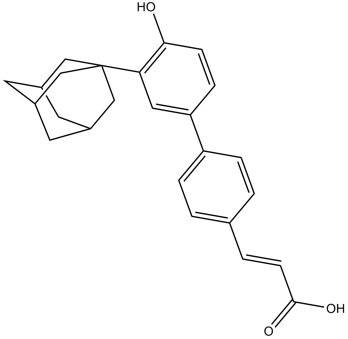
GC13959
Adarotene
非典型レチノイド
-
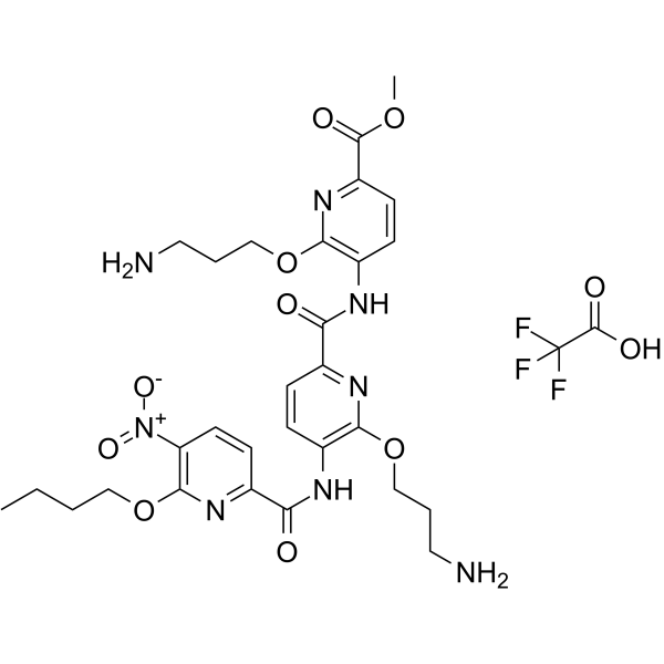
GC65880
ADH-6 TFA
ADH-6 TFA はトリピリジルアミド化合物です。 ADH-6 は、変異型 p53 DBD の凝集核形成サブドメインの自己組織化を無効にします。 ADH-6 TFA は、ヒト癌細胞の変異型 p53 凝集体を標的にして解離し、p53' の転写活性を回復させ、細胞周期の停止とアポトーシスを引き起こします。 ADH-6 TFA は、がん疾患の研究の可能性を秘めています。
-
GC42735
Adipostatin A
アジポスタチン A (アジポスタチン A) は、4.1 μM の IC50 を持つグリセロール-3-リン酸脱水素酵素 (GPDH) 阻害剤です。

-
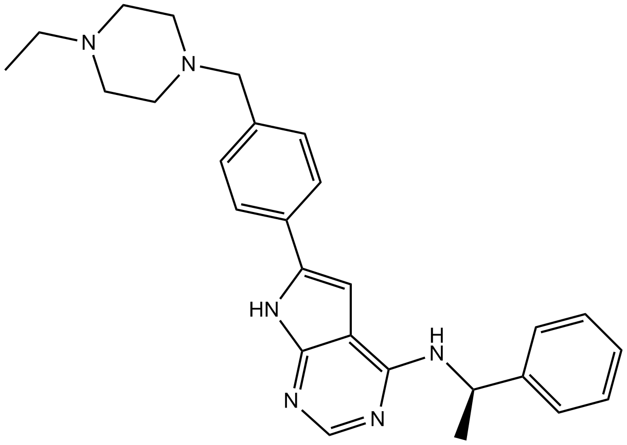
GC11892
AEE788 (NVP-AEE788)
AEE788 (NVP-AEE788) は、それぞれ 2 および 6 nM の IC50 値を持つ EGFR および ErbB2 の阻害剤です。
-
GC42743
AEM1
AEM1 は Nrf2 阻害剤です。 AEM1 は、A549 細胞における Nrf2 依存性遺伝子の発現を低下させ、in vitro および in vivo で A549 細胞の増殖を阻害します。

-
GC13168
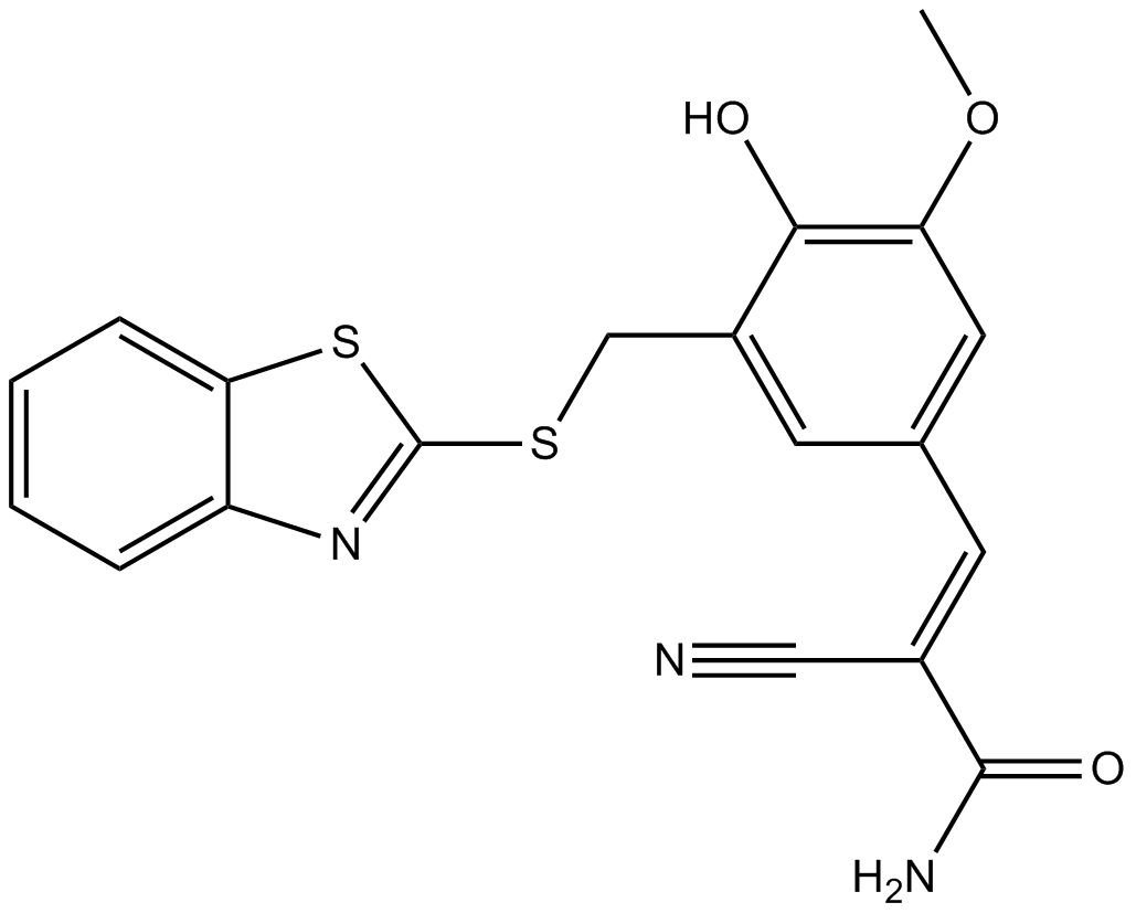
AG 825
AG 825 (Tyrphostin AG 825) は、0.35 μM の IC50 でチロシンリン酸化を抑制する、選択的かつ ATP 競合的な ErbB2 阻害剤です。
-
GC13697
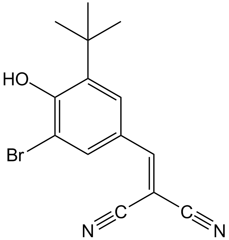
AG-1024
AG-1024 (Tyrphostin AG 1024) は可逆的で競合的かつ選択的な IGF-1R 阻害剤であり、IC50 は 7 μM です。
-
GC17881
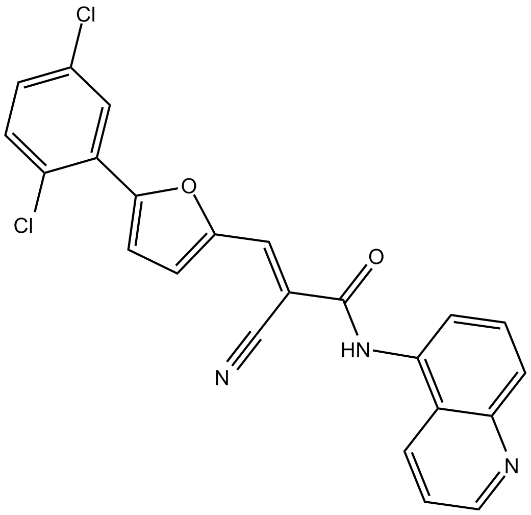
AGK 2
AGK 2 は、IC50 が 3.5 μM の選択的 SIRT2 阻害剤です。 AGK 2 は、SIRT1 および SIRT3 をそれぞれ 30 および 91 μM の IC50 で阻害します。
-
GC39584
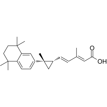
AGN194204
AGN194204(IRX4204)は、RXRα、RXRβ、およびRXRγに対してKd値がそれぞれ0.4 nM、3.6 nM、および3.8 nMであり、EC50値が分別で0.2 nM、0.8 nM、および0.08 nMの経口投与可能な選択的なRXRアゴニストです。
-
GC16120
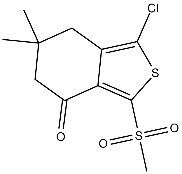
AI-3
Nrf2/Keap1およびKeap1/Cul3相互作用阻害剤
-
GC46821
Ajoene
ニンニク由来の化合物であるアホエンは、抗血栓剤および抗真菌剤です.

-
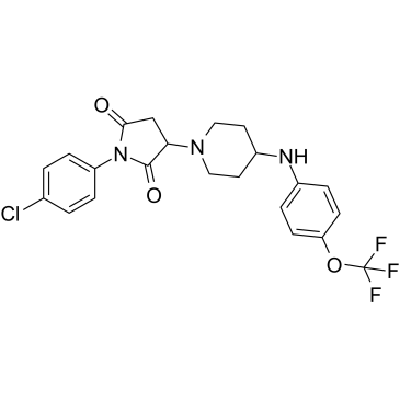
GC39620
AKOS-22
-
GC11589
AKT inhibitor VIII
Akt1とAkt2の強力な阻害剤

-
GC35275
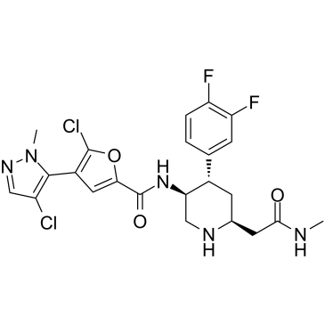
AKT-IN-3
AKT-IN-3 (化合物 E22) は、Akt1、Akt2、および Akt3 に対してそれぞれ 1.4 nM、1.2 nM、および 1.7 nM の強力な経口活性の低 hERG 遮断 Akt 阻害剤です。 AKT-IN-3 (化合物 E22) は、PKA、PKC、ROCK1、RSK1、P70S6K、および SGK などの他の AGC ファミリー キナーゼに対しても優れた阻害活性を示します。 AKT-IN-3 (化合物 E22) はアポトーシスを誘導し、癌細胞の転移を阻害します。
-
GC49773
Albendazole sulfone-d3
アルベンダゾールスルホンの定量化のための内部標準

-
GC48848
Albendazole-d7
アルベンダゾール-d7 (SKF-62979-d7) は、アルベンダゾールで標識された重水素です。

-
GC41080
Albofungin
アルボフンギンは、Aから分離されたキサントンです。

-
GC16597
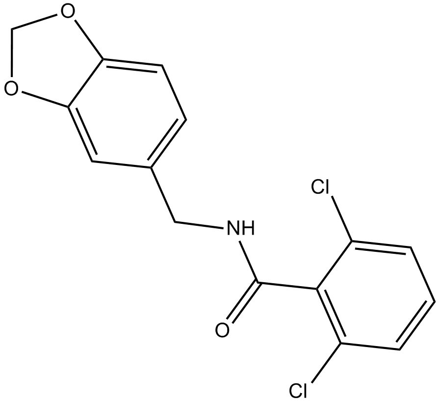
Alda 1
Alda 1 は強力で選択的な ALDH2 アゴニストであり、野生型 ALDH2 を活性化し、野生型に近い活性を ALDH2*2 に回復させます。
-
GC35288
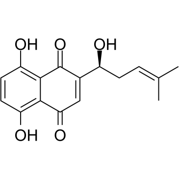
Alkannin
アルカンニンは、腫瘍特異的ピルビン酸キナーゼ-M2 (PKM2) の強力かつ特異的な阻害剤です。
-
GC49393
all-trans-13,14-Dihydroretinol
全トランスレチノイン酸の代謝物

-
GC32127
Alofanib (RPT835)
アロファニブ (RPT835) (RPT835) は、線維芽細胞増殖因子受容体 2 (FGFR2) の強力かつ選択的なアロステリック阻害剤です。

-
GC14314
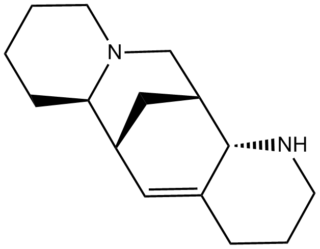
Aloperine
アロペリンは、ソフォラ alopecuroides L などのソフォラ植物に含まれるアルカロイドで、抗がん、抗炎症、抗ウイルスの特性を示しています 。
-
GC35306
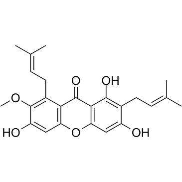
alpha-Mangostin
α-マンゴスチン (α-マンゴスチン) は、抗酸化作用、抗アレルギー作用、抗ウイルス作用、抗菌作用、抗炎症作用、抗がん作用などの幅広い生物学的活性を持つ食事性キサントンです。これは、2.85 μM の Ki を持つ変異型 IDH1 (IDH1-R132H) の阻害剤です。
-
GC18437
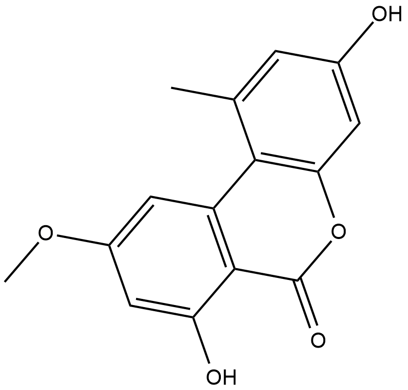
Alternariol monomethyl ether
Anthocleista djalonensis (Loganiaceae) の根から分離された Alternariol モノメチル エーテルは、植物種の重要な分類学的マーカーです。
-
GC33356
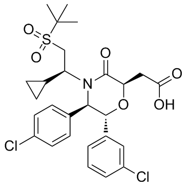
AM-8735
AM-8735 は、IC50 が 25 nM の強力で選択的な MDM2 阻害剤です。
-
GC42776
Amarogentin
アマロゲンチンは、主にセンブリとリンドウの根から抽出されるセコイリドイド配糖体です。

-
GN10484
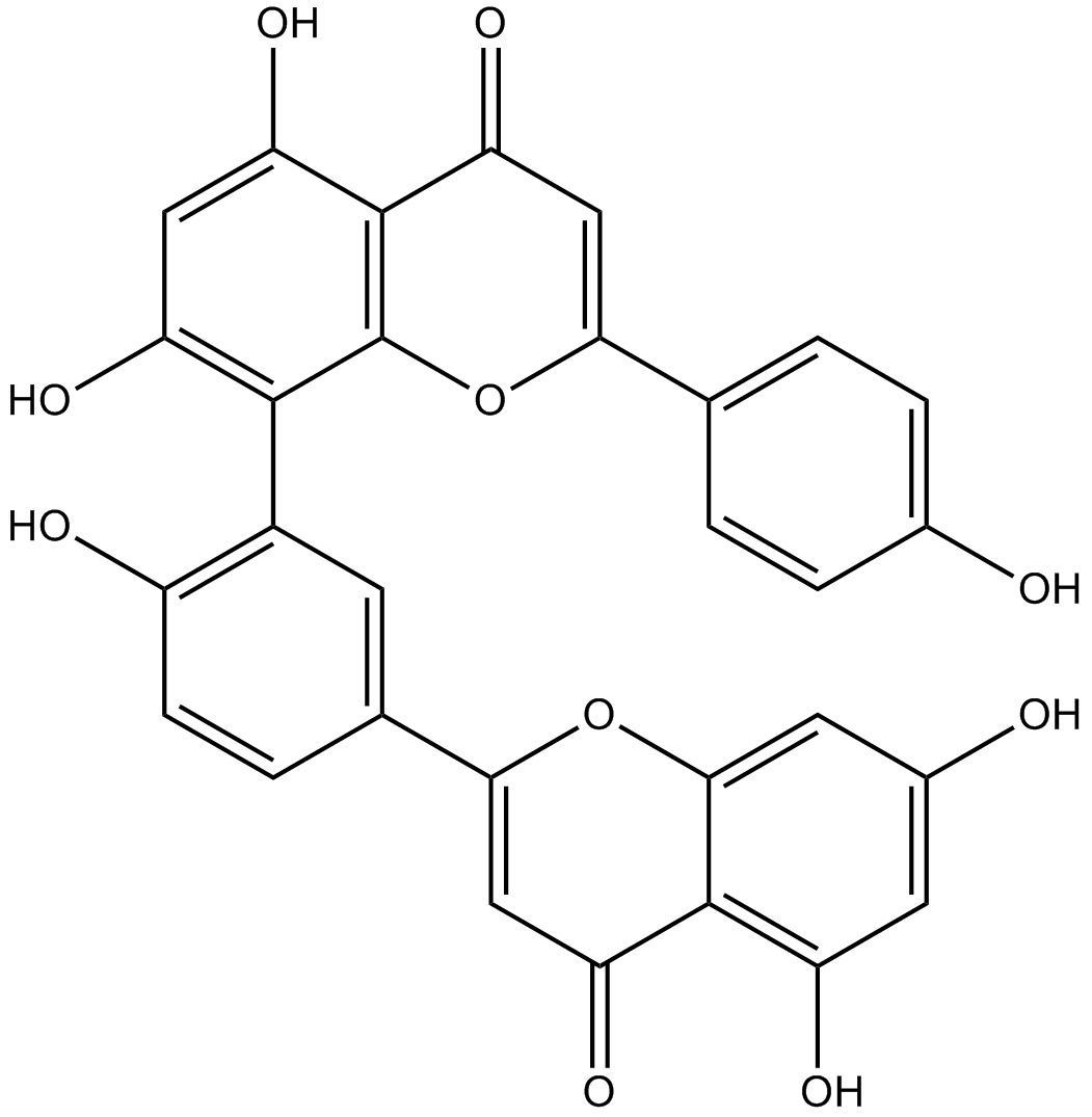
Amentoflavone
-
GC42783
Ametantrone
アメタントロン (NSC 196473) は、DNA にインターカレートし、トポイソメラーゼ II (TOP2) を介した DNA 切断を誘導する抗腫瘍剤です。

-
GC19452
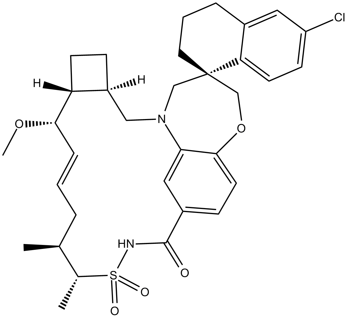
AMG-176
AMG-176 (AMG-176) は、0.13 nM の Ki を持つ強力で選択的な経口活性 MCL-1 阻害剤です。
-
GC15828
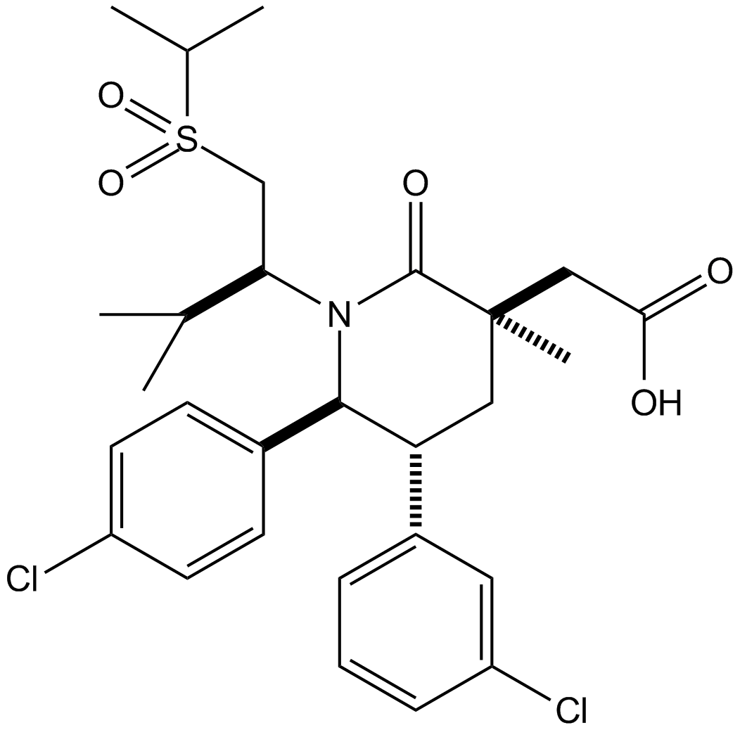
AMG232
AMG232 (AMG 232) は、p53-MDM2 相互作用の強力で選択的な経口利用可能な阻害剤であり、IC50 は 0.6 nM です。 AMG232 は 0.045 nM の Kd で MDM2 に結合します。
-
GC42785
Amifostine (hydrate)
アミフォスチン (水和物) (WR2721 三水和物) は、広域スペクトルの細胞保護剤および放射線防護剤です。アミフォスチン (水和物) は、放射線や化学療法による損傷から正常な組織を選択的に保護します。アミフォスチン (水和物) は、強力な低酸素誘導因子-α1 (HIF-α1) および p53 インデューサーです。アミフォスチン (水和物) は、酸素由来のフリーラジカルを除去することにより、細胞を損傷から保護します。アミフォスチン(水和物)は、腎毒性を軽減し、血管新生阻害作用があります。

-
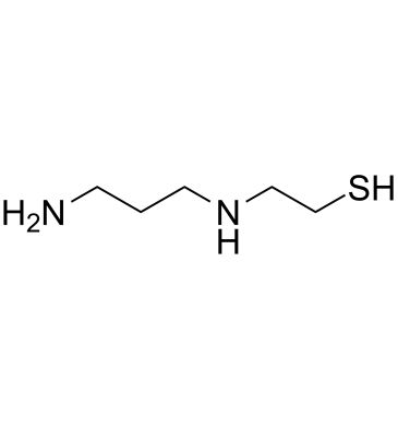
GC61804
Amifostine thiol
アミフォスチン チオール (WR-1065) は、細胞保護剤であるアミフォスチンの活性代謝物です。アミフォスチン チオールは、放射線防護能力を持つ細胞保護剤です。アミフォスチン チオールは、JNK 依存性シグナル伝達経路を介して p53 を活性化します。
-
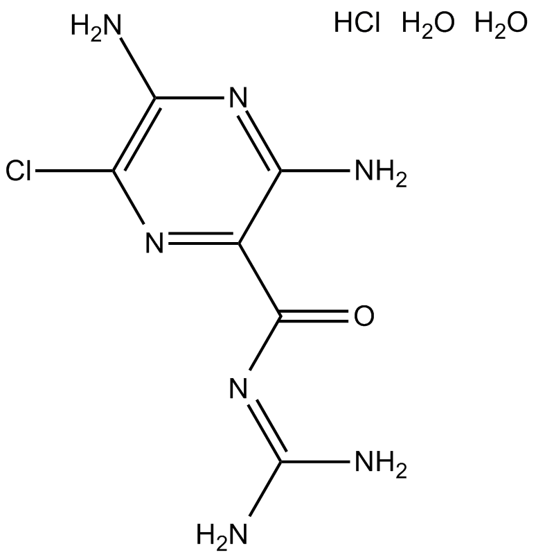
GC12051
Amiloride HCl dihydrate
アミロリド HCl 二水和物 (MK-870 塩酸塩二水和物) は、上皮ナトリウム チャネル (ENaC[1]) とウロキナーゼ型プラスミノーゲン活性化因子受容体 (uTPA[2]) の両方の阻害剤です。
-
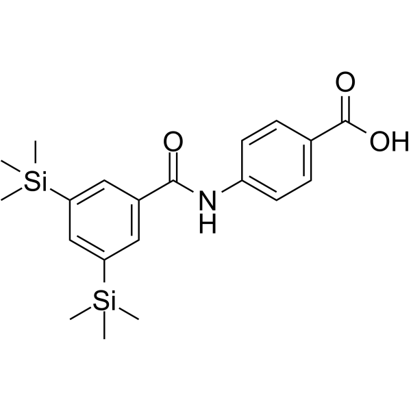
GC63932
Amsilarotene
アムシラロテン (TAC-101; Am 555S) は、経口で活性な合成レチノイドであり、レチノイン酸受容体 α に対して選択的な親和性があります。 (RAR-α) RAR-α に対して 2.4、400 nM の Ki で結合。 RAR-β.アムシラロテンは、ヒトの胃癌、肝細胞癌、卵巣癌細胞のアポトーシスを誘導します。アムシラロテンは癌の研究に使用できます。
-
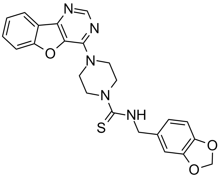
GC16391
Amuvatinib (MP-470, HPK 56)
多標的RTK阻害剤
-
GC48339
Amycolatopsin A
抗マイコバクテリアおよび抗がん作用を持つマクロライドポリケチド。

-
GC48341
Amycolatopsin B
細菌代謝産物

-
GC48350
Amycolatopsin C
抗マイコバクテリアおよび抗がん作用を持つポリケチド・マクロライド。

-
GC42806
Andrastin A
アンドラスチンAは、メロテルペノイド・ファルネシルトランスフェラーゼ阻害剤です。

-
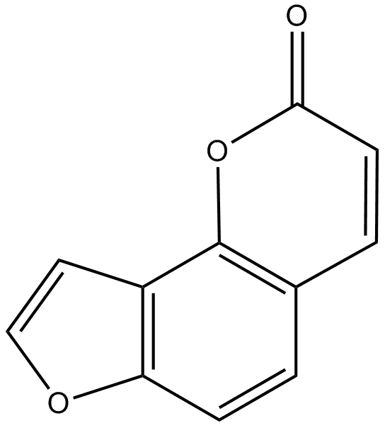
GN10045
Angelicin
-
GC60584
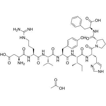
Angiotensin II human acetate
アンジオテンシン II ヒト (アンジオテンシン II) アセテートは、血管収縮剤であり、レニン/アンギオテンシン系の主要な生理活性ペプチドです。
-
GC42813
Anguinomycin A
アングイノマイシンAは、最初にストレプトマイセス属から単離された抗生物質です。

-
GC40614
Anhydroepiophiobolin A
オフィオボリン A の類似体であるアンヒドロエピオフィボリン A は、光合成の強力な阻害剤です (クロレラとほうれん草の光合成の I50 はそれぞれ 6.1 mM と 1 mM)。

-
GC40214
Anhydroophiobolin A
アンヒドロオフィオボリン A は、クロレラとほうれん草の光合成における IC50 がそれぞれ 77 mM と 14 mM の強力な光合成阻害剤です。

-
GC11559
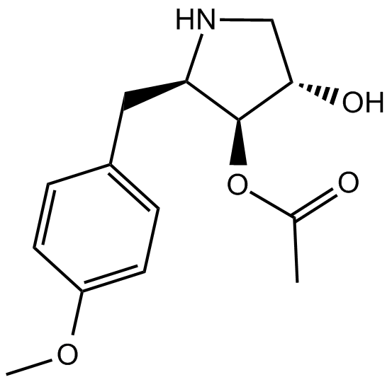
Anisomycin
JNKアゴニスト、強力かつ特異的
-
GC49259
Antagonist G (trifluoroacetate salt)
神経ペプチド拮抗剤

-
GC66337
Anti-Mouse PD-L1 Antibody
Anti-Mouse PD-L1 Antibody は、宿主ラット由来の抗マウス PD-L1 IgG2b 抗体阻害剤です。
-
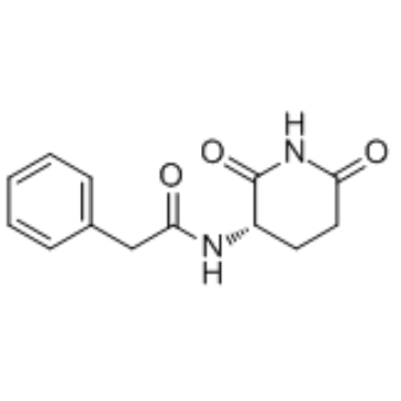
GC35361
Antineoplaston A10
アンチネオプラストン A10 は、人体に自然に存在する物質であり、神経膠腫、リンパ腫、星状細胞腫、および乳癌の治療に潜在的な Ras 阻害剤です。
-
GC34172
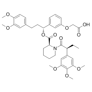
AP1867
AP1867 は合成 FKBP12F36V 指向性リガンドです。
-
GC61745
AP1867-2-(carboxymethoxy)
AP1867-2-(カルボキシメトキシ)、AP1867 (合成 FKBP12F36V 指向リガンド) ベースの部分は、リンカーを介して CRBN リガンドに結合し、dTAG 分子を形成します。

-
GC15586
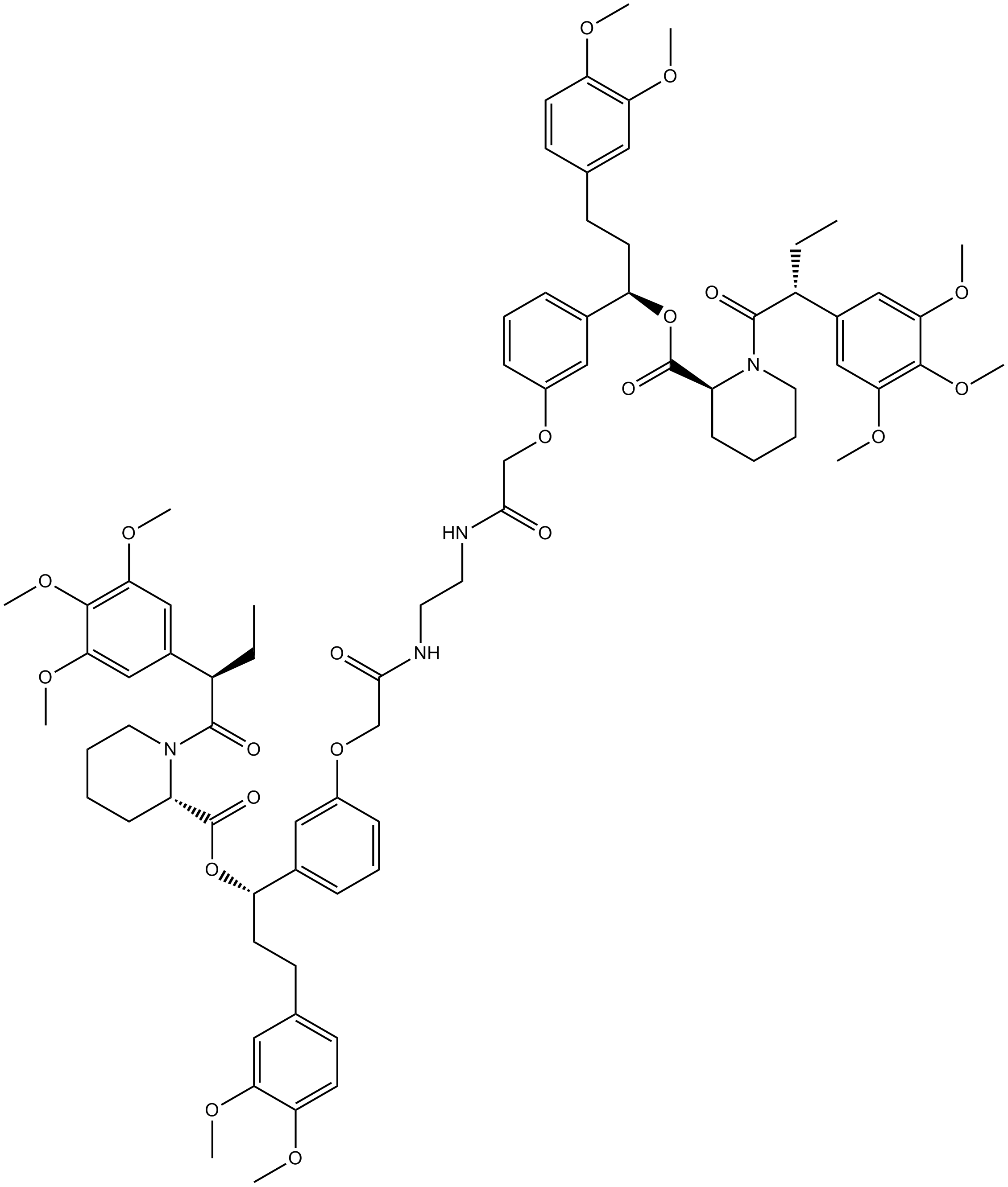
AP1903
AP1903 (AP1903) は、FKBP ドメインを架橋することによって作用するダイマー化剤です。 AP1903 (AP1903) は、カスパーゼ 9 自殺スイッチを二量体化し、アポトーシスを急速に誘導します。
-
GC14498
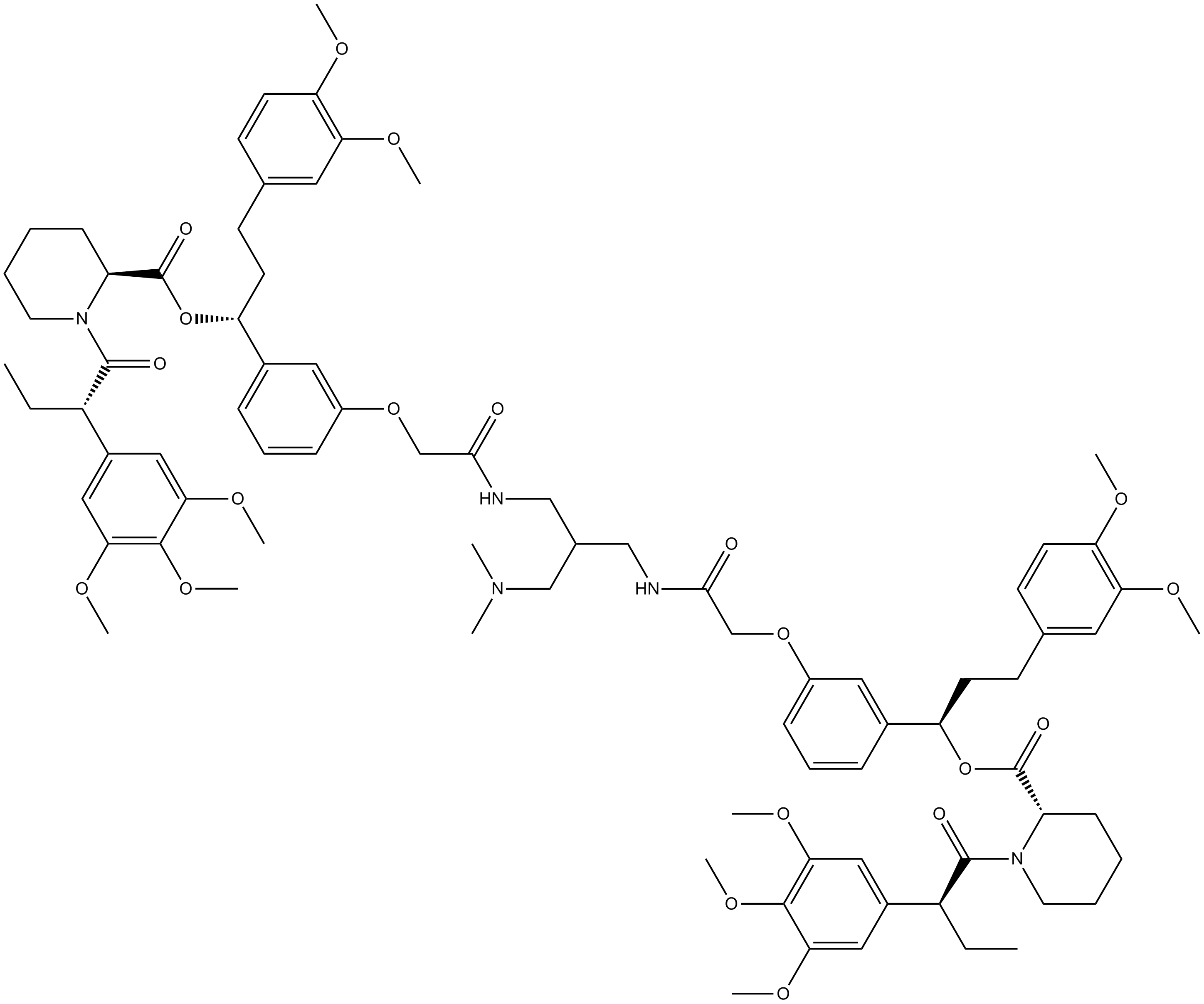
AP20187
AP20187 (B/B Homodimerizer) は、FK506 結合タンパク質 (FKBP) 融合タンパク質を二量体化し、生物学的シグナル伝達カスケードおよび遺伝子発現を開始するか、タンパク質間相互作用を破壊するために使用される細胞透過性リガンドです。
-
GC18518
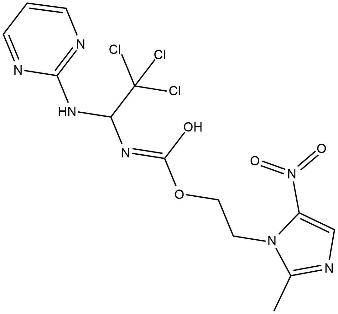
Apcin
Cdc20 のリガンドであるアプシンは、強力で競争力のある後期促進複合体/サイクロソーム (APC/C(Cdc20)) E3 リガーゼ活性阻害剤です。 Apcin は、Cdc20 に結合して基質認識を阻害することにより、APC/C 依存性ユビキチン化を競合的に阻害します。アプシンは、WD40 ドメインの側面にある D ボックス結合ポケットを占有し、有糸分裂を延長することができます。
-
GC62419
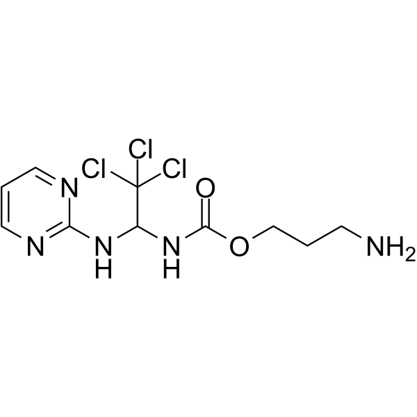
Apcin-A
アプシン誘導体であるアプシン-Aは、後期促進複合体(APC)阻害剤です。 Apcin-A は Cdc20 と強く相互作用し、Cdc20 基質のユビキチン化を阻害します。 Apcin-A は、PROTAC CP5V の合成に使用できます。
-
GC35367
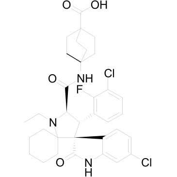
APG-115
APG-115 (APG-115) は、それぞれ 3.8 nM および 1 nM の IC50 および Ki 値で MDM2 タンパク質に結合する経口活性 MDM2 タンパク質阻害剤です。 APG-115 は MDM2 と p53 の相互作用を遮断し、p53 依存的に細胞周期の停止とアポトーシスを誘導します。
-
GC62640
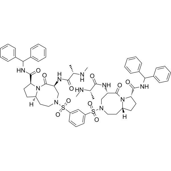
APG-1387
APG-1387 は、二価の SMAC 模倣薬であり、IAP アンタゴニストであり、IAP ファミリータンパク質 (XIAP、cIAP-1、cIAP-2、および ML-IAP) の活性をブロックします。 APG-1387 は、cIAP-1 および XIAP タンパク質の分解、ならびにカスパーゼ-3 活性化および PARP 切断を誘導し、アポトーシスを引き起こします。 APG-1387 は、肝細胞がん、卵巣がん、鼻咽頭がんの研究に使用できます。
-
GC12961
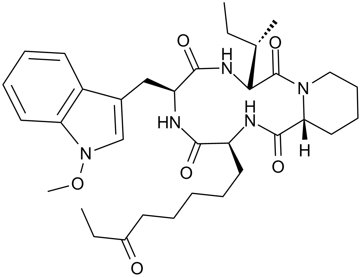
Apicidin
細胞膜透過性のHDAC阻害剤
-
GC46862
Apigenin-d5
アピゲニンの定量化のための内部標準

-
GC16237
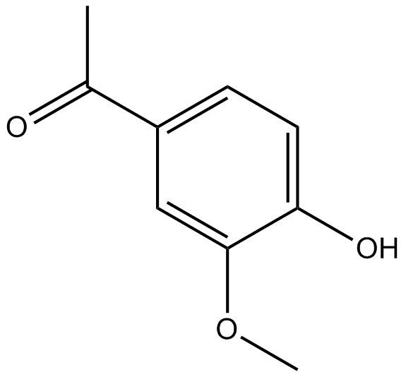
Apocynin
アポシニンは、IC50 が 10 μM の選択的 NADPH オキシダーゼ阻害剤です。
-
GC14080
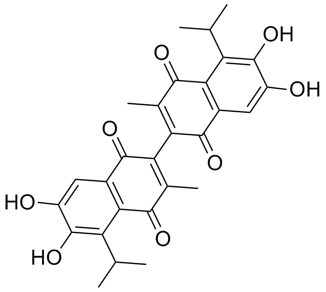
Apogossypolone (ApoG2)
-
GC42827
Apoptolidin
アポトリジンは、ノカルジオプシス菌から分離されたポリケチドです。アポトリジンは、選択的なミトコンドリア F1FO ATPase 阻害剤です。アポトリジンはアポトーシス誘導因子であり、ElA (IC50=10-17 ng/ml) を含むアデノウイルス 12 型癌遺伝子で形質転換された細胞ではアポトーシス細胞死を誘導しますが、正常細胞では誘導しません。

-
GC14209
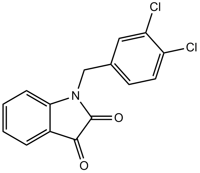
Apoptosis Activator 2
カスパーゼの活性化剤
-
GC14411
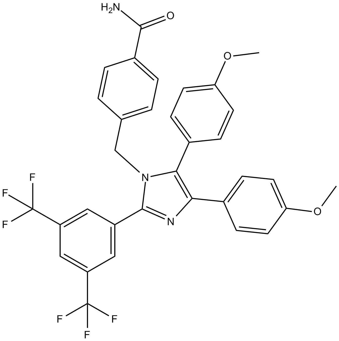
Apoptozole
Apoptozole (Apoptosis Activator VII) は、Hsc70 および Hsp70 の ATPase ドメインの阻害剤であり、Kds はそれぞれ 0.21 および 0.14 μM であり、アポトーシスを誘導することができます。
-
GC65004
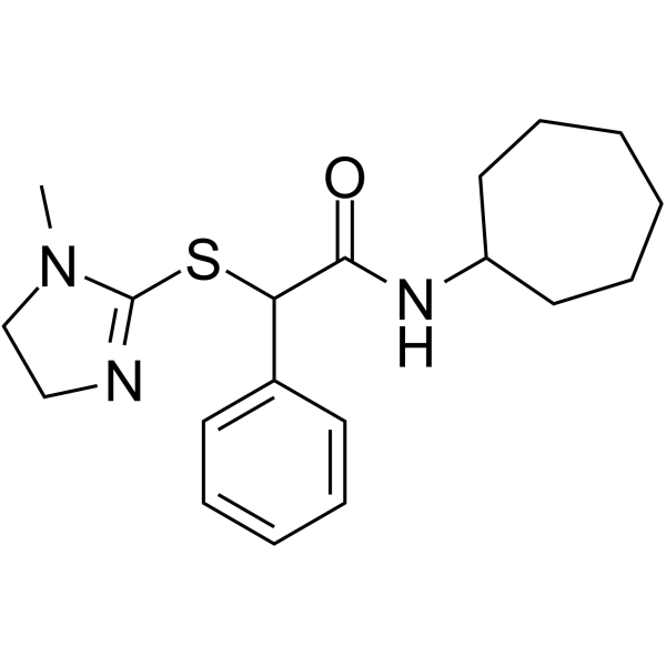
Apostatin-1
アポスタチン-1 (Apt-1) は強力な TRADD 阻害剤です。
-
GC35377
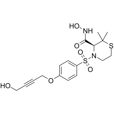
Apratastat
Apratastat (TMI-005) は、経口で活性な、非選択的で可逆的な TACE/MMPs 阻害剤であり、TNF-α の放出を阻害することができます。 Apratastat は、非小細胞肺癌 (NSCLC) の放射線治療抵抗性を克服する可能性を秘めています。
-
GC10420
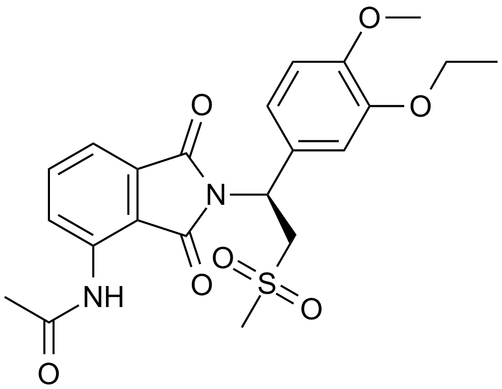
Apremilast (CC-10004)
経口投与可能なPDE4阻害剤
-
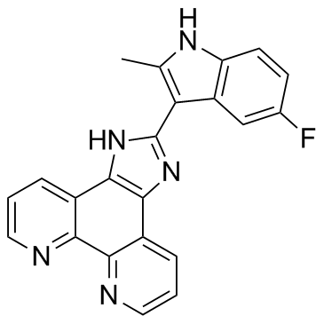
GC32692
APTO-253 (LOR-253)
APTO-253 (LOR-253) (LOR-253) は、c-Myc 発現を阻害し、G-四重鎖 DNA を安定化し、急性骨髄性白血病細胞の細胞周期停止とアポトーシスを誘導する低分子です。
-
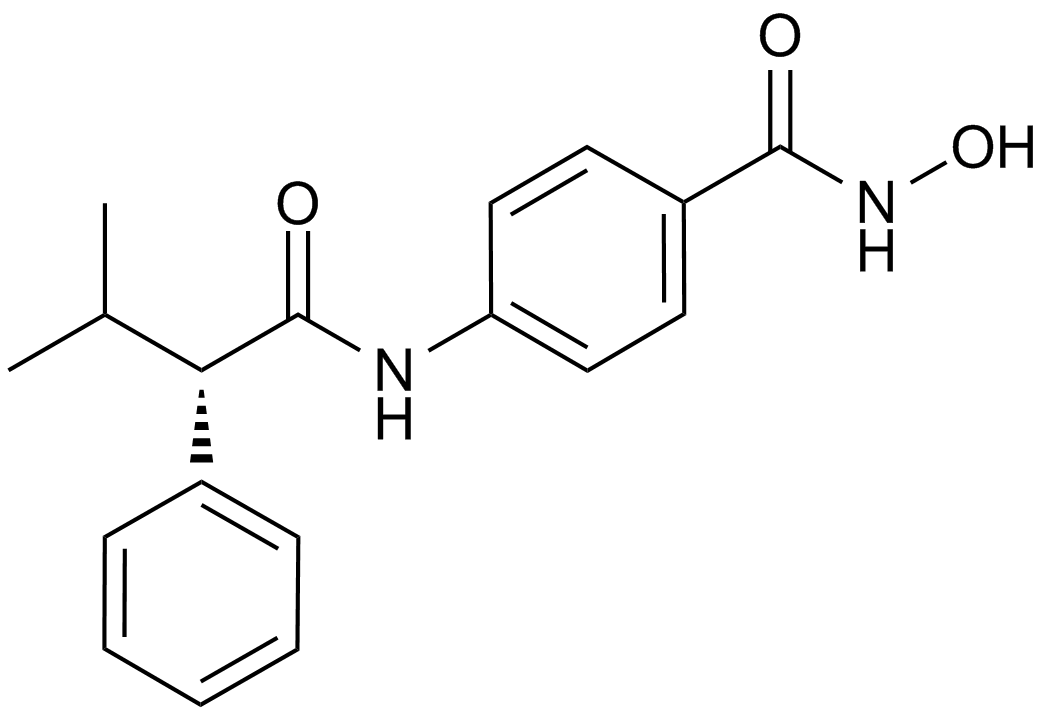
GC14590
AR-42 (OSU-HDAC42)
HDAC阻害剤、新規かつ強力な
-
GC45385
Ara-G

-
GC46878
Aranciamycin
多様な生物学的活性を持つ真菌代謝産物

-
GC40116
Aranorosin
強力な抗真菌性抗生物質であるアラノロシンは、Pseudoarachniotus roseus Kuehn 株の培養濾液と菌糸体から分離されました 。

-
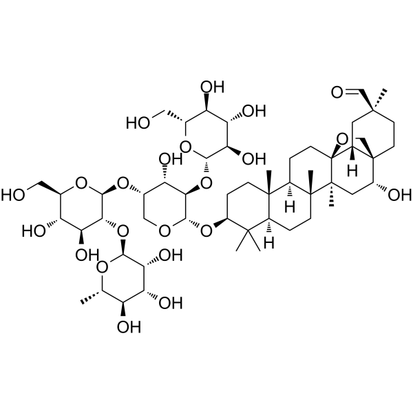
GC65163
Ardisiacrispin B
Ardisiacrispin B は、フェロトーシスおよびアポトーシス細胞死を介して、多因子薬剤耐性癌細胞に細胞毒性効果を示します。
-
GC49314
Arecaidine propargyl ester (hydrobromide)
ムスカリン性M2アゴニスト

-
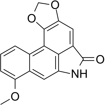
GC35388
Aristolactam I
アリストロラクタム I (AL-I) は、アリストロク酸 I (AA-I) の主要な代謝産物であり、腎障害を引き起こすプロセスに関与しています。
-
GC35395
Arnicolide D
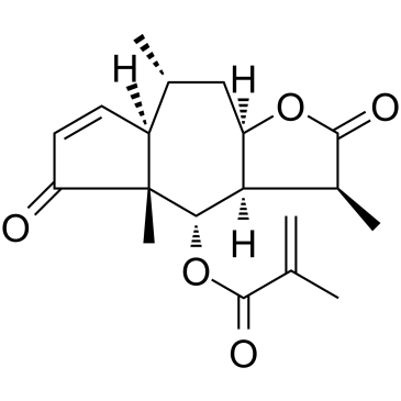
アルニコリド D は、ムカデ ミニマから分離されたセスキテルペン ラクトンです。アルニコライド D は、細胞周期を調節し、カスパーゼ シグナル伝達経路を活性化し、PI3K/AKT/mTOR および STAT3 シグナル伝達経路を阻害します。アルニコライド D は、鼻咽頭癌 (NPC) の細胞生存率を濃度および時間依存的に阻害します。
-
GC19037
ARS-853
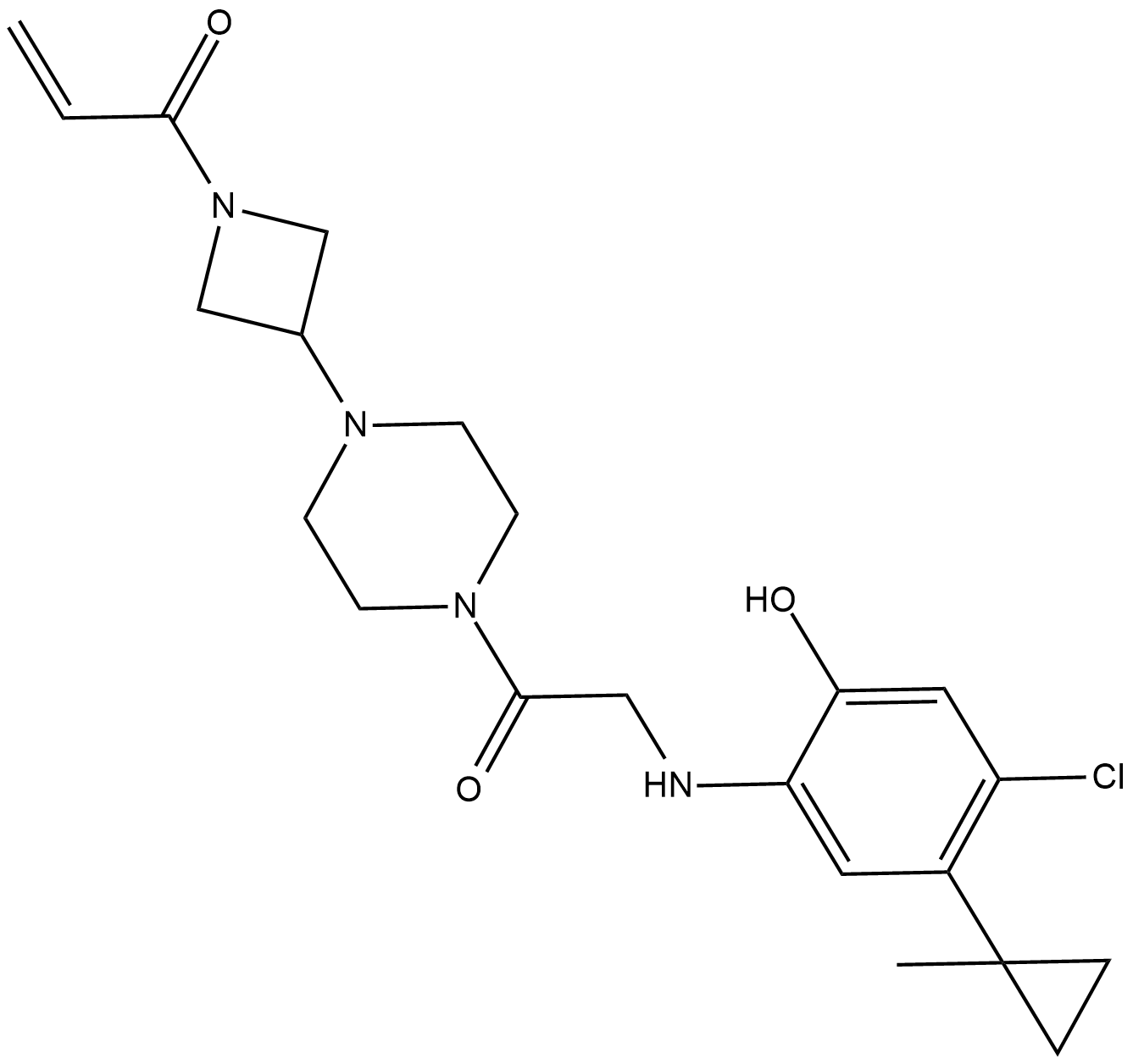
ARS-853 は、2.5 μM の IC50 を持つ細胞活性、選択的、共有結合の KRAS G12C 阻害剤です。 ARS-853 は、GDP 結合癌タンパク質に結合して活性化を防止することにより、変異型 KRAS 駆動シグナル伝達を阻害します。
-
GC46882
Artemisinin-d3
アルテミシニンの定量化のための内部標準

-
GC10040
Arylquin 1
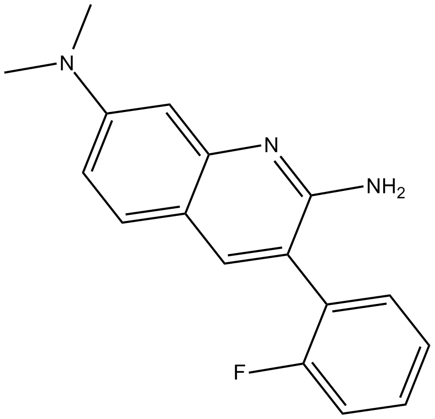
前立腺アポトーシス応答 4 (Par-4) 分泌促進物質である Arylquin 1 は、ビメンチンを標的として Par-4 分泌を誘導します。 Arylquin 1 は、リソソーム膜透過処理 (LMP) の誘導を通じて、癌細胞の非アポトーシス細胞死を誘導します。
-
GC62615
AS-99
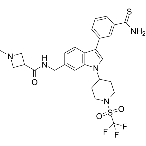
AS-99 は、抗白血病活性を有するファーストインクラスの強力かつ選択的な ASH1L ヒストンメチルトランスフェラーゼ阻害剤 (IC50=0.79μM、Kd=0.89μM) です。 AS-99 は、細胞増殖をブロックし、アポトーシスと分化を誘導し、MLL 融合標的遺伝子をダウンレギュレートし、in vivo での白血病の負担を軽減します。
-
GC40715
Ascochlorin
イソプレノイド抗生物質であるアスコクロリン (イリシコリン D) は、主に STAT3 シグナル伝達カスケードの抑制を介してその抗腫瘍効果を媒介します。

-
GC13215
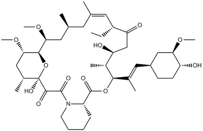
Ascomycin(FK 520)
強力なマクロライド免疫抑制剤
-
GC12070
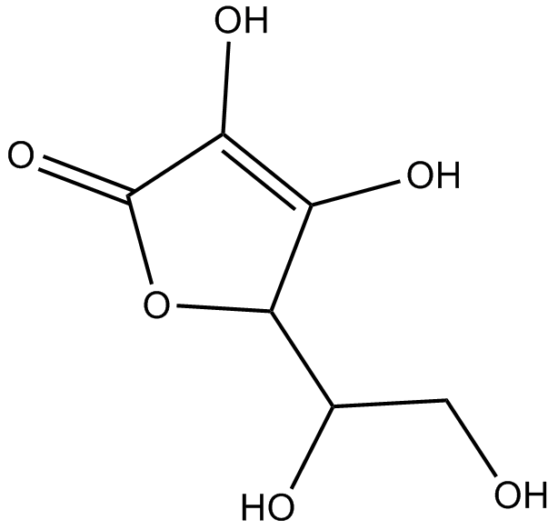
Ascorbic acid
電子供与体
-
GN10702
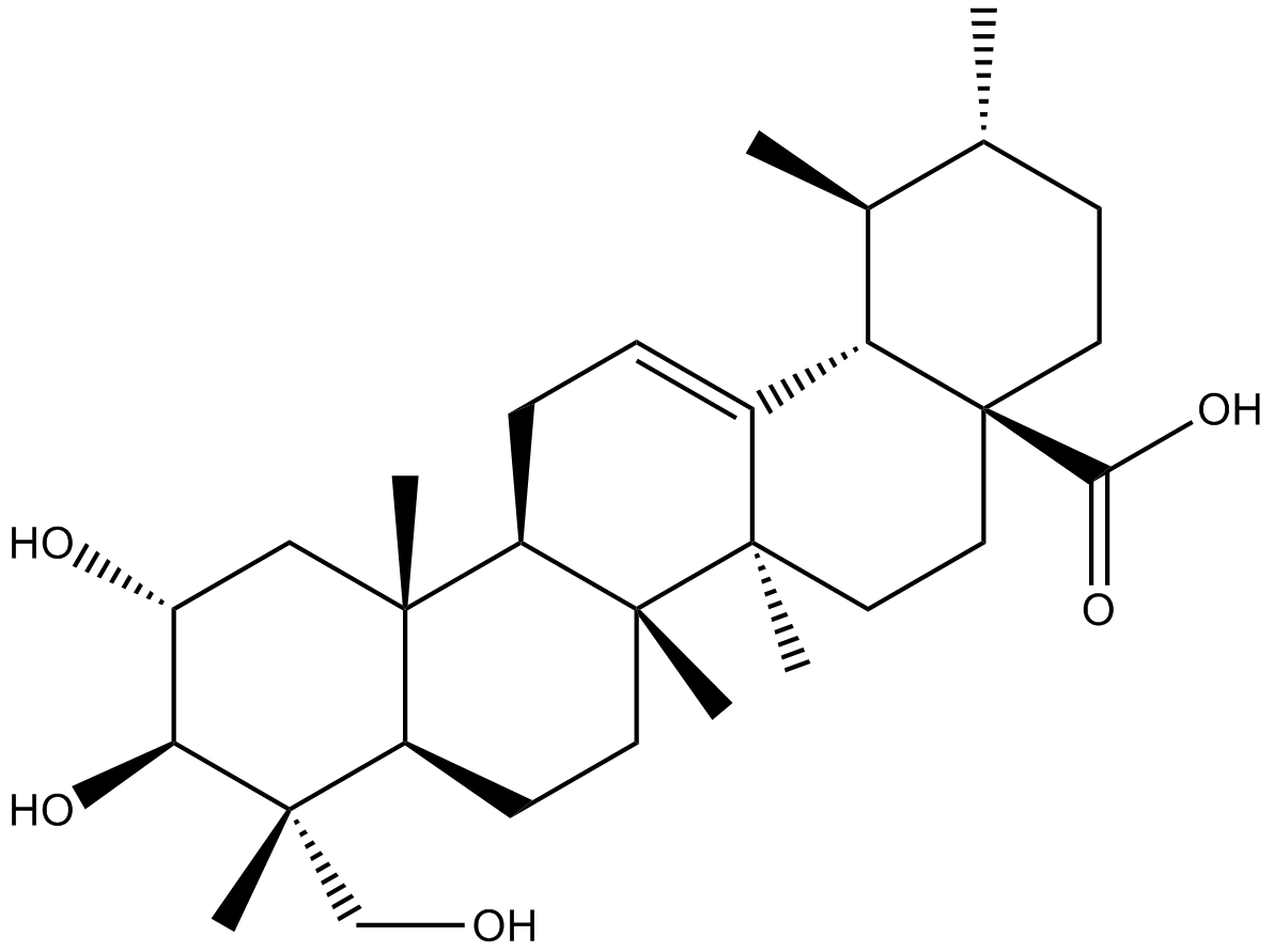
Asiatic acid
-
GN10534
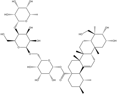
Asiaticoside
-
GC19041
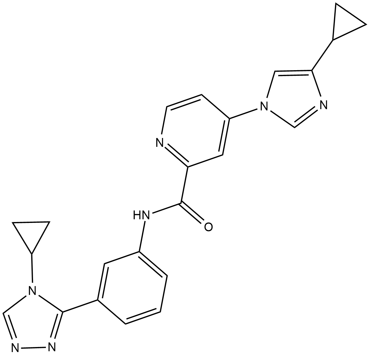
ASK1-IN-1
ASK1-IN-1 は、2.87 nM の IC50 を持つアポトーシスシグナル調節キナーゼ 1 (ASK1) の強力な経口投与可能な選択的 ATP 競合阻害剤です。
-
GC62426
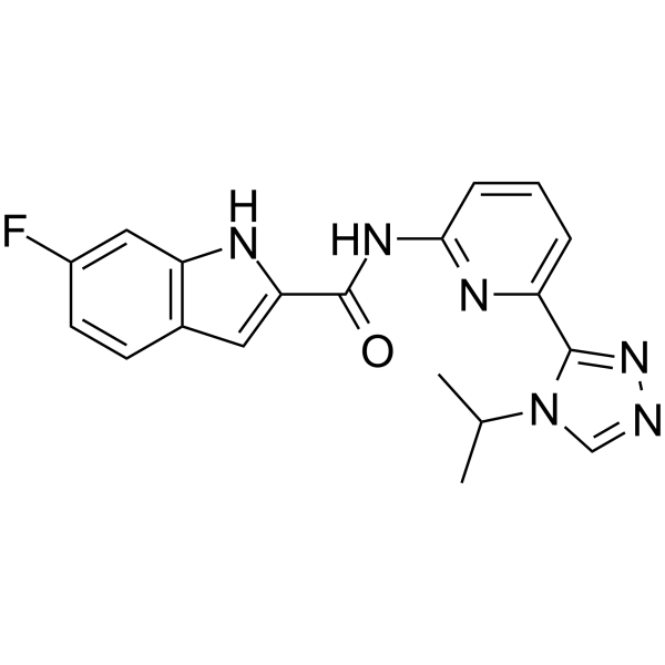
ASK1-IN-2
ASK1-IN-2 は、アポトーシスシグナル調節キナーゼ 1 (ASK1) の強力な経口活性阻害剤であり、IC50 は 32.8 nM です。
-
GC42858
Aspergillin PZ
Aspergillin PZ は、Aspergillus awamori 由来の新規イソインドールアルカロイドです。

-
GN10064
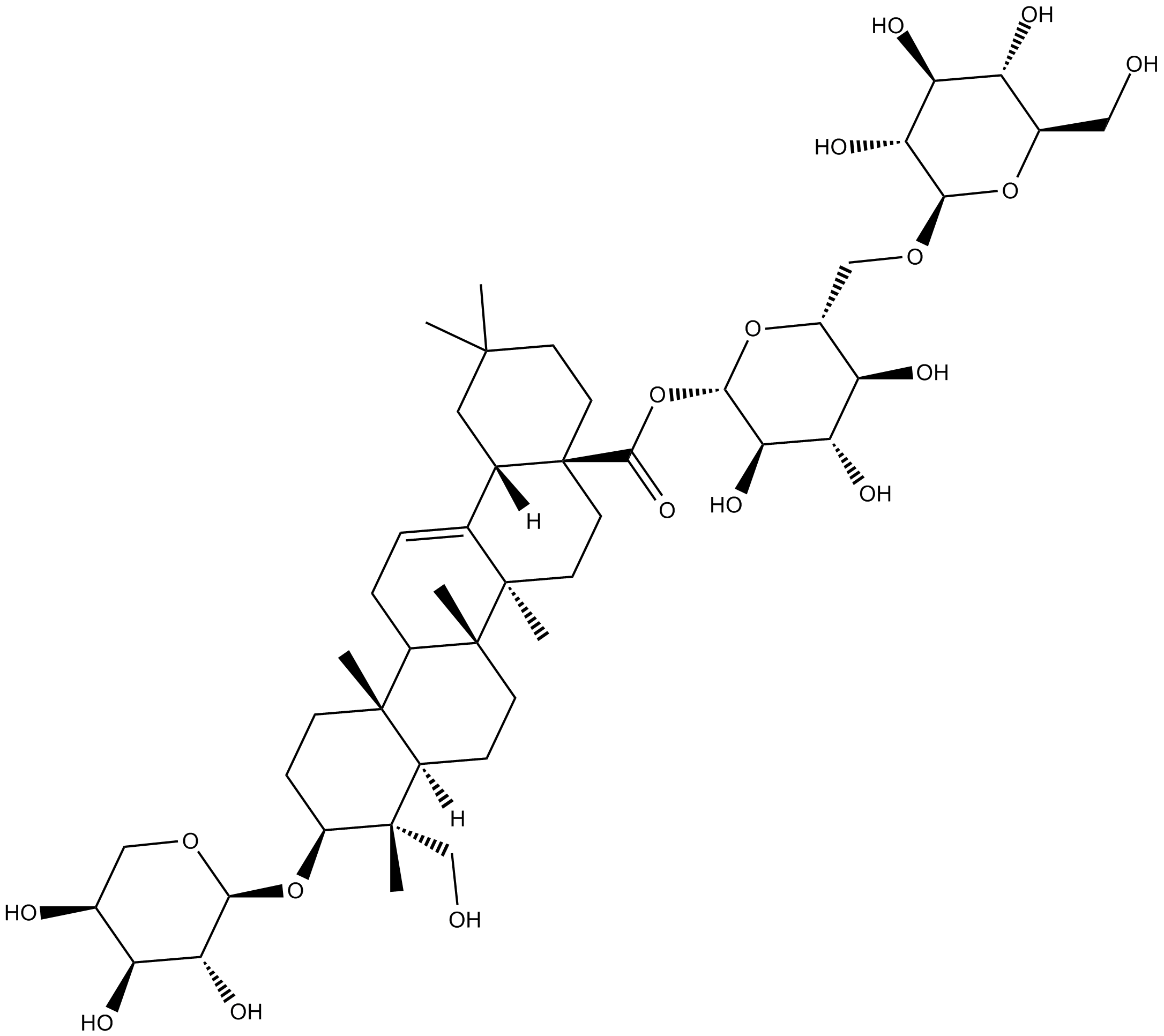
Asperosaponin VI
-
GC60603
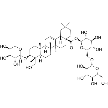
Asperosaponin VI
多様な生物学的活性を持つトリテルペノイドサポニン
-
GC42860
Aspochalasin D
アスポカラシンDは、元々Aから分離された共代謝物質です。

-
GC41640
Asterriquinol D dimethyl ether
アステリキノール D ジメチル エーテルは真菌代謝産物であり、マウス骨髄腫 NS-1 細胞株を 28 μg/mL の IC50 で阻害できます。

-
GN10415
Astilbin
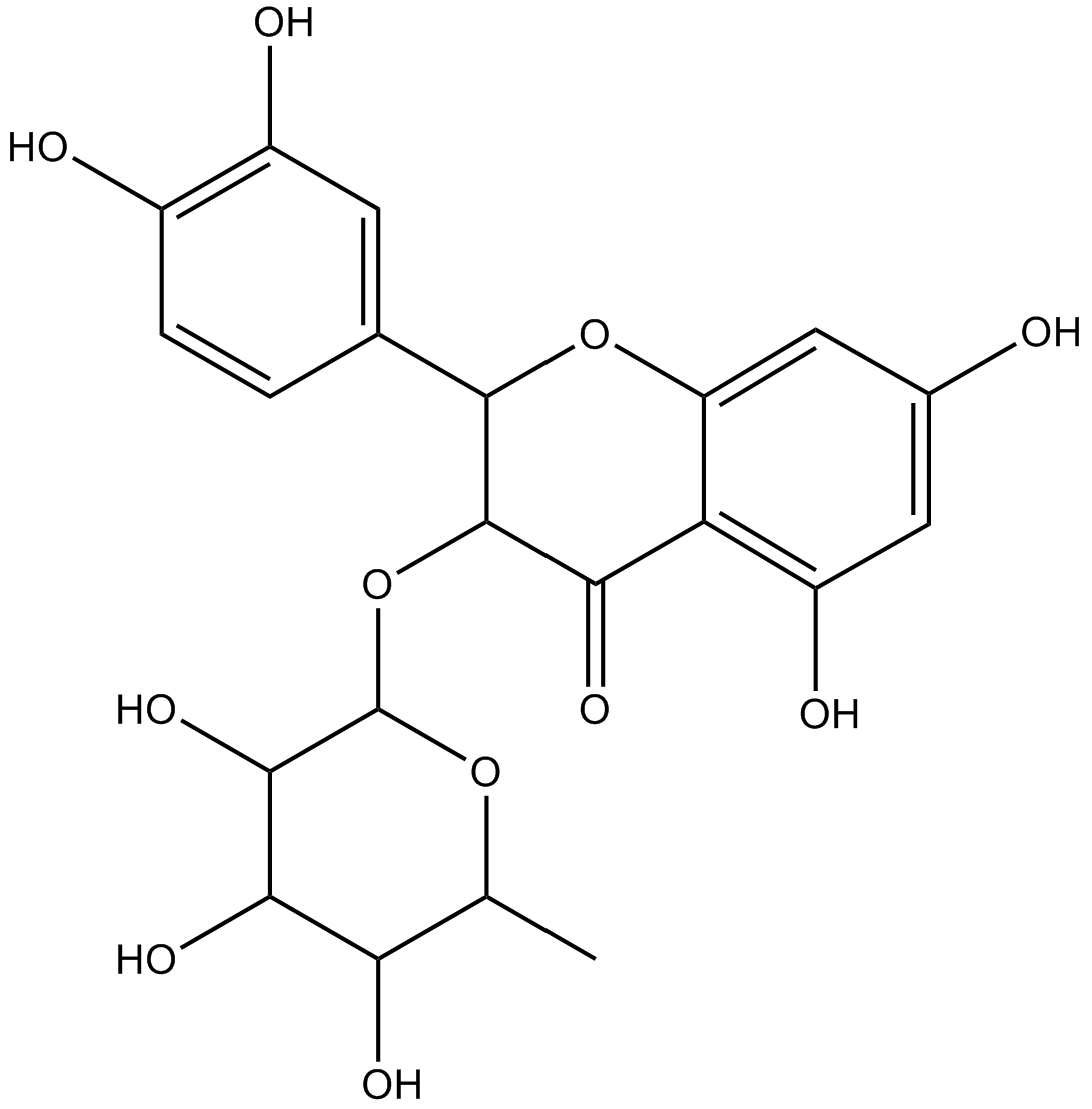
-
GN10561
astragalin
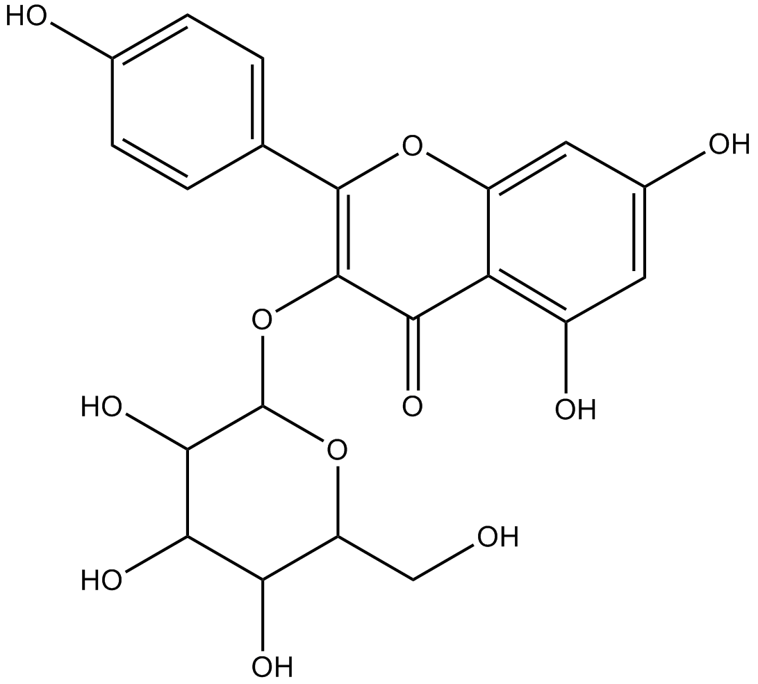
-
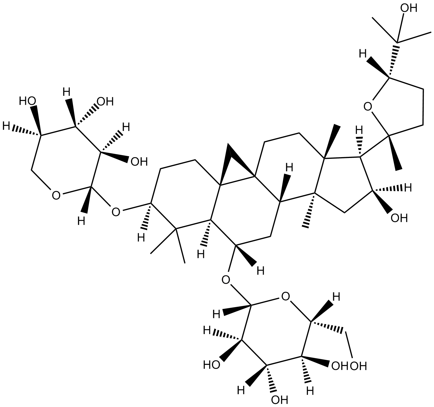
GC18109
Astragaloside A
抗高血圧作用、陽性変力作用、抗炎症作用、および心筋損傷の防止
-
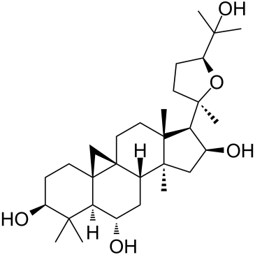
GC35415
Astramembrangenin
-
GC32803
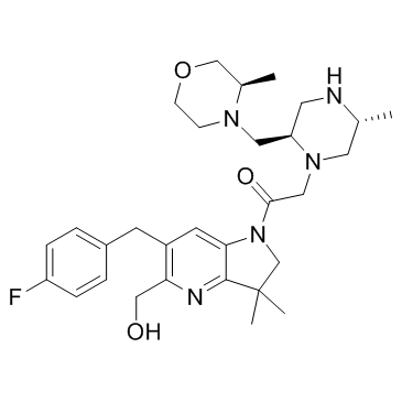
ASTX660
ASTX660 は、細胞内アポトーシス抑制タンパク質 (cIAP) と X リンク型アポトーシス抑制タンパク質 (XIAP) の経口投与可能なデュアル アンタゴニストです。
-
GC42863
Asukamycin
アスカマイシンは、Sから分離されたポリケチドです。

-
GC11106
AT-101
