Bafilomycin A1 (Synonyms: NSC 381866) |
Catalog No.GC17597 |
Bafilomycin A1은 Streptomyces로부터 분리된 큰 고리형 항생제로, 액포 H ATPase (V-ATPase)의 억제제이다.
Products are for research use only. Not for human use. We do not sell to patients.
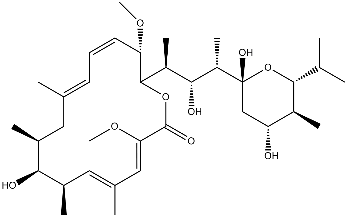
Cas No.: 88899-55-2
Sample solution is provided at 25 µL, 10mM.
Bafilomycin A1은 Streptomyces로부터 분리된 큰 고리형 항생제로, 액포 H ATPase (V-ATPase)의 억제제이다.Bafilomycin A1은 오토파지 (autophagy)를 연구하기 위해 오토파지-리소솜 융합 억제제 및 리소솜 분해 억제제로 사용되어 왔다.V-ATPase 복합체의 V0 sector subunit c와 결합하여 처리된 세포의 세포질에 H의 축적을 일으키는 H 전이를 억제한다. Bafilomycin A1은 세포의 성장을 억제하여 세포사멸과 세포분화를 일으킨다. Bafilomycin A1의 이러한 항암 작용은 V-ATPase의 억제로 인한 세포 내 산증 때문인 것으로 알려져 있다.
Bafilomycin A1의 체외 연구에 따르면Bafilomycin A1이 소아 b 세포 급성 림프모구 백혈병 세포의 In vitro 성장을 우선적으로 억제함을 나타내었다.B-ALL (697, Nalm-6, RS4;11, NB4, HL-60, K562, BV173)을 나타내는 697, Nalm-6, RS4;11, NB4, HL-60), 급성 골수성 백혈병 (NB4, HL-60), 만성 골수성 백혈병 (K562, BV173)을 포함하는 다양한 백혈병 세포주를 이용하여 Bafilomycin A1의 효과를 조사하였다.Bafilomycin A1 (0 nM, 0.5 nM, 1 nM)의 농도가 증가하는 상태에서 세포를 배양하였다.세포증식은 MTT assay를 이용하여 측정하였다.그 결과 Bafilomycin A1은 자주 장애물 로서 사용 되어 왔 autophagosomes-lysosomes 융합을 차단, 또는에서 lysosomal 활동을 억제 하기 위해 고용량 (m 0.1-1 μ)다.낮은 농도의 Bafilomycin A1 (1 nM) cpuld 가 효과적이고 구체적으로 오토파지를 불활성화시키고 소아 β-all 세포의 여러 표적에서 apoptosis를 특이하게 억제하며 여러 표적에 대한 세포사멸을 활성화시킨다.
체내 연구 결과, 대조군에 비해 Bafilomycin A1이 B-ALL 말기 생쥐의 생존기간을 연장시켰다. B-ALL 모델군의 적혈구 수, 헤모글로빈, 혈소판 수는 현저하게 감소하였으나 Bafilomycin A1치료군의 적혈구 수, 헤모글로빈, 혈소판 수는 정상으로 회복되었다. B-ALL모델 쥐의 간 및 비대 비대는 대조군에 비해 심각하였다.질병모델 그룹과 비교해 Bafilomycin A1치료 그룹은 간, 비장의 크기와 무게가 모두 정상으로 나타났다. 그 결과 Bafilomycin A1은 생체 내 백혈병 세포를 표적으로 하여 소아 B-ALL 세포 생성을 극적으로 억제하는 것으로 나타났다.
References:
[1]. Yamamoto A, et al. Bafilomycin A1 prevents maturation of autophagic vacuoles by inhibiting fusion between autophagosomes and lysosomes in rat hepatoma cell line, H-4-II-E cells. Cell Struct Funct. 1998;23(1):33–42.
[2] Bowman EJ, et al. The bafilomycin/concanamycin binding site in subunit c of the V-ATPases from Neurospora crassa and Saccharomyces cerevisiae. J Biol Chem. 2004;279(32):33131–33138.
[3]. Yuan N, et al. Bafilomycin A1 targets both autophagy and apoptosis pathways in pediatric B-cell acute lymphoblastic leukemia. Haematologica. 2015 Mar;100(3):345-56.
세포 실험[1, 2]: | |
세포 라인 | 호산 구 |
제조 방법 | 혈액에서 호산 구 성분이 35 per μL을 초과하는 기증자로부터 순화하였습니다. 사용된 이오시닌 준비물의 순수도는 98% 이상이고 생존률은 약 98%입니다. |
반응 조건 | 호산 구은 완전한 매디엄 (RPMI 1640에 10% 태소축혈청을 추가)에서 1 × 10⁶/mL의 농도로 배양되었으며 IL3 (2 ng/mL)또는 IL5 (2 ng/mL)를 추가하여 BD Biosciences에서 제공하는 두 가지 사이토카인을 20시간 동안 배양했습니다. 그리고 이전 설명대로(HA-IgG 또는 IgG) 또는 IgG를 코팅한 완전 매디엄 (IL3이나 IL5를 추가하지 않음)에서 세포를 씻어서 다시 배양했습니다. |
응용 분야 | Bafilomycin A1은 자소포사체 형성이 호산 구 세포용액에 미치는 영향을 보여줄 수 있습니다. IgG와의 상호작용 전에 IL3 및 IL5를 사용하여 프라임 처리된 호산 구에 Bafilomycin A1을 처리한 후 로다민-110(R110)을 사용하여 세포용액을 측정하였습니다. Bafilomycin A1 처리는 IL3 프라임 처리 및 IL5 프라임 처리된 호산 구 라이시스를 각각 50%와 100% 증가시켜 자소포사체 형성이 IL3 및 IL5 프라임 처리된 호산 구 모두의 세포용액을 방어한다는 것을 의미합니다. |
동물 실험 [3]: | |
동물 모형 | Lck-mTOR + CLP 쥐, WT + CLP 쥐, Lck-TSC1 쥐 |
제조 방법 | TSC1loxp/loxp와 mTORloxp/loxp 쥐를 Lck-Cre 쥐 (림프구 특이성 단백질 틴오신킨아제 Lck에 의한 Cre 표현)와 교배하여 각각 F1 세대 Lck-Cre:mTORloxp/− 쥐와 Lck-Cre:TSC1loxp/− 쥐를 얻었습니다. F1 세대 쥐를 각 그룹 내에서 교배하여 F2 세대 Lck-Cre: mTORloxp/loxp와 Lck-Cre;TSC1loxp/loxp 쥐를 얻었습니다. Lck-Cre를 결여한 동란 쥐를 대조군으로 사용하였습니다. 다음으로 쥐를 마취하고 복부를 수술로 열고 쇠창의 중간에서 멀리 떨어진 극과 기저부 사이에서 절반으로 쇠창을 결졍하였습니다. 22호 바늘로 쇠창 끊김을 한 번 찌르고 쇠창을 되돌려 넣고 복부를 닫았습니다. 각 동물에게 1mL 소금수를 피부하 주사로 주입하여 유체 재수생을 진행했습니다. sham 그룹의 동물은 결졍하거나 찌르지 않고 동일한 수술 절차를 거쳤습니다. CLP 수술 1시간 후에 복강 내에 Bafilomycin A1을 투여하였습니다. |
제형 | 1 mg/kg |
응용 분야 | Bafilomycin A1은 자소포섬과 라이소섬의 합성 특이적으로 차단하고 mTOR 삭제로 인한 CD4+ T세포의 조상 효과를 완화시킬 수 있습니다. Bafilomycin A1을 치료한 Lck-mTOR+CLP 쥐에서 CD4+ T 세포의 조상률은 Bafilomycin A1을 받지 않은 쥐보다 높고 WT+CLP+ Bafilomycin A1 그룹의 쥐보다 낮았습니다. |
참고문헌: [1]. Esnault S, et al. IL-3 up-regulates and activates human eosinophil CD32 and αMβ2 integrin causing degranulation. Clin Exp Allergy. 2017 Apr;47(4):488-498. [2]. Esnault S, et al. Autophagy Protects against Eosinophil Cytolysis and Release of DNA. Cells. 2022 Jun 2;11(11):1821. [3]. Wang H, et al. mTOR deletion ameliorates CD4 + T cell apoptosis during sepsis by improving autophagosome-lysosome fusion. Apoptosis. 2022 Jun;27(5-6):401-408. |
Cas No. | 88899-55-2 | SDF | |
Synonyms | NSC 381866 | ||
Chemical Name | (3Z,5E,7R,8S,9S,11E,13E,15S,16R)-16-[(2S,3R,4S)-4-[(2R,4R,5S,6R)-2,4-dihydroxy-5-methyl-6-propan-2-yloxan-2-yl]-3-hydroxypentan-2-yl]-8-hydroxy-3,15-dimethoxy-5,7,9,11-tetramethyl-1-oxacyclohexadeca-3,5,11,13-tetraen-2-one | ||
Canonical SMILES | CC1CC(=CC=CC(C(OC(=O)C(=CC(=CC(C1O)C)C)OC)C(C)C(C(C)C2(CC(C(C(O2)C(C)C)C)O)O)O)OC)C | ||
Formula | C35H58O9 | M.Wt | 622.84 |
Solubility | 5 mg/ml in DMSO or in menthanol | Storage | Desiccate at -20°C, protect from light |
General tips | Please select the appropriate solvent to prepare the stock solution according to the
solubility of the product in different solvents; once the solution is prepared, please store it in
separate packages to avoid product failure caused by repeated freezing and thawing.Storage method
and period of the stock solution: When stored at -80°C, please use it within 6 months; when stored
at -20°C, please use it within 1 month. To increase solubility, heat the tube to 37°C and then oscillate in an ultrasonic bath for some time. |
||
Shipping Condition | Evaluation sample solution: shipped with blue ice. All other sizes available: with RT, or with Blue Ice upon request. |
Prepare stock solution | |||
![]() |
1 mg | 5 mg | 10 mg |
1 mM | 1.6055 mL | 8.0277 mL | 16.0555 mL |
5 mM | 0.3211 mL | 1.6055 mL | 3.2111 mL |
10 mM | 0.1606 mL | 0.8028 mL | 1.6055 mL |
Step 1: Enter information below (Recommended: An additional animal making an allowance for loss during the experiment)
Step 2: Enter the in vivo formulation (This is only the calculator, not formulation. Please contact us first if there is no in vivo formulation at the solubility Section.)
Calculation results:
Working concentration: mg/ml;
Method for preparing DMSO master liquid: mg drug pre-dissolved in μL DMSO ( Master liquid concentration mg/mL, Please contact us first if the concentration exceeds the DMSO solubility of the batch of drug. )
Method for preparing in vivo formulation: Take μL DMSO master liquid, next addμL PEG300, mix and clarify, next addμL Tween 80, mix and clarify, next add μL ddH2O, mix and clarify.
Method for preparing in vivo formulation: Take μL DMSO master liquid, next add μL Corn oil, mix and clarify.
Note: 1. Please make sure the liquid is clear before adding the next solvent.
2. Be sure to add the solvent(s) in order. You must ensure that the solution obtained, in the previous addition, is a clear solution before proceeding to add the next solvent. Physical methods such as vortex, ultrasound or hot water bath can be used to aid dissolving.
3. All of the above co-solvents are available for purchase on the GlpBio website.
Quality Control & SDS
- View current batch:
- Purity: >95.00%
- COA (Certificate Of Analysis)
- SDS (Safety Data Sheet)
- Datasheet
-
Related Biological Data
Autophagy is activated in GLY-treated TM3 cells. (A) Cells were incubated with 1 mM GLY for 24 h and then the protein levels of LC3-Ⅱ and ATG5 were determined by immunoblotting (n = 3). (B) Cells were pretreated with BA for 4 h and then treated with GLY for 24 h to detect the protein levels of LC3-Ⅱ (n = 3).
Cells were pretreated with BA for 4 h and then treated with GLY for 24 h to detect the protein levels of LC3-Ⅱ (n = 3).
Science of The Total Environment (2024): 169927. PMID: 38199345 IF: 9.8003 -
Related Biological Data
Mitophagy is activated in low-dose GLY-treated TM3 cells.Cells were pretreated with 25 nM BA for 4 h, and then incubated with 10 μM GLY for another 24 h to analyze the protein levels of HSP60, COX IV and LC3.
Cells were pretreated with 25 nM BA for 4 h, and then incubated with 10 μM GLY for another 24 h to analyze the protein levels of HSP60, COX IV and LC3.
Environmental Pollution 314 (2022): 120314. PMID: 36183875 IF: 8.9 -
Related Biological Data
The enhanced autophagic activity promoted NLRP3 degradation. (E) The levels of NLRP3 and cleaved Casp1 (p10) in BMDMs after treatment with LPS/ATP/RAPA in presence or absence of BafA1 determined by western blot. n=4.
To further evaluate the effect of autophagy on corneal transplantation, other autophagy inducers 5-Aminoimidazole-4-carboxamide ribonucleotide (AICAR, 6.5 µg/µL, 6µL, MCE) and inhibitor Bafilomycin-A1 (BafA1, 0.25 µg/µL, 6µL, GLPBIO) was subconjunctivally injected into the recipient mice.
American Journal of Transplantation. PMID: 35092164 IF: 8.086 -
Related Biological Data
Knockout of TNPO3 does not interfere with endocytosis and acidification of IAV. (C). Baf-A1 was used as a specific inhibitor of vacuolar-type H(+)- ATPase to inhibit acidification and protein degradation in lysosomes of cultured cells. WT cells and TNPO3 KO cells were incubated either with or without Baf-A1.
The cells were treated with Bafilomycin A1 (100 nM) or DMSO at 37 ◦C in an incubator for 2 h and then washed with PBS three times.
Int J Mol Sci 23.8 (2022): 4128. PMID: 35456945 IF: 5.9238 -
Related Biological Data
RGS20 promotes NSCLC cell proliferation and activates autophagy in vitro. CCK-8 assay was performed to assess the proliferation of H1299 and Anip973 cells in the RGS20-OE group treated with Baf-A1 (50 nM, 2 h).
Representative western blot results show the protein levels of autophagy related proteins after blockage of autophagy flux by Baf-A1 (50 nM, 2 h) in three groups (CTRL, RGS20-KD, and RGS20-OE) of H1299 and Anip973 cells.
Cancer Cell International 24.1 (2024): 1-13. PMID: 38431606 IF: 5.8 -
Related Biological Data
Resveratrol controlled autophagosome-lysosome fusion. (A–C) The protein levels of P62 and Lc3B II/I were detected usingWestern blotting and normalized to GAPDH (n = 3).
Luteinized granulosa cells were treated with 100 nM Baf A1 with or without 200 µM H2O2 and 20 µM resveratrol, and the dose of Baf A1 was based on a previous study
Int J Mol Sci, 2023, 24(13): 10914. PMID: 37446088 IF: 5.5999
Average Rating: 5
(Based on Reviews and 39 reference(s) in Google Scholar.)GLPBIO products are for RESEARCH USE ONLY. Please make sure your review or question is research based.
Required fields are marked with *