Apoptosis
As one of the cellular death mechanisms, apoptosis, also known as programmed cell death, can be defined as the process of a proper death of any cell under certain or necessary conditions. Apoptosis is controlled by the interactions between several molecules and responsible for the elimination of unwanted cells from the body.
Many biochemical events and a series of morphological changes occur at the early stage and increasingly continue till the end of apoptosis process. Morphological event cascade including cytoplasmic filament aggregation, nuclear condensation, cellular fragmentation, and plasma membrane blebbing finally results in the formation of apoptotic bodies. Several biochemical changes such as protein modifications/degradations, DNA and chromatin deteriorations, and synthesis of cell surface markers form morphological process during apoptosis.
Apoptosis can be stimulated by two different pathways: (1) intrinsic pathway (or mitochondria pathway) that mainly occurs via release of cytochrome c from the mitochondria and (2) extrinsic pathway when Fas death receptor is activated by a signal coming from the outside of the cell.
Different gene families such as caspases, inhibitor of apoptosis proteins, B cell lymphoma (Bcl)-2 family, tumor necrosis factor (TNF) receptor gene superfamily, or p53 gene are involved and/or collaborate in the process of apoptosis.
Caspase family comprises conserved cysteine aspartic-specific proteases, and members of caspase family are considerably crucial in the regulation of apoptosis. There are 14 different caspases in mammals, and they are basically classified as the initiators including caspase-2, -8, -9, and -10; and the effectors including caspase-3, -6, -7, and -14; and also the cytokine activators including caspase-1, -4, -5, -11, -12, and -13. In vertebrates, caspase-dependent apoptosis occurs through two main interconnected pathways which are intrinsic and extrinsic pathways. The intrinsic or mitochondrial apoptosis pathway can be activated through various cellular stresses that lead to cytochrome c release from the mitochondria and the formation of the apoptosome, comprised of APAF1, cytochrome c, ATP, and caspase-9, resulting in the activation of caspase-9. Active caspase-9 then initiates apoptosis by cleaving and thereby activating executioner caspases. The extrinsic apoptosis pathway is activated through the binding of a ligand to a death receptor, which in turn leads, with the help of the adapter proteins (FADD/TRADD), to recruitment, dimerization, and activation of caspase-8 (or 10). Active caspase-8 (or 10) then either initiates apoptosis directly by cleaving and thereby activating executioner caspase (-3, -6, -7), or activates the intrinsic apoptotic pathway through cleavage of BID to induce efficient cell death. In a heat shock-induced death, caspase-2 induces apoptosis via cleavage of Bid.
Bcl-2 family members are divided into three subfamilies including (i) pro-survival subfamily members (Bcl-2, Bcl-xl, Bcl-W, MCL1, and BFL1/A1), (ii) BH3-only subfamily members (Bad, Bim, Noxa, and Puma9), and (iii) pro-apoptotic mediator subfamily members (Bax and Bak). Following activation of the intrinsic pathway by cellular stress, pro‑apoptotic BCL‑2 homology 3 (BH3)‑only proteins inhibit the anti‑apoptotic proteins Bcl‑2, Bcl-xl, Bcl‑W and MCL1. The subsequent activation and oligomerization of the Bak and Bax result in mitochondrial outer membrane permeabilization (MOMP). This results in the release of cytochrome c and SMAC from the mitochondria. Cytochrome c forms a complex with caspase-9 and APAF1, which leads to the activation of caspase-9. Caspase-9 then activates caspase-3 and caspase-7, resulting in cell death. Inhibition of this process by anti‑apoptotic Bcl‑2 proteins occurs via sequestration of pro‑apoptotic proteins through binding to their BH3 motifs.
One of the most important ways of triggering apoptosis is mediated through death receptors (DRs), which are classified in TNF superfamily. There exist six DRs: DR1 (also called TNFR1); DR2 (also called Fas); DR3, to which VEGI binds; DR4 and DR5, to which TRAIL binds; and DR6, no ligand has yet been identified that binds to DR6. The induction of apoptosis by TNF ligands is initiated by binding to their specific DRs, such as TNFα/TNFR1, FasL /Fas (CD95, DR2), TRAIL (Apo2L)/DR4 (TRAIL-R1) or DR5 (TRAIL-R2). When TNF-α binds to TNFR1, it recruits a protein called TNFR-associated death domain (TRADD) through its death domain (DD). TRADD then recruits a protein called Fas-associated protein with death domain (FADD), which then sequentially activates caspase-8 and caspase-3, and thus apoptosis. Alternatively, TNF-α can activate mitochondria to sequentially release ROS, cytochrome c, and Bax, leading to activation of caspase-9 and caspase-3 and thus apoptosis. Some of the miRNAs can inhibit apoptosis by targeting the death-receptor pathway including miR-21, miR-24, and miR-200c.
p53 has the ability to activate intrinsic and extrinsic pathways of apoptosis by inducing transcription of several proteins like Puma, Bid, Bax, TRAIL-R2, and CD95.
Some inhibitors of apoptosis proteins (IAPs) can inhibit apoptosis indirectly (such as cIAP1/BIRC2, cIAP2/BIRC3) or inhibit caspase directly, such as XIAP/BIRC4 (inhibits caspase-3, -7, -9), and Bruce/BIRC6 (inhibits caspase-3, -6, -7, -8, -9).
Any alterations or abnormalities occurring in apoptotic processes contribute to development of human diseases and malignancies especially cancer.
References:
1.Yağmur Kiraz, Aysun Adan, Melis Kartal Yandim, et al. Major apoptotic mechanisms and genes involved in apoptosis[J]. Tumor Biology, 2016, 37(7):8471.
2.Aggarwal B B, Gupta S C, Kim J H. Historical perspectives on tumor necrosis factor and its superfamily: 25 years later, a golden journey.[J]. Blood, 2012, 119(3):651.
3.Ashkenazi A, Fairbrother W J, Leverson J D, et al. From basic apoptosis discoveries to advanced selective BCL-2 family inhibitors[J]. Nature Reviews Drug Discovery, 2017.
4.McIlwain D R, Berger T, Mak T W. Caspase functions in cell death and disease[J]. Cold Spring Harbor perspectives in biology, 2013, 5(4): a008656.
5.Ola M S, Nawaz M, Ahsan H. Role of Bcl-2 family proteins and caspases in the regulation of apoptosis[J]. Molecular and cellular biochemistry, 2011, 351(1-2): 41-58.
What is Apoptosis? The Apoptotic Pathways and the Caspase Cascade
Targets for Apoptosis
- Pyroptosis(15)
- Caspase(77)
- 14.3.3 Proteins(3)
- Apoptosis Inducers(71)
- Bax(15)
- Bcl-2 Family(136)
- Bcl-xL(13)
- c-RET(15)
- IAP(32)
- KEAP1-Nrf2(73)
- MDM2(21)
- p53(137)
- PC-PLC(6)
- PKD(8)
- RasGAP (Ras- P21)(2)
- Survivin(8)
- Thymidylate Synthase(12)
- TNF-α(141)
- Other Apoptosis(1145)
- Apoptosis Detection(0)
- Caspase Substrate(0)
- APC(6)
- PD-1/PD-L1 interaction(60)
- ASK1(4)
- PAR4(2)
- RIP kinase(47)
- FKBP(22)
Products for Apoptosis
- Cat.No. 상품명 정보
-
GC15355
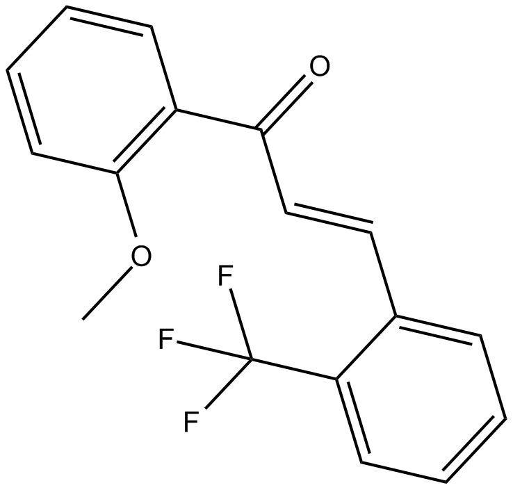
2-Trifluoromethyl-2'-methoxychalcone
Nrf2 activator
-
GN10800
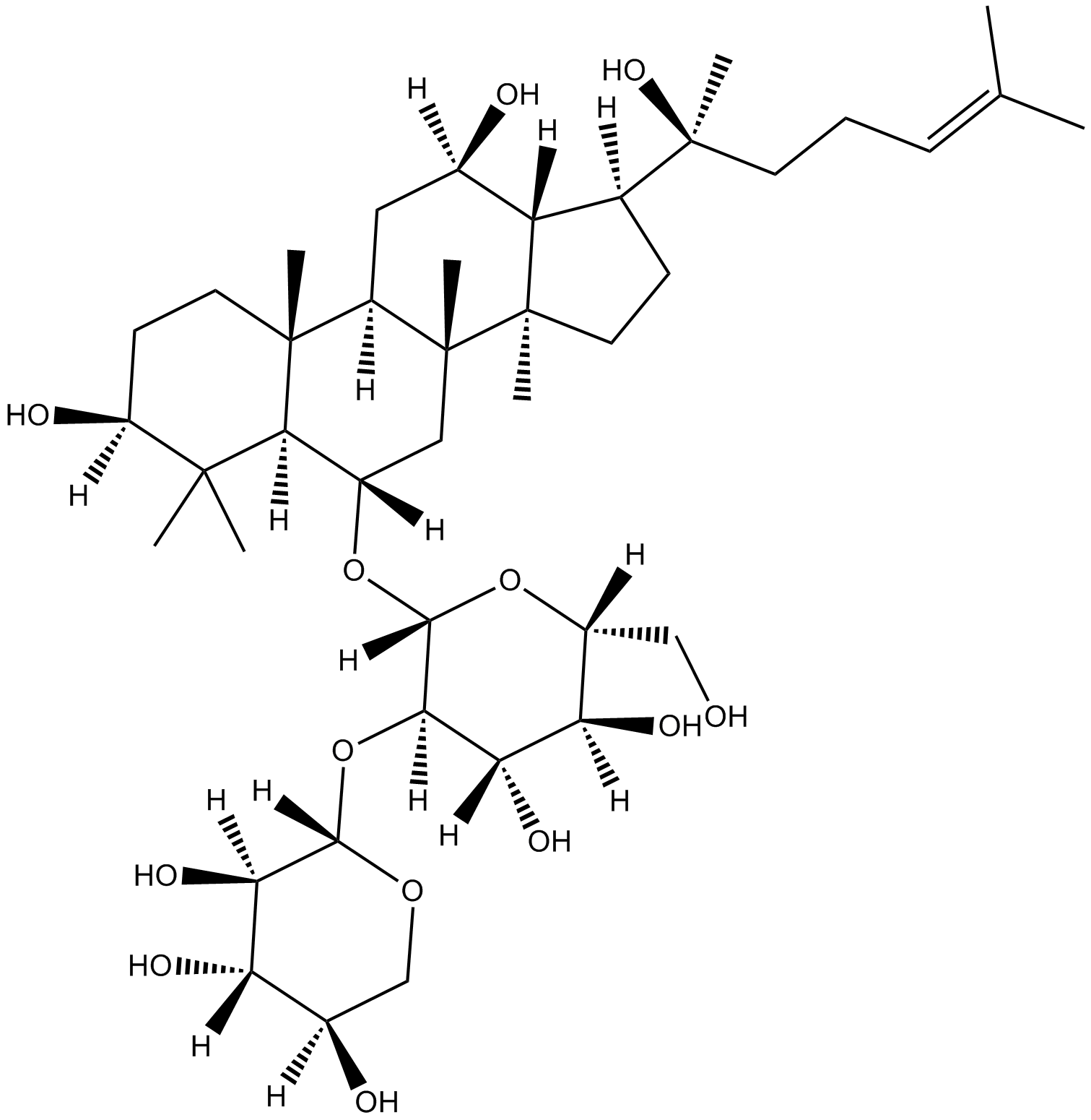
20(S)-NotoginsenosideR2
-
GC46528
25-hydroxy Cholesterol-d6
An internal standard for the quantification of 25hydroxy cholesterol

-
GC48482
28-Acetylbetulin
A lupane triterpenoid with anti-inflammatory and anticancer activities

-
GC35112
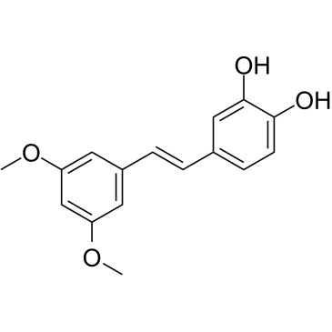
3'-Hydroxypterostilbene
3'-Hydroxypterostilbene은 Pterostilbene 유사체입니다. 3'-Hydroxypterostilbene은 각각 9.0, 40.2 및 70.9μM의 IC50으로 COLO 205, HCT-116 및 HT-29 세포의 성장을 억제합니다. 3'-Hydroxypterostilbene은 PI3K/Akt 및 MAPK 신호 전달 경로를 유의하게 하향 조절하고 세포자멸사 및 자가포식을 유도하여 인간 결장암 세포의 성장을 효과적으로 억제합니다. 3'-Hydroxypterostilbene은 암 연구에 사용할 수 있습니다.
-
GC12791
3,3'-Diindolylmethane
A phytochemical with antiradiation and chemopreventative effects

-
GC42237
3,5-dimethyl PIT-1
PtdIns-(3,4,5)-P3 (PIP3) serves as an anchor for the binding of signal transduction proteins bearing pleckstrin homology (PH) domains such as phosphatidylinositol 3-kinase (PI3K) or PTEN.

-
GC64762
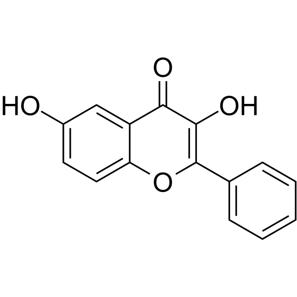
3,6-Dihydroxyflavone
3,6-Dihydroxyflavone은 항암제입니다. 3,6-디히드록시플라본은 용량 및 시간에 따라 세포 생존력을 감소시키고 카스파제 캐스케이드를 활성화하여 폴리(ADP-리보스) 폴리머라제(PARP)를 절단함으로써 세포자멸사를 유도합니다. 3,6-Dihydroxyflavone은 세포 내 산화 스트레스와 지질 과산화를 증가시킵니다.
-
GC46583
3-Amino-2,6-Piperidinedione
An active metabolite of (±)-thalidomide

-
GC49849
3-Aminosalicylic Acid
A salicylic acid derivative

-
GC35106
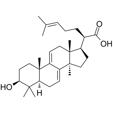
3-Dehydrotrametenolic acid
Poria cocos의 공막에서 분리된 3-디하이드로트라메테놀산은 젖산 탈수소효소(LDH) 억제제입니다. 3-디하이드로트라메테놀산은 시험관 내에서 지방세포 분화를 촉진하고 생체 내에서 인슐린 감작제로 작용합니다. 3-디히드로트라메테놀산은 세포자살을 유도하고 항암 활성이 있습니다.
-
GC68537
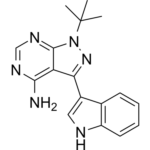
3-IN-PP1
3-IN-PP1은 단백질 키나제 D (PKD) 억제제입니다. 3-IN-PP1은 PKD1, PKD2 및 PKD3에 대해 유효한 광범위한 PKD 억제 활성을 가지며, 각각의 IC50 값은 108, 94 및 108 nM입니다. 또한, 3-IN-PP1은 폭넓게 항암제이며 다양한 종류의 암세포 성장을 억제하는 작용이 있습니다. 따라서, 3-IN-PP1은 암 연구에 사용될 수 있습니다.
-
GC17394
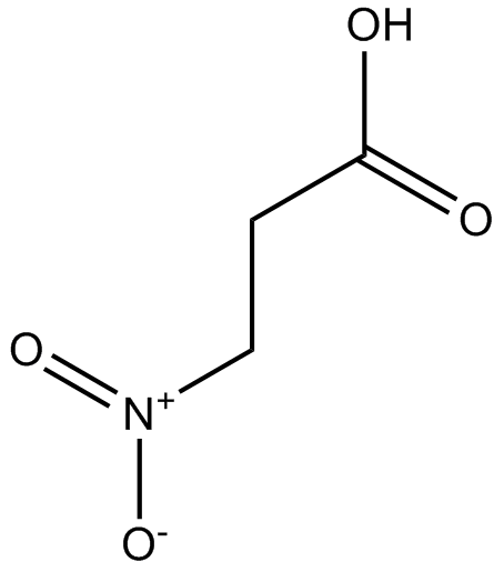
3-Nitropropionic acid
3-니트로프로피온산(β-니트로프로피온산)은 숙신산 탈수소효소의 비가역적 억제제입니다.
-
GC35099
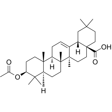
3-O-Acetyloleanolic acid
Vigna sinensis K.의 종자에서 분리된 올레아놀산 유도체인 3-O-아세틸올레아놀산(3AOA)은 암을 유도하고 항혈관신생 활성을 나타냅니다.
-
GC60507
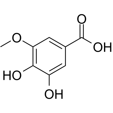
3-O-Methylgallic acid
3-O-Methylgallic acid(3,4-Dihydroxy-5-methoxybenzoic acid)는 안토시아닌 대사 산물이며 강력한 항산화 능력을 가지고 있습니다. 3-O-methylgallic acid는 24.1μM의 IC50 값으로 Caco-2 세포 증식을 억제합니다. 3-O-methylgallic acid는 또한 세포 사멸을 유도하고 항암 효과가 있습니다.
-
GC32767
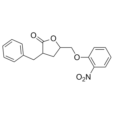
3BDO
3BDO는 자가포식을 억제할 수 있는 새로운 mTOR 활성제입니다.
-
GC45354
4β-Hydroxywithanolide E
4β, Physalis peruviana L.에서 분리된 하이드록시위타놀라이드 E.

-
GC48437
4'-Acetyl Chrysomycin A
A bacterial metabolite with antibacterial and anticancer activities

-
GC42346
4-bromo A23187
4-Bromo A23187은 고선택성 칼슘 이온 전달 물질 A-23187의 할로겐화 유사체입니다.

-
GC42401
4-hydroperoxy Cyclophosphamide
사이클로포스파미드의 활성화된 아날로그

-
GC30896
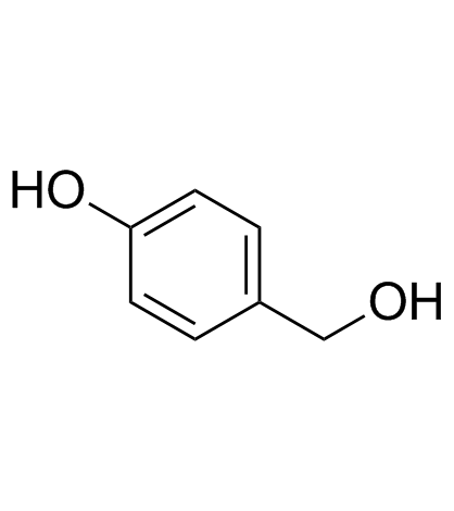
4-Hydroxybenzyl alcohol
4-하이드록시벤질 알코올은 다양한 종류의 식물에 널리 분포되어 있는 페놀 화합물입니다.
-
GC33815
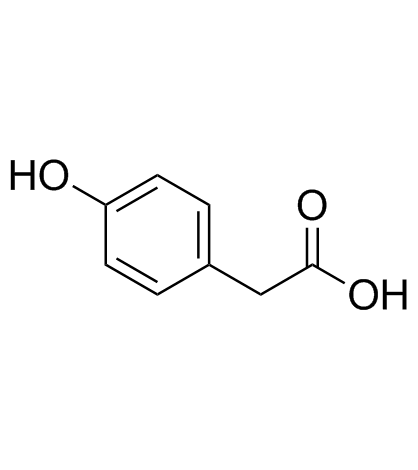
4-Hydroxyphenylacetic acid
4-하이드록시페닐아세트산은 폴리페놀의 주요 미생물 유래 대사산물이며 항산화 작용에 관여합니다.
-
GC35138
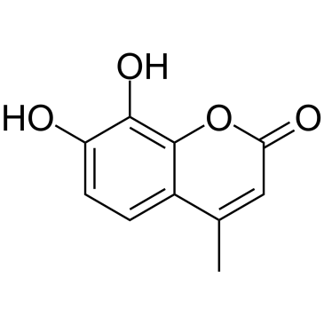
4-Methyldaphnetin
4-메틸다프네틴은 4-메틸 쿠마린 유도체 합성의 전구체입니다. 4-Methyldaphnetin은 여러 암세포주에 강력하고 선택적인 항증식 및 세포자멸사 유도 효과가 있습니다. 4-Methyldaphnetin은 라디칼 소거성을 가지고 있으며 막 지질 과산화를 강력하게 억제합니다.
-
GC68231
4-Methylsalicylic acid

-
GC31648
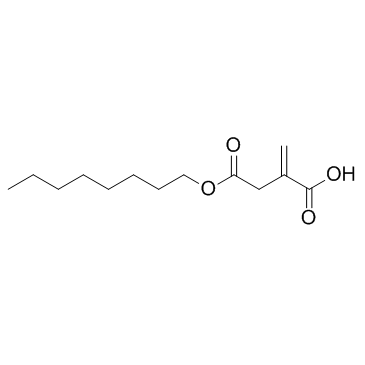
4-Octyl Itaconate
4-옥틸 이타콘산 (4-OI)은 세포막 침투성을 가진 이타콘산 유도체입니다. 이타콘산과 4-옥틸 이타콘산은 비슷한 티올 반응성을 보이므로, 생물학적 기능 연구를 위해 적절한 이타콘산 대용체인 4-옥틸 이타콘산입니다.
-
GC49127
4-oxo Cyclophosphamide
An inactive metabolite of cyclophosphamide

-
GC45352
4-oxo Withaferin A
4-옥소 Withaferin A는 withaferin A의 유사체입니다. Withaferin A는 Withania somnifera에서 분리된 withanolide입니다. 4-옥소 Withaferin A는 다발성 골수종 연구의 가능성이 있습니다.

-
GC45353
4-oxo-27-TBDMS Withaferin A
4-oxo-27-TBDMS Withaferin A 유도체인 Withaferin A는 종양 세포에 강력한 항증식 효과를 나타냅니다. 4-oxo-27-TBDMS Withaferin A는 종양 세포의 세포 사멸을 유도합니다. 4-oxo-27-TBDMS Withaferin A는 항암제입니다.

-
GC60525
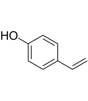
4-Vinylphenol (10%w/w in propylene glycol)
4-Vinylphenol은 약초인 Hedyotis diffusa Willd, 야생 쌀에서 발견되며, 와인의 유산균에 의한 p-쿠마르산과 페룰산의 대사산물이기도 합니다. 4-비닐페놀은 세포 사멸을 유도하고 혈관 형성을 억제하며 생체 내 침습성 유방 종양 성장을 억제합니다.
-
GC10468
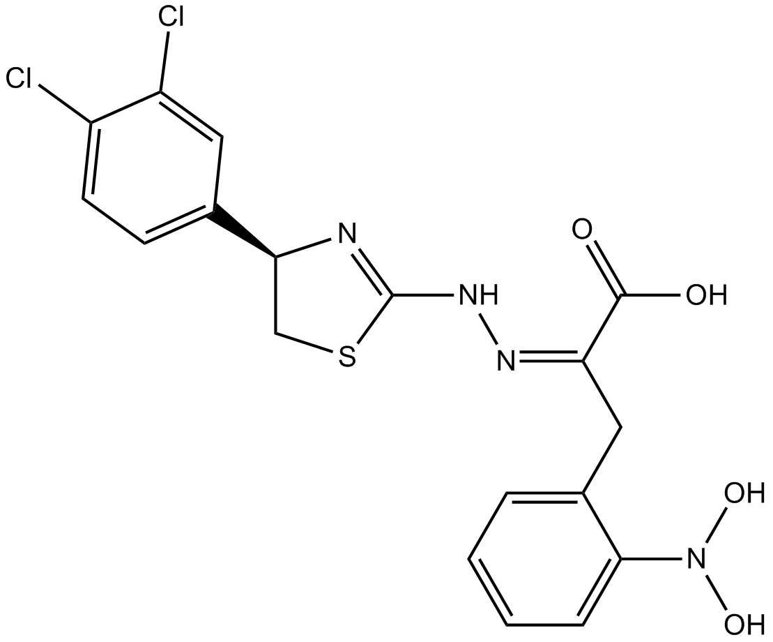
4EGI-1
4EGI-1은 eIF4E 결합에 대한 Kd가 25μM인 eIF4E/eIF4G 상호작용의 억제제입니다.
-
GC35150
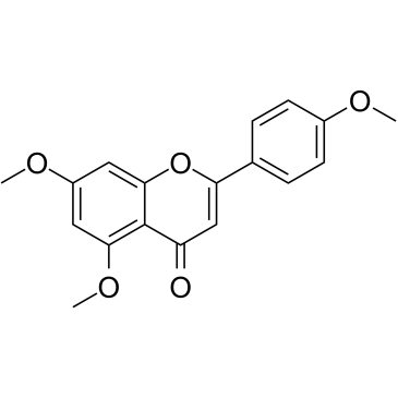
5,7,4'-Trimethoxyflavone
5,7,4'-Trimethoxyflavone은 태국의 유명한 약용 식물인 Kaempferia parviflora(KP)에서 분리됩니다. 5,7,4'-Trimethoxyflavone은 sub-G1 단계의 증가, DNA 단편화, annexin-V/PI 염색, Bax/Bcl-xL 비율, caspase-3의 단백질 분해 활성화 및 폴리 (ADP-ribose) polymerase (PARP) protein.5,7,4'-Trimethoxyflavone은 농도 의존적 \u200b\u200b방식으로 SNU-16 인간 위암 세포의 증식을 억제하는 데 상당히 효과적입니다.
-
GN10629
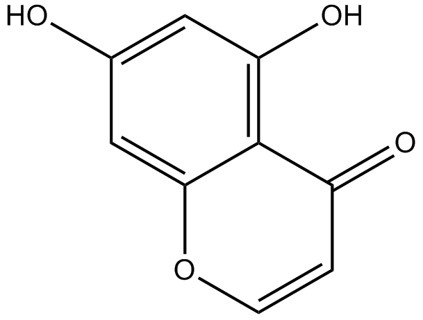
5,7-dihydroxychromone
-
GC63972
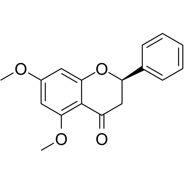
5,7-Dimethoxyflavanone
5,7-Dimethoxyflavanone은 S.
-
GC52227
5-(3',4'-Dihydroxyphenyl)-γ-Valerolactone
An active metabolite of various polyphenols

-
GC35147
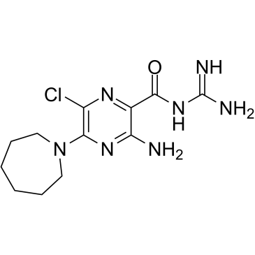
5-(N,N-Hexamethylene)-amiloride
5-(N,N-Hexamethylene)-amiloride(Hexamethylene amiloride)는 amiloride에서 파생되며 강력한 Na+/H+ 교환기 억제제로, 백혈병에서 세포 내 pH(pHi)를 낮추고 세포자멸사를 유도합니다. 세포.
-
GC45356
5-Aminolevulinic Acid (hydrochloride)

-
GC46681
5-Bromouridine
A brominated uridine analog

-
GC42545
5-Fluorouracil-13C,15N2
5-Fluorouracil-13C,15N2 is intended for use as an internal standard for the quantification of 5-flurouracil by GC- or LC-MS.

-
GC46705
5-Methoxycanthinone
5-Methoxycanthinone은 Leishmania 균주의 경구 활성 억제제입니다.

-
GC42586
6α-hydroxy Paclitaxel
6α-하이드록시 파클리탁셀은 파클리탁셀의 1차 대사산물입니다. 6α-하이드록시 파클리탁셀은 파클리탁셀과 유사한 억제 효능으로 유기 음이온 수송 폴리펩타이드 1B1/SLCO1B1(OATP1B1)에 대한 시간 의존적 효과를 유지하지만, 더 이상 OATP1B3의 시간 의존적 억제를 나타내지 않습니다. 6α-hydroxy Paclitaxel은 암 연구에 사용할 수 있습니다.

-
GC45772
6(5H)-Phenanthridinone
An inhibitor of PARP1 and 2

-
GN10093
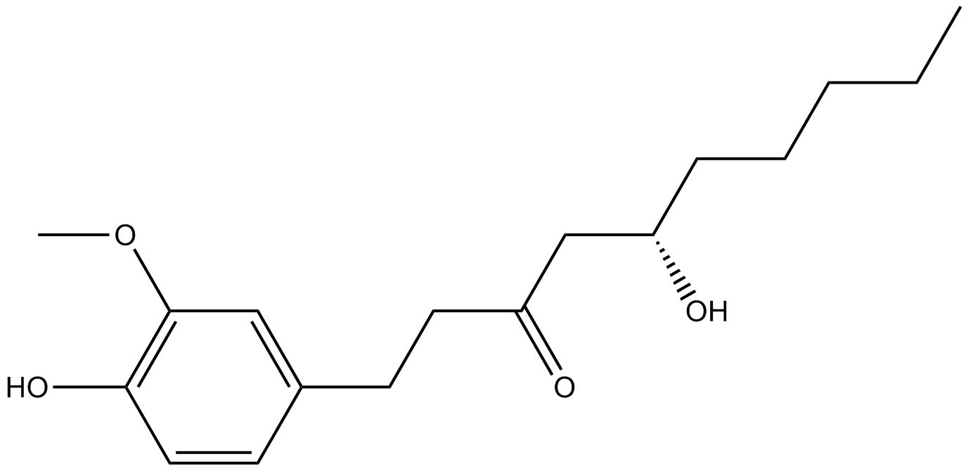
6-gingerol
-
GC49429
6-keto Lithocholic Acid
A metabolite of lithocholic acid
-
GC35184
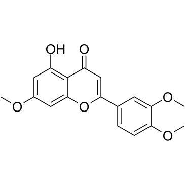
7,3',4'-Tri-O-methylluteolin
플라보노이드 화합물인 7,3',4'-Tri-O-methylluteolin(5-Hydroxy-3',4',7-trimethoxyflavone)은 염증 매개체, NO, PGE2 및 전염증성 사이토카인의 방출.
-
GC45673
7,8-Dihydroneopterin
염증 마커인 7,8-디히드로네옵테린은 산화질소 합성효소(iNOS) 발현의 향상을 통해 성상교세포와 뉴런에서 세포 사멸을 유도합니다.

-
GC16853
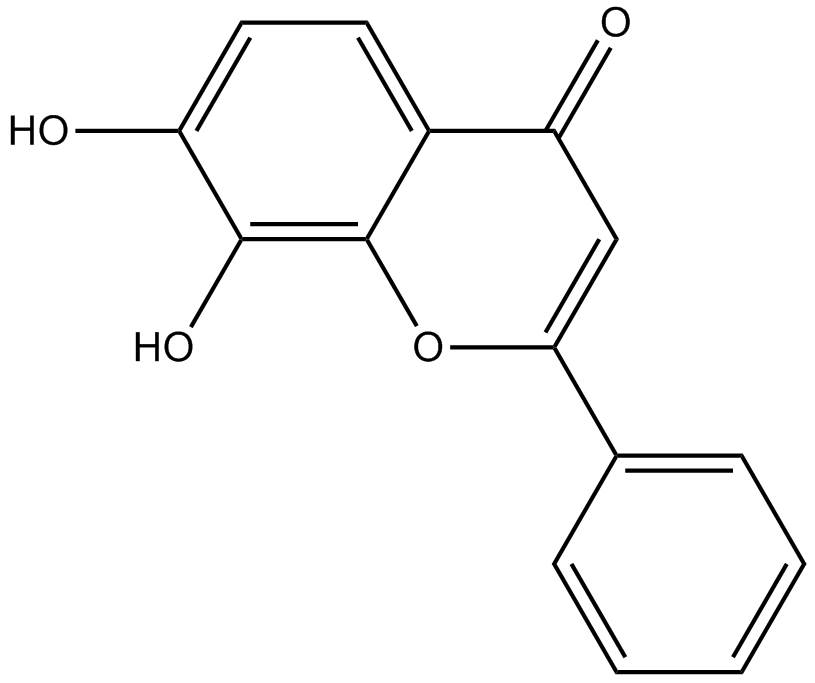
7,8-Dihydroxyflavone
7,8-Dihydroxyflavone은 뇌 유래 신경영양 인자(BDNF)의 생리학적 작용을 모방하는 강력하고 선택적인 TrkB 작용제입니다.
-
GC42616
7-oxo Staurosporine
7-oxo Staurosporine is an antibiotic originally isolated from S.

-
GC16037
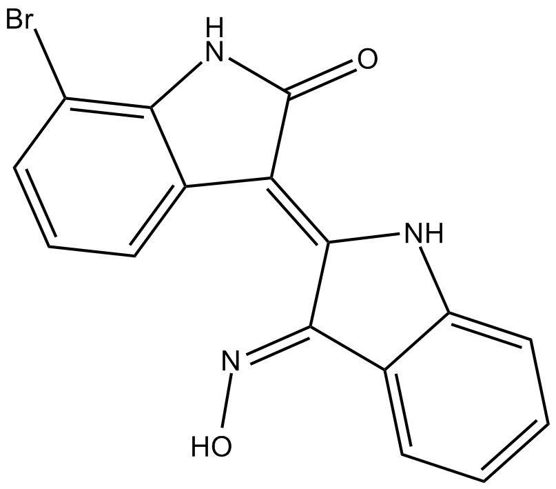
7BIO
7BIO(7-브로모인디루빈-3-옥심)는 인디루빈의 유도체입니다.
-
GC46741
8(E),10(E),12(Z)-Octadecatrienoic Acid
A conjugated PUFA

-
GC42622
8-bromo-Cyclic AMP
8-bromo-Cyclic AMP is a brominated derivative of cAMP that remains long-acting due to its resistance to degradation by cAMP phosphodiesterase.

-
GC49275
8-Oxycoptisine
8-옥시콥티신은 항암 활성이 있는 천연 프로토베르베린 알칼로이드입니다.

-
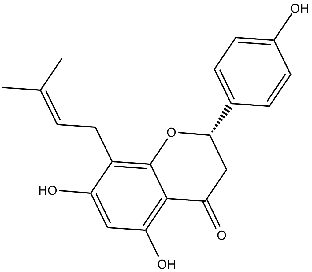
GC17119
8-Prenylnaringenin
8-prenylnaringenin은 세포독성이 있는 홉 콘(Humulus lupulus)에서 분리된 프레닐플라보노이드입니다. 8-프레닐나린제닌은 내인성 및 외인성 경로 매개 세포자멸사 유도를 통해 HCT-116 결장암 세포에 대해 항증식 활성을 갖는다. 8-Prenylnaringenin은 또한 쥐에서 Akt 인산화 경로의 활성화를 통해 고정화로 인한 근육 위축의 회복을 촉진합니다.
-
GC41642
9(E),11(E),13(E)-Octadecatrienoic Acid
9(E),11(E),13(E)-Octadecatrienoic acid (β-ESA) is a conjugated polyunsaturated fatty acid that is found in plant seed oils and in mixtures of conjugated linolenic acids synthesized by the alkaline isomerization of linolenic acid.

-
GC41643
9(Z),11(E),13(E)-Octadecatrienoic Acid
9(Z),11(E),13(E)-Octadecatrienoic Acid (α-ESA) is a conjugated polyunsaturated fatty acid commonly found in plant seed oil.

-
GC40785
9(Z),11(E),13(E)-Octadecatrienoic Acid ethyl ester
9(Z),11(E),13(E)-Octadecatrienoic Acid ethyl ester (α-ESA) is a conjugated polyunsaturated fatty acid commonly found in plant seed oil.

-
GC40710
9(Z),11(E),13(E)-Octadecatrienoic Acid methyl ester
9Z,11E,13E-octadecatrienoic acid (α-ESA) is a conjugated polyunsaturated fatty acid commonly found in plant seed oil.

-
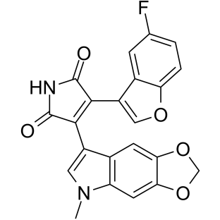
GC39152
9-ING-41
9-ING-41은 IC50이 0.71μM인 말레이미드 기반 ATP 경쟁적이고 선택적 글리코겐 합성효소 키나제-3β(GSK-3β) 억제제입니다. 9-ING-41은 암세포에서 세포 주기 정지, 자가포식 및 세포자멸사를 유의하게 유도합니다. 9-ING-41은 항암 활성이 있으며 화학 요법 약물의 항종양 효과를 향상시킬 가능성이 있습니다.
-
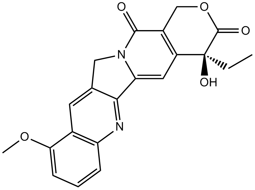
GN10035
9-Methoxycamptothecin
-
GC45960
9c(i472)
9c(i472)는 IC50 값이 0.19 μ인 15-LOX-1(15-리폭시게나제-1)의 강력한 억제제입니다.

-
GC50465
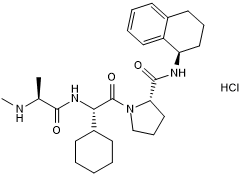
A 410099.1
High affinity XIAP antagonist; active in vivo
-
GC17512
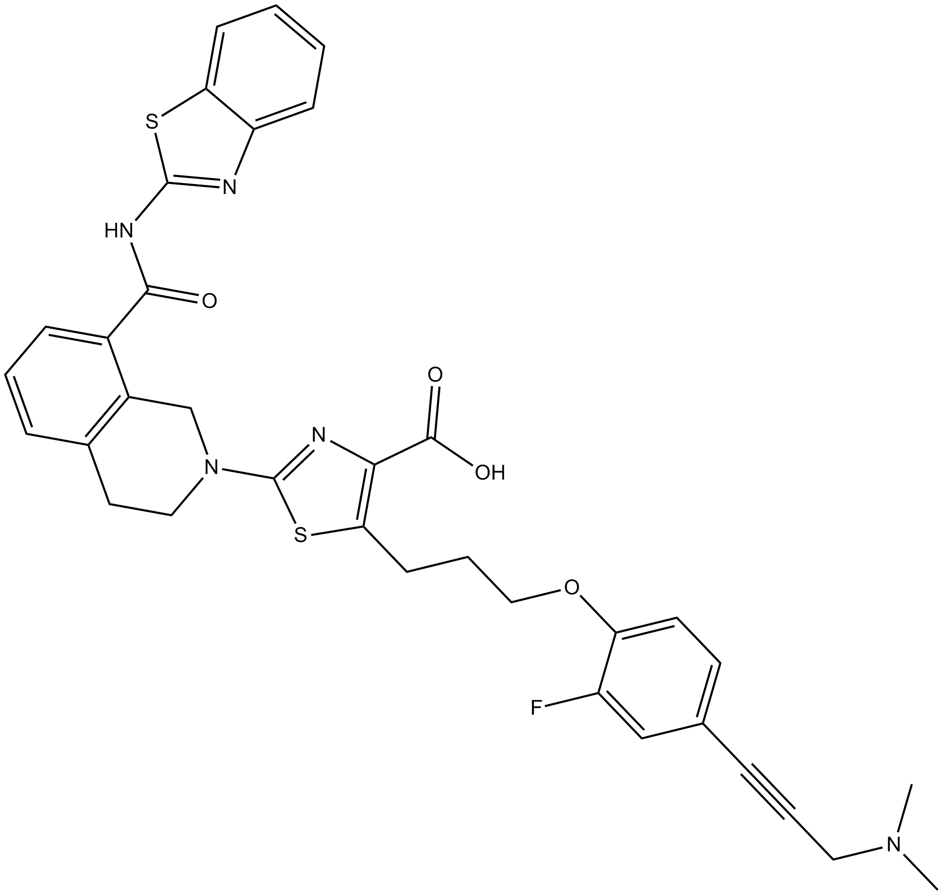
A-1155463
A-1155463은 Molt-4 세포에서 EC50이 70nM인 매우 강력하고 선택적인 BCL-XL 억제제입니다.
-
GC16278
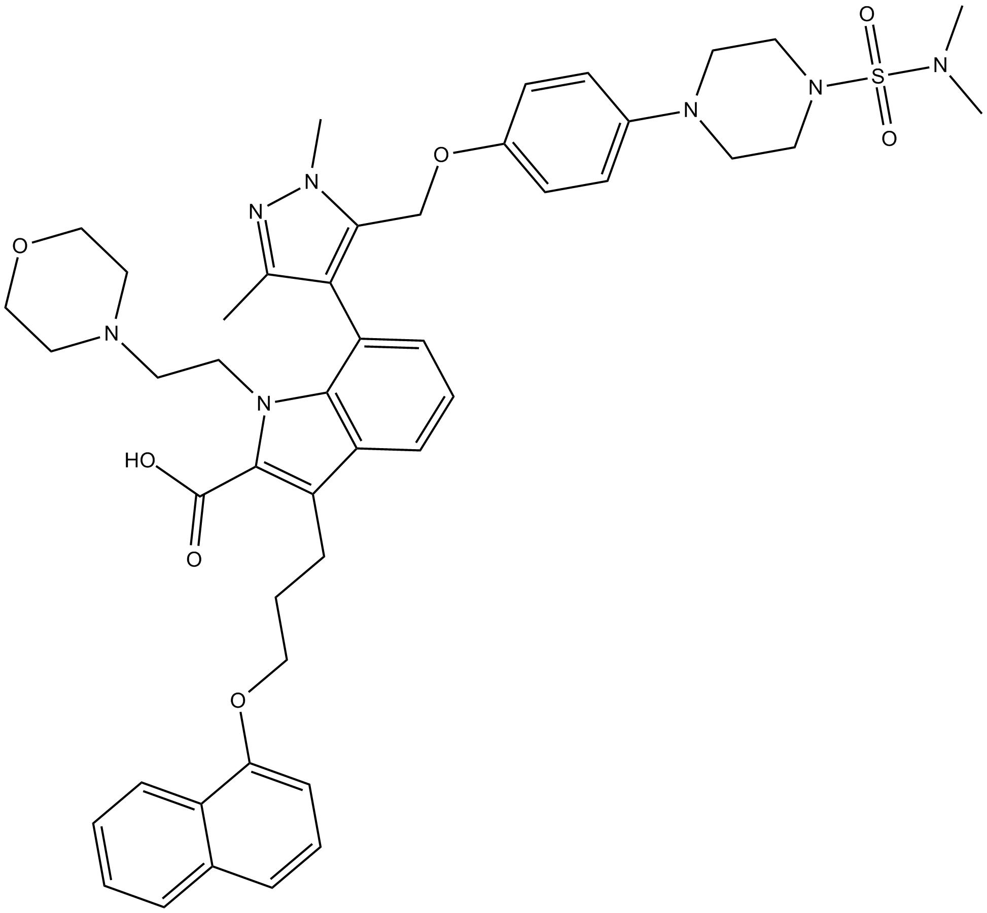
A-1210477
A-1210477은 0.45nM의 Ki를 갖는 MCL-1의 강력하고 선택적인 억제제입니다. A-1210477은 MCL-1에 특이적으로 결합하고 MCL-1 의존적 방식으로 암세포의 세포자멸사를 촉진한다.
-
GC17513
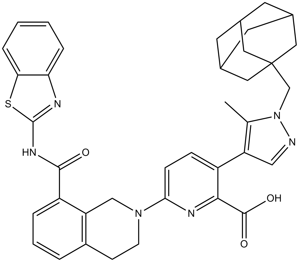
A-1331852
A-1331852는 Ki가 10pM 미만인 경구용 BCL-XL 선택적 억제제입니다.
-
GC60544
A-192621
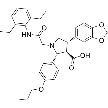
A-192621은 IC50이 4.5nM이고 Ki가 8.8nM인 강력한 비펩타이드성 경구 활성 및 선택적 엔도텔린 B(ETB) 수용체 길항제입니다.
-
GC32981
A-385358
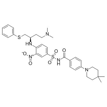
A-385358은 Bcl-XL 및 Bcl-2에 대해 각각 0.80 및 67nM의 Kis를 갖는 Bcl-XL의 선택적 억제제입니다.
-
GC11200
A23187
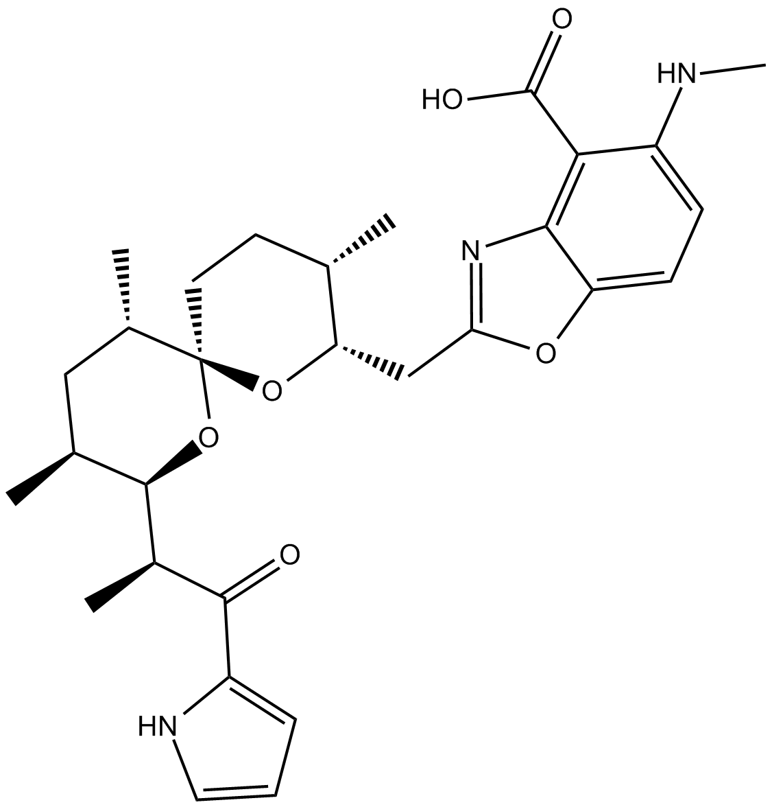
A23187 (A-23187)은 항생제이자 칼슘과 마그네슘과 같은 독특한 이중양이온 이온포어입니다.
-
GC42659
A23187 (calcium magnesium salt)
A23187 is a divalent cation ionophore.

-
GC35216
AAPK-25
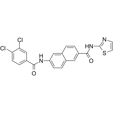
AAPK-25는 항종양 활성이 있는 강력하고 선택적인 Aurora/PLK 이중 억제제로, 생체지표 히스톤 H3Ser10 인산화에 따라 세포자멸사 급증이 뒤따르는 전중기 단계에서 유사분열 지연 및 세포 정지를 유발할 수 있습니다. AAPK-25는 Kd 값이 23-289nM인 Aurora-A, -B 및 -C와 Kd 값이 55-456nM인 PLK-1, -2 및 -3을 대상으로 합니다.
-
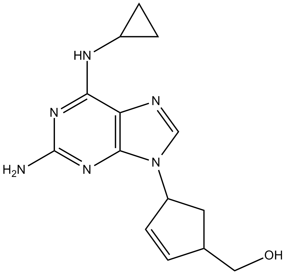
GC13805
Abacavir
-
GC64674
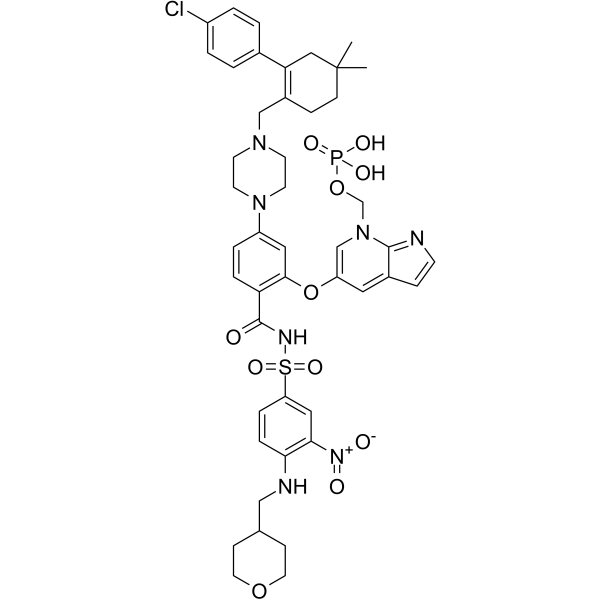
ABBV-167
ABBV-167은 BCL-2 억제제 베네토클락스의 인산염 전구약물이다.
-
GC60548
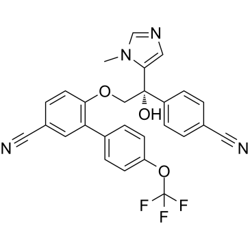
ABT-100
ABT-100은 강력하고 선택성이 높으며 경구 활성인 파르네실트랜스퍼라제 억제제입니다. ABT-100은 세포 증식을 억제합니다. 2, PC-3 및 DU-145 세포)는 세포자멸사를 증가시키고 혈관신생을 감소시킨다. ABT-100은 광범위한 항종양 활성을 가지고 있습니다.
-
GC14069
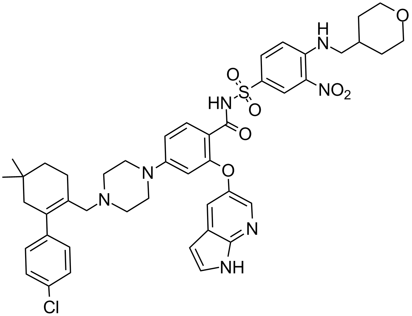
ABT-199
Bcl-2 억제제
-
GC12405
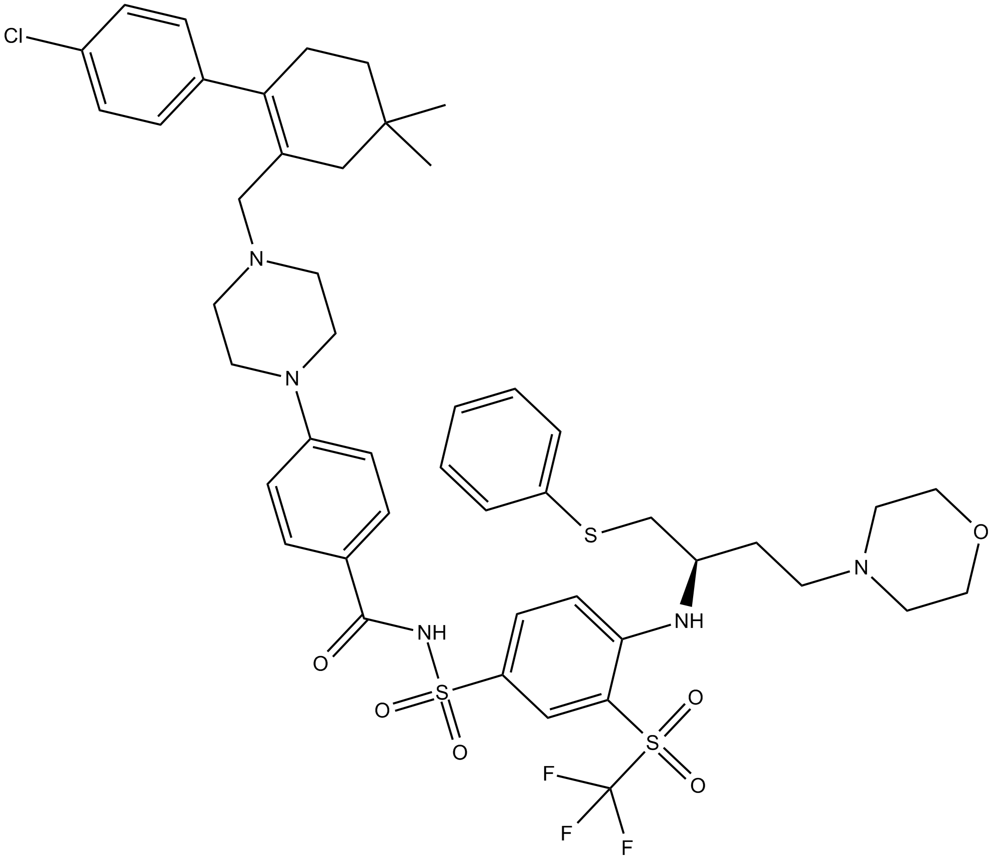
ABT-263 (Navitoclax)
Bcl-2 패밀리 단백질의 억제제
-
GC49745
ABT-263-d8
ABT-263-d8은 Navitoclax라고 표시된 중수소입니다. Navitoclax(ABT-263)는 Bcl-xL, Bcl-2 및 Bcl-w와 같은 여러 항-세포자멸사 Bcl-2 계열 단백질에 결합하는 강력하고 경구 활성인 Bcl-2 계열 단백질 억제제입니다. 1nM 이상.

-
GC17234
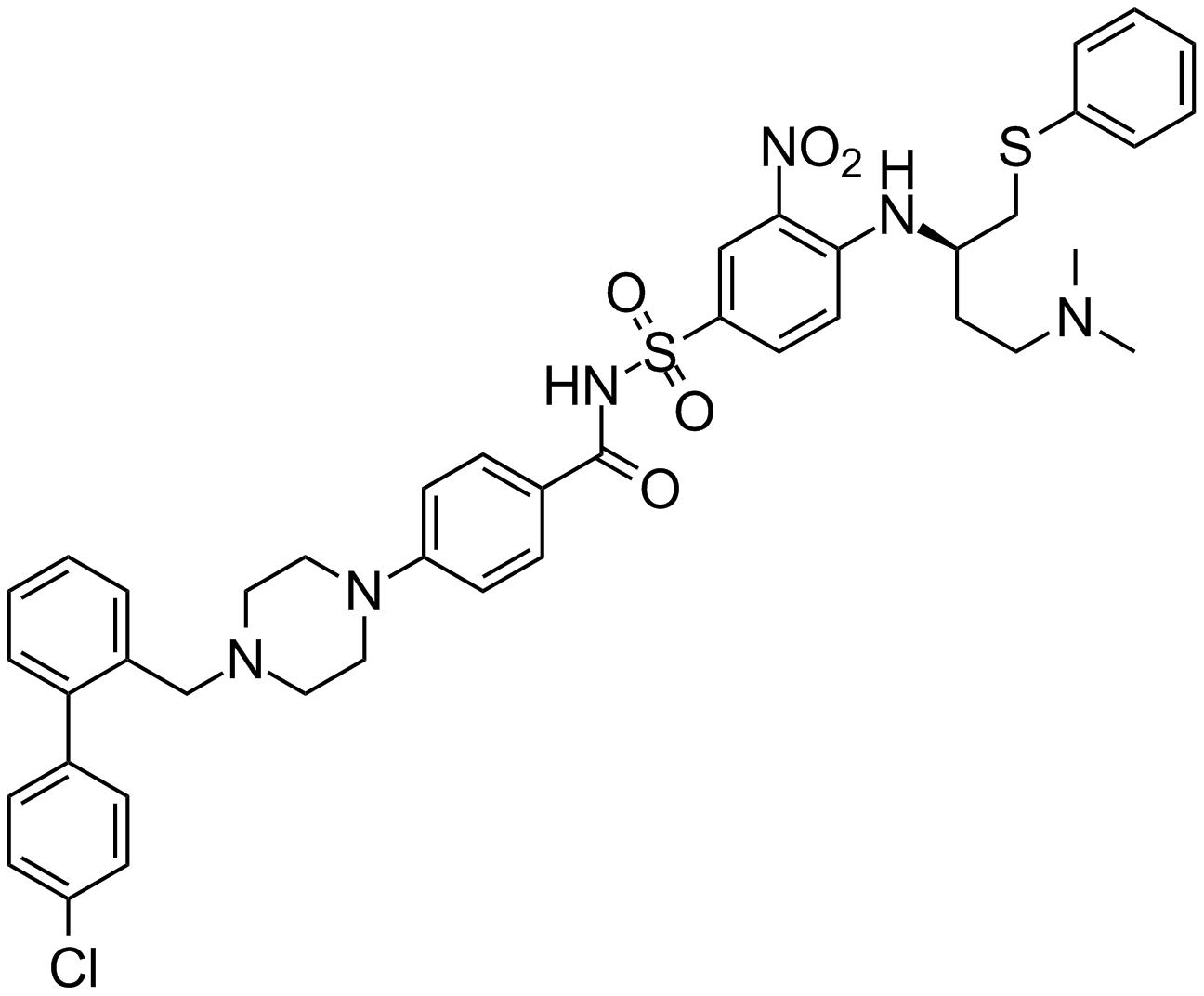
ABT-737
An inhibitor of anti-apoptotic Bcl-2 proteins
-
GA20494
Ac-Asp-Glu-Val-Asp-pNA
The cleavage of the chromogenic caspase-3 substrate Ac-DEVD-pNA can be monitored at 405 nm.
-
GC17602
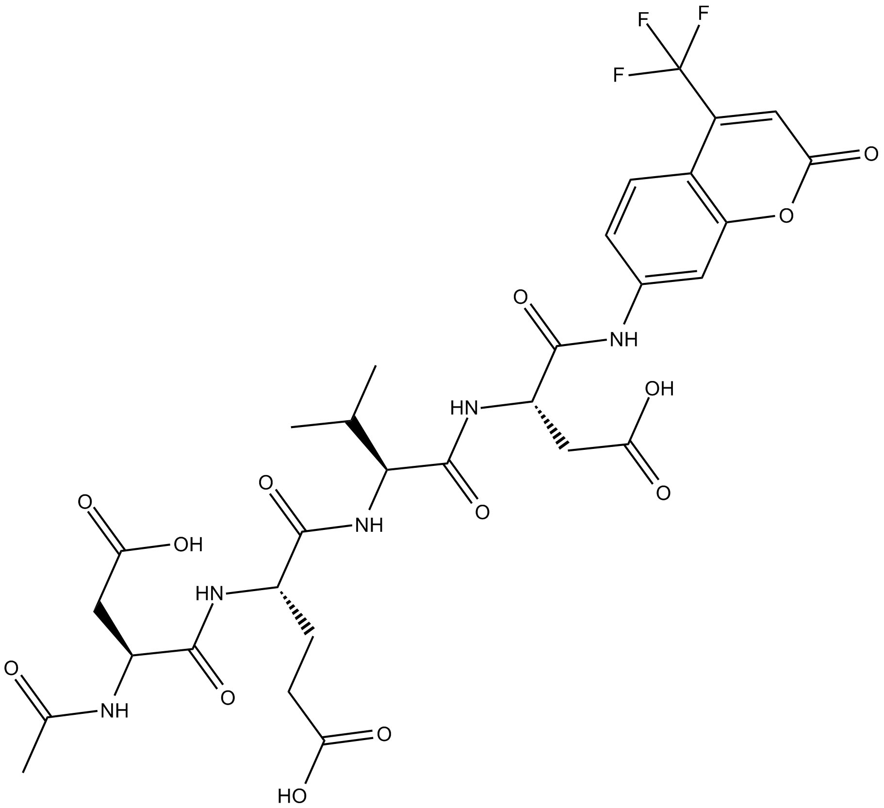
Ac-DEVD-AFC
Ac-DEVD-AFC는 형광 기질입니다(Λex=400 nm, Λem=530 nm).
-
GC32695
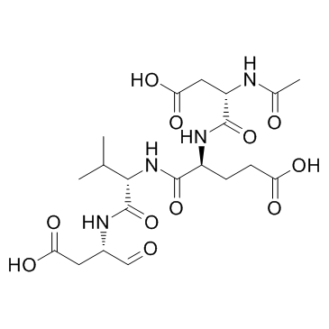
Ac-DEVD-CHO
Ac-DEVD-CHO는 230pM의 Ki 값을 갖는 특정 Caspase-3 억제제입니다.
-
GC48470
Ac-DEVD-CHO (trifluoroacetate salt)
A dual caspase3/caspase7 inhibitor

-
GC10951
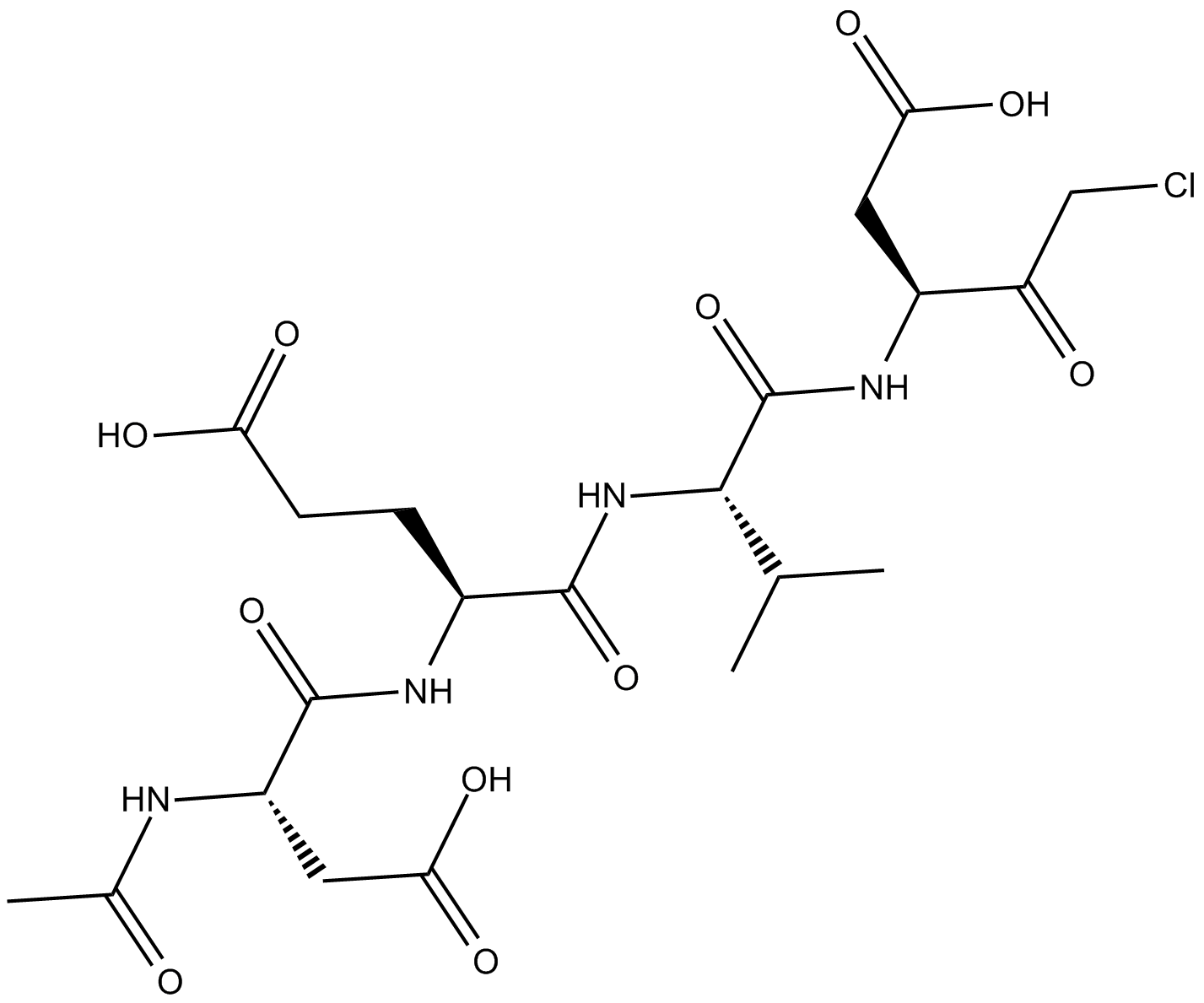
Ac-DEVD-CMK
cell-permeable, and irreversible inhibitor of caspase
-
GC42689
Ac-DNLD-AMC
Ac-WLA-AMC는 caspase-3의 형광 기질입니다.

-
GC65107
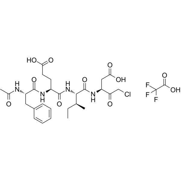
Ac-FEID-CMK TFA
Ac-FEID-CMK TFA는 강력한 제브라피쉬 특이적 GSDMEb 유래 펩타이드 억제제입니다.
-
GC60558
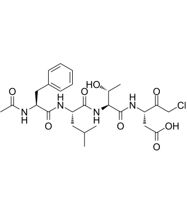
Ac-FLTD-CMK
Gasdermin D(GSDMD) 유래 억제제인 Ac-FLTD-CMK는 특정 염증성 카스파제 억제제입니다.
-
GC49704
Ac-FLTD-CMK (trifluoroacetate salt)
An inhibitor of caspase-1, -4, -5, and -11

-
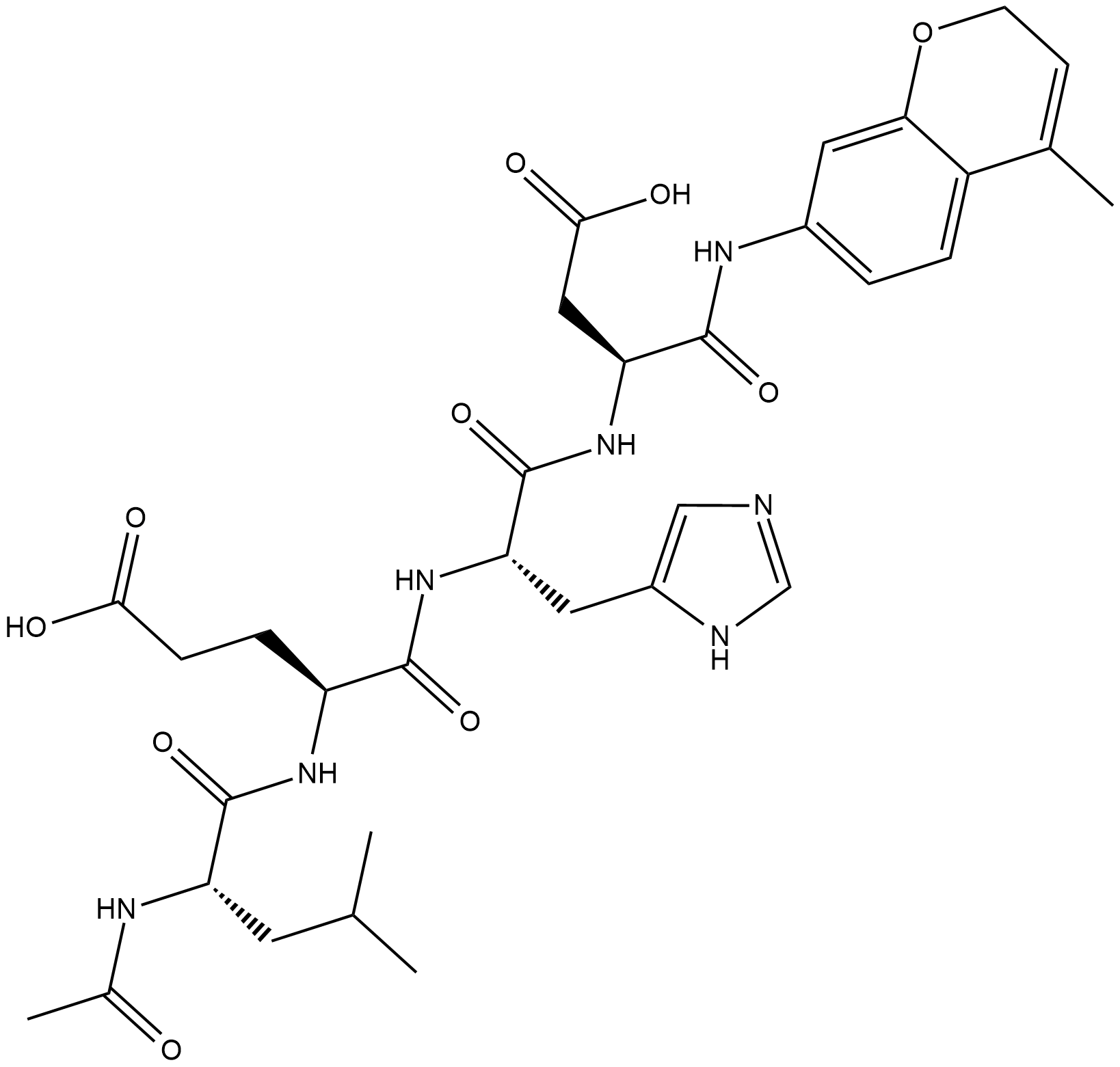
GC18226
Ac-LEHD-AMC (trifluoroacetate salt)
Ac-LEHD-AMC(트리플루오로아세테이트 염)는 카스파제-9에 대한 형광 기질입니다(여기: 341 nm, 방출: 441 nm).
-
GC40556
Ac-LETD-AFC
Ac-LETD-AFC는 카스파제-8 형광 기질입니다.

-
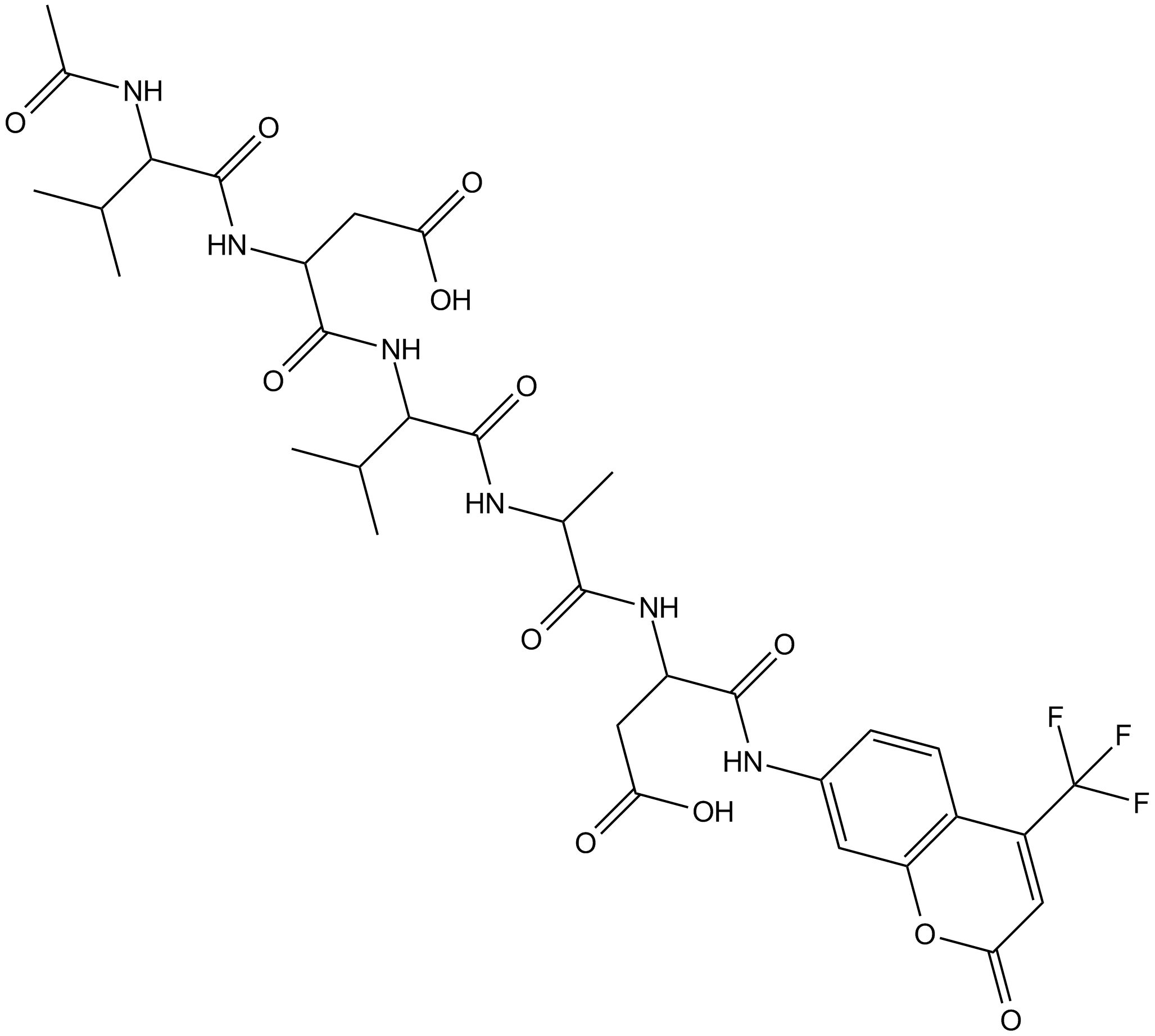
GC13400
Ac-VDVAD-AFC
Ac-VDVAD-AFC는 카스파제 특이적 형광 기질입니다. Ac-VDVAD-AFC는 caspase-3-like 활성 및 caspase-2 활성을 측정할 수 있으며 종양 및 암 연구에 사용할 수 있습니다.
-
GC52372
Ac-VDVAD-AFC (trifluoroacetate salt)
A fluorogenic substrate for caspase-2

-
GC48974
Ac-VEID-AMC (ammonium acetate salt)
A caspase-6 fluorogenic substrate

-
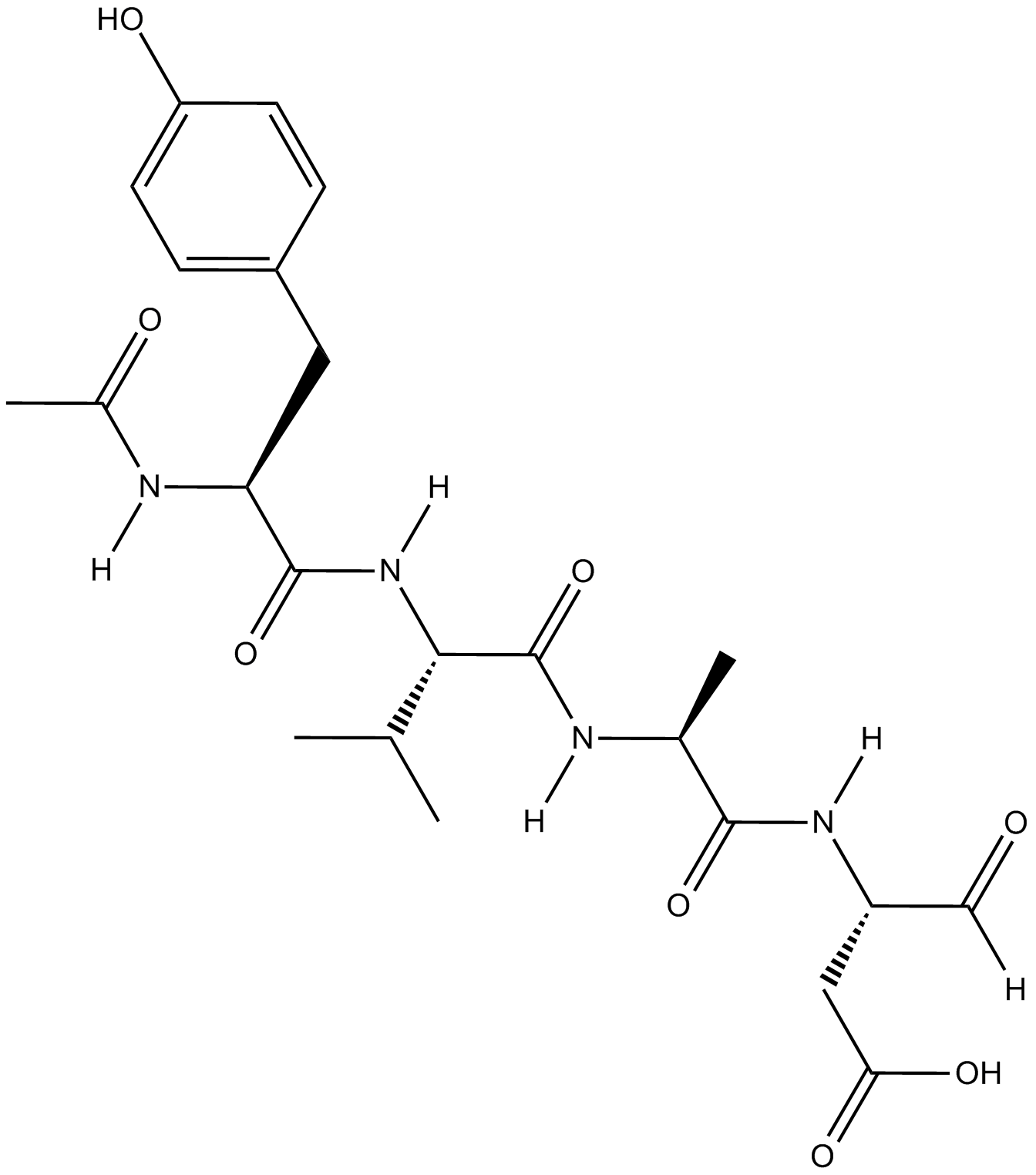
GC18021
Ac-YVAD-CHO
Ac-YVAD-CHO(L-709049)는 3.0 및 0.76nM의 마우스 및 인간 Ki 값을 갖는 강력하고 가역적인 특이적 테트라펩타이드 인터루킨-1β 전환 효소(ICE) 억제제입니다.
-
GC42721
Ac-YVAD-CMK
Ac-YVAD-CMK는 카스패이스-1의 선택적인 불가역 억제제입니다(Ki=0.8nM). 이것은 염증 유발 사이토카인 IL-1β 활성화를 방지할 수 있습니다. Ac-YVAD-CMK는 염증 반응을 감소시키고 장기간 신경 보호 효과를 유도할 수 있습니다.

-
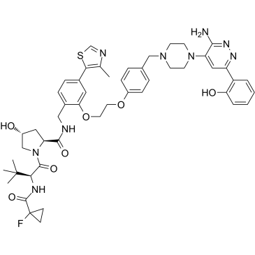
GC35227
ACBI1
ACBI1은 각각 6, 11 및 32nM의 DC50을 갖는 강력하고 협력적인 SMARCA2, SMARCA4 및 PBRM1 분해기입니다. ACBI1은 PROTAC 분해제입니다. ACBI1은 항증식 활성을 나타냅니다. ACBI1은 세포 사멸을 유도합니다.
-
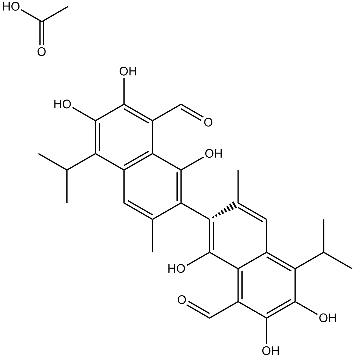
GN10341
Acetate gossypol
-
GC11786
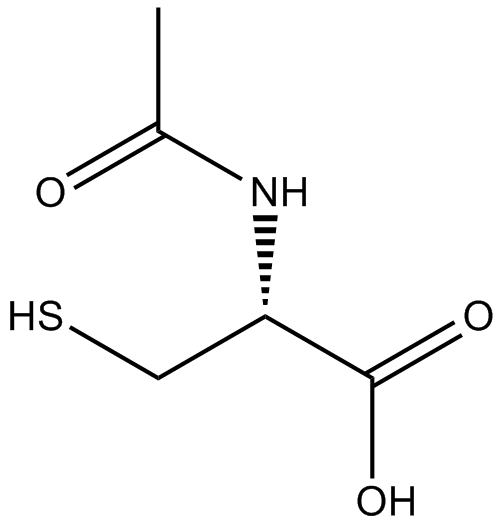
Acetylcysteine
아세틸시스테인은 시스테인의 N-아세틸 유도체입니다.
-
GC17094
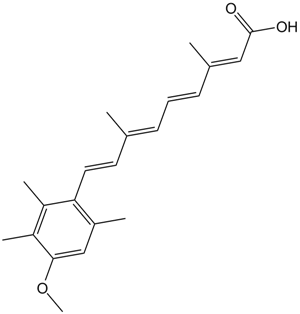
Acitretin
아시트레틴(Ro 10-1670)은 건선 치료에 사용된 2세대 전신 레티노이드입니다.
-
GC35242
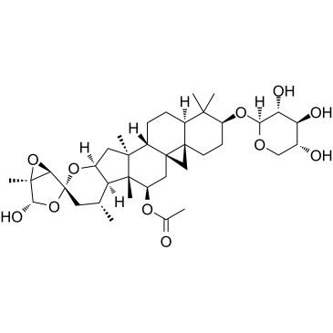
Actein
악테인은 Cimicifuga foetida의 뿌리줄기에서 분리된 트리테르펜 배당체입니다. 액테인은 인간 방광암에서 ROS/JNK 활성화를 촉진하고 AKT 경로를 둔화시켜 세포 증식을 억제하고 자가포식 및 세포자멸사를 유도합니다. 액테인은 생체 내 독성이 거의 없습니다.
-
GC16866
Actinomycin D
항암 활성을 가진 DNA와 상호작용하는 전사 차단제

-
GC16350
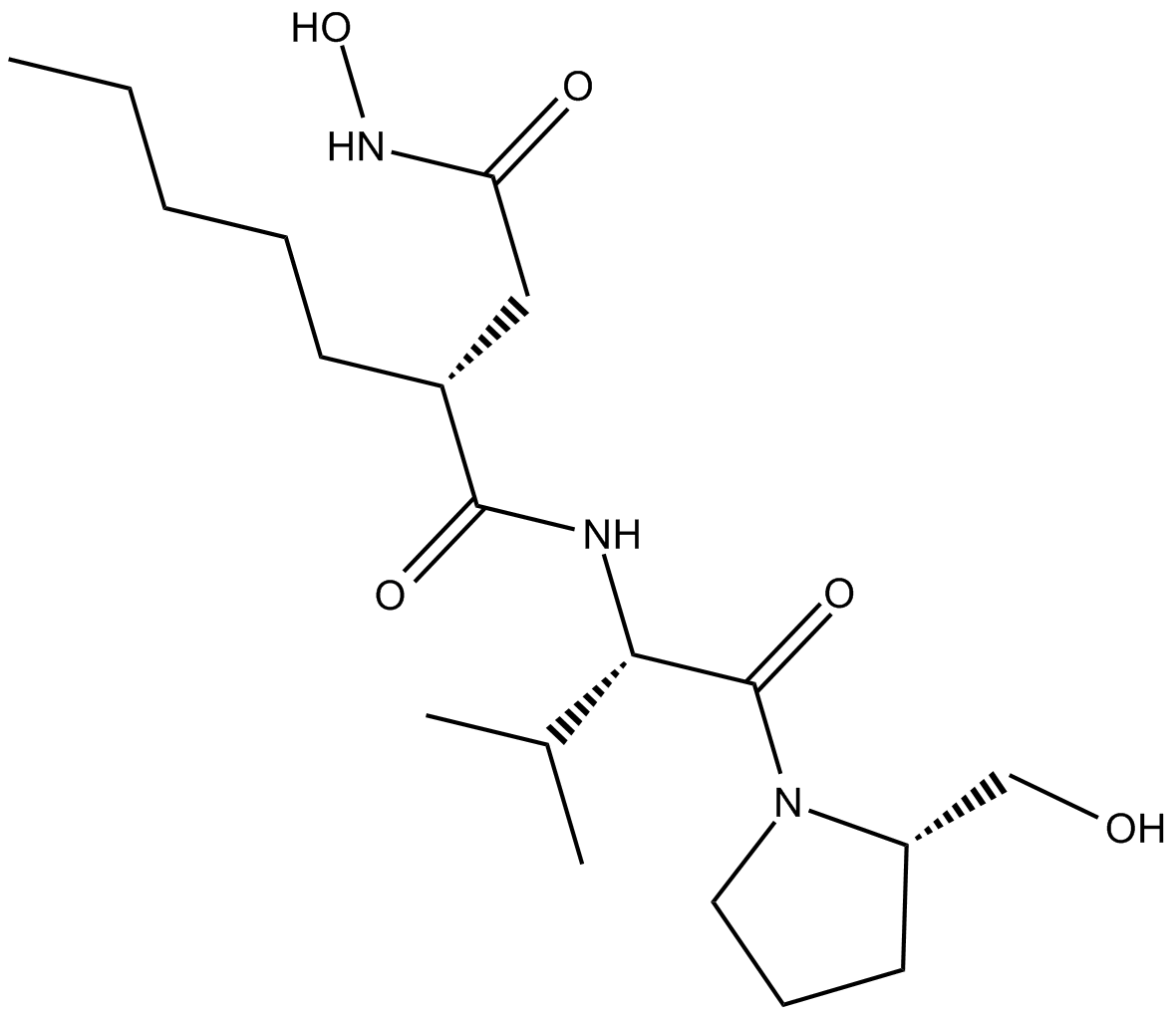
Actinonin
Actinonin ((-)-Actinonin)은 Actinomyces에 의해 생성되는 자연 발생 항균제입니다.
-
GC16362
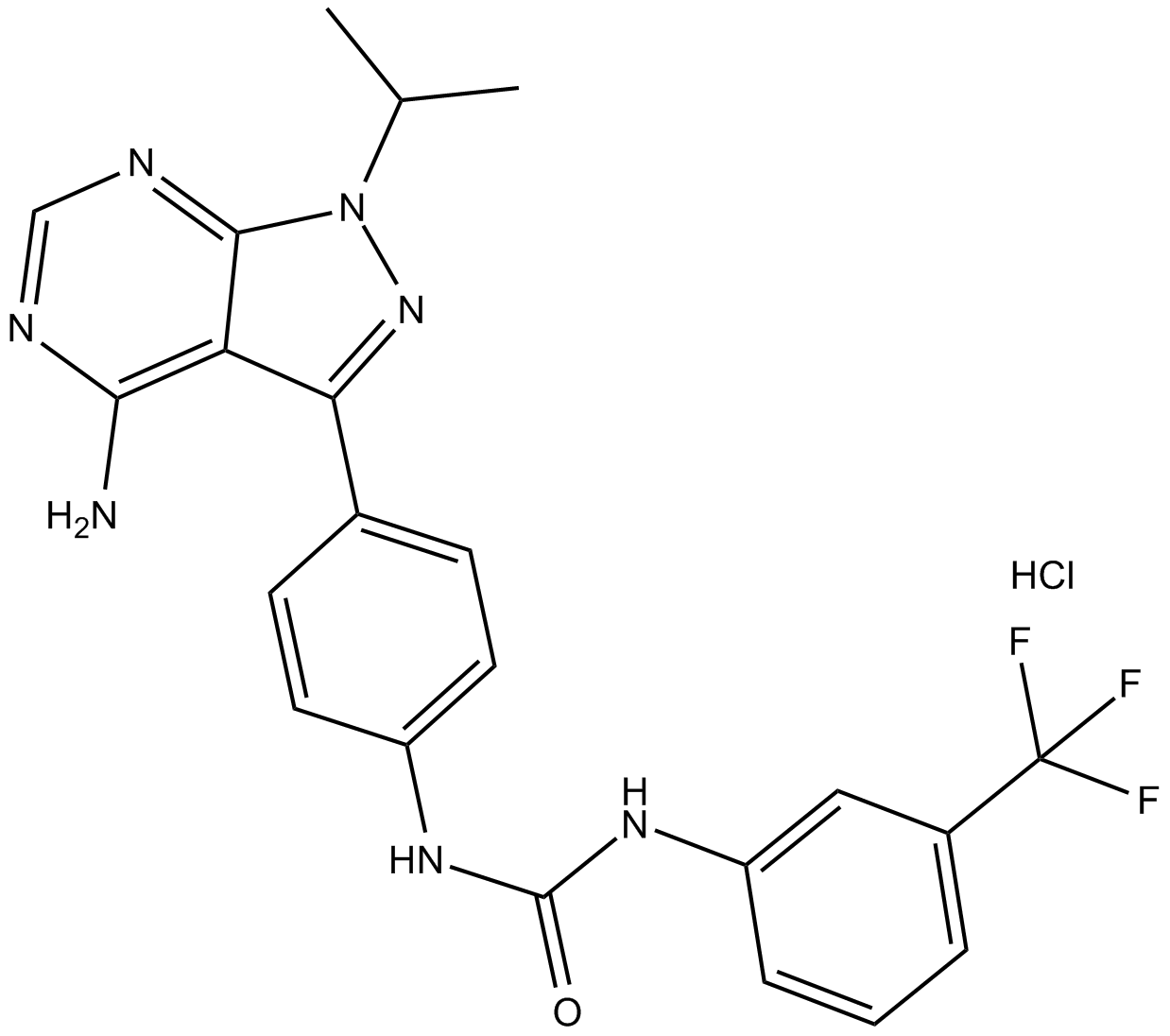
AD57 (hydrochloride)
polypharmacological cancer therapeutic that inhibits RET.
-
GC34214
Adalimumab (Anti-Human TNF-alpha, Human Antibody)
Adalimumab (안티-인간 TNF-알파, 인간 항체)는 종양 괴사 인자 알파(TNF-α)를 대상으로 하는 인간 모노클로널 IgG1 항체입니다.


