Apoptosis
As one of the cellular death mechanisms, apoptosis, also known as programmed cell death, can be defined as the process of a proper death of any cell under certain or necessary conditions. Apoptosis is controlled by the interactions between several molecules and responsible for the elimination of unwanted cells from the body.
Many biochemical events and a series of morphological changes occur at the early stage and increasingly continue till the end of apoptosis process. Morphological event cascade including cytoplasmic filament aggregation, nuclear condensation, cellular fragmentation, and plasma membrane blebbing finally results in the formation of apoptotic bodies. Several biochemical changes such as protein modifications/degradations, DNA and chromatin deteriorations, and synthesis of cell surface markers form morphological process during apoptosis.
Apoptosis can be stimulated by two different pathways: (1) intrinsic pathway (or mitochondria pathway) that mainly occurs via release of cytochrome c from the mitochondria and (2) extrinsic pathway when Fas death receptor is activated by a signal coming from the outside of the cell.
Different gene families such as caspases, inhibitor of apoptosis proteins, B cell lymphoma (Bcl)-2 family, tumor necrosis factor (TNF) receptor gene superfamily, or p53 gene are involved and/or collaborate in the process of apoptosis.
Caspase family comprises conserved cysteine aspartic-specific proteases, and members of caspase family are considerably crucial in the regulation of apoptosis. There are 14 different caspases in mammals, and they are basically classified as the initiators including caspase-2, -8, -9, and -10; and the effectors including caspase-3, -6, -7, and -14; and also the cytokine activators including caspase-1, -4, -5, -11, -12, and -13. In vertebrates, caspase-dependent apoptosis occurs through two main interconnected pathways which are intrinsic and extrinsic pathways. The intrinsic or mitochondrial apoptosis pathway can be activated through various cellular stresses that lead to cytochrome c release from the mitochondria and the formation of the apoptosome, comprised of APAF1, cytochrome c, ATP, and caspase-9, resulting in the activation of caspase-9. Active caspase-9 then initiates apoptosis by cleaving and thereby activating executioner caspases. The extrinsic apoptosis pathway is activated through the binding of a ligand to a death receptor, which in turn leads, with the help of the adapter proteins (FADD/TRADD), to recruitment, dimerization, and activation of caspase-8 (or 10). Active caspase-8 (or 10) then either initiates apoptosis directly by cleaving and thereby activating executioner caspase (-3, -6, -7), or activates the intrinsic apoptotic pathway through cleavage of BID to induce efficient cell death. In a heat shock-induced death, caspase-2 induces apoptosis via cleavage of Bid.
Bcl-2 family members are divided into three subfamilies including (i) pro-survival subfamily members (Bcl-2, Bcl-xl, Bcl-W, MCL1, and BFL1/A1), (ii) BH3-only subfamily members (Bad, Bim, Noxa, and Puma9), and (iii) pro-apoptotic mediator subfamily members (Bax and Bak). Following activation of the intrinsic pathway by cellular stress, pro‑apoptotic BCL‑2 homology 3 (BH3)‑only proteins inhibit the anti‑apoptotic proteins Bcl‑2, Bcl-xl, Bcl‑W and MCL1. The subsequent activation and oligomerization of the Bak and Bax result in mitochondrial outer membrane permeabilization (MOMP). This results in the release of cytochrome c and SMAC from the mitochondria. Cytochrome c forms a complex with caspase-9 and APAF1, which leads to the activation of caspase-9. Caspase-9 then activates caspase-3 and caspase-7, resulting in cell death. Inhibition of this process by anti‑apoptotic Bcl‑2 proteins occurs via sequestration of pro‑apoptotic proteins through binding to their BH3 motifs.
One of the most important ways of triggering apoptosis is mediated through death receptors (DRs), which are classified in TNF superfamily. There exist six DRs: DR1 (also called TNFR1); DR2 (also called Fas); DR3, to which VEGI binds; DR4 and DR5, to which TRAIL binds; and DR6, no ligand has yet been identified that binds to DR6. The induction of apoptosis by TNF ligands is initiated by binding to their specific DRs, such as TNFα/TNFR1, FasL /Fas (CD95, DR2), TRAIL (Apo2L)/DR4 (TRAIL-R1) or DR5 (TRAIL-R2). When TNF-α binds to TNFR1, it recruits a protein called TNFR-associated death domain (TRADD) through its death domain (DD). TRADD then recruits a protein called Fas-associated protein with death domain (FADD), which then sequentially activates caspase-8 and caspase-3, and thus apoptosis. Alternatively, TNF-α can activate mitochondria to sequentially release ROS, cytochrome c, and Bax, leading to activation of caspase-9 and caspase-3 and thus apoptosis. Some of the miRNAs can inhibit apoptosis by targeting the death-receptor pathway including miR-21, miR-24, and miR-200c.
p53 has the ability to activate intrinsic and extrinsic pathways of apoptosis by inducing transcription of several proteins like Puma, Bid, Bax, TRAIL-R2, and CD95.
Some inhibitors of apoptosis proteins (IAPs) can inhibit apoptosis indirectly (such as cIAP1/BIRC2, cIAP2/BIRC3) or inhibit caspase directly, such as XIAP/BIRC4 (inhibits caspase-3, -7, -9), and Bruce/BIRC6 (inhibits caspase-3, -6, -7, -8, -9).
Any alterations or abnormalities occurring in apoptotic processes contribute to development of human diseases and malignancies especially cancer.
References:
1.Yağmur Kiraz, Aysun Adan, Melis Kartal Yandim, et al. Major apoptotic mechanisms and genes involved in apoptosis[J]. Tumor Biology, 2016, 37(7):8471.
2.Aggarwal B B, Gupta S C, Kim J H. Historical perspectives on tumor necrosis factor and its superfamily: 25 years later, a golden journey.[J]. Blood, 2012, 119(3):651.
3.Ashkenazi A, Fairbrother W J, Leverson J D, et al. From basic apoptosis discoveries to advanced selective BCL-2 family inhibitors[J]. Nature Reviews Drug Discovery, 2017.
4.McIlwain D R, Berger T, Mak T W. Caspase functions in cell death and disease[J]. Cold Spring Harbor perspectives in biology, 2013, 5(4): a008656.
5.Ola M S, Nawaz M, Ahsan H. Role of Bcl-2 family proteins and caspases in the regulation of apoptosis[J]. Molecular and cellular biochemistry, 2011, 351(1-2): 41-58.
What is Apoptosis? The Apoptotic Pathways and the Caspase Cascade
Targets for Apoptosis
- Pyroptosis(15)
- Caspase(77)
- 14.3.3 Proteins(3)
- Apoptosis Inducers(71)
- Bax(15)
- Bcl-2 Family(136)
- Bcl-xL(13)
- c-RET(15)
- IAP(32)
- KEAP1-Nrf2(73)
- MDM2(21)
- p53(137)
- PC-PLC(6)
- PKD(8)
- RasGAP (Ras- P21)(2)
- Survivin(8)
- Thymidylate Synthase(12)
- TNF-α(141)
- Other Apoptosis(1145)
- Apoptosis Detection(0)
- Caspase Substrate(0)
- APC(6)
- PD-1/PD-L1 interaction(60)
- ASK1(4)
- PAR4(2)
- RIP kinase(47)
- FKBP(22)
Products for Apoptosis
- Cat.No. 상품명 정보
-
GC17448
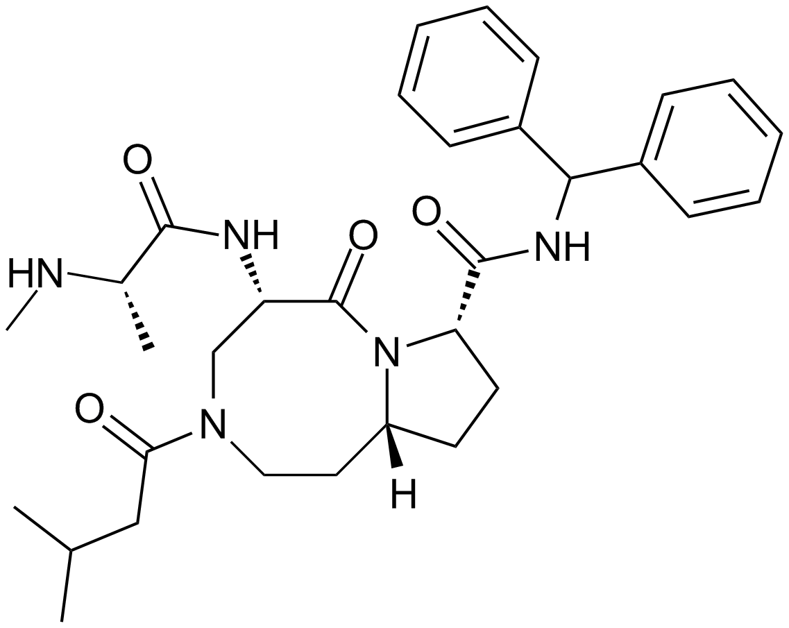
AT-406 (SM-406)
AT-406(SM-406)(AT-406)은 강력하고 경구 생체이용 가능한 Smac 모방체이자 IAP의 길항제이며, 각각 66.4, 1.9 및 5.1nM의 Ki로 XIAP, cIAP1 및 cIAP2 단백질에 결합합니다. .
-
GC15870
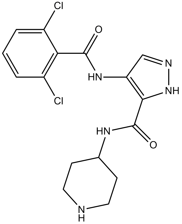
AT7519
AT7519(AT7519M)는 CDK의 강력한 억제제로서, CDK1, CDK2, CDK4에서 CDK6 및 CDK9에 대해 IC50이 각각 210, 47, 100, 13, 170 및 <10 nM입니다.
-
GC13998
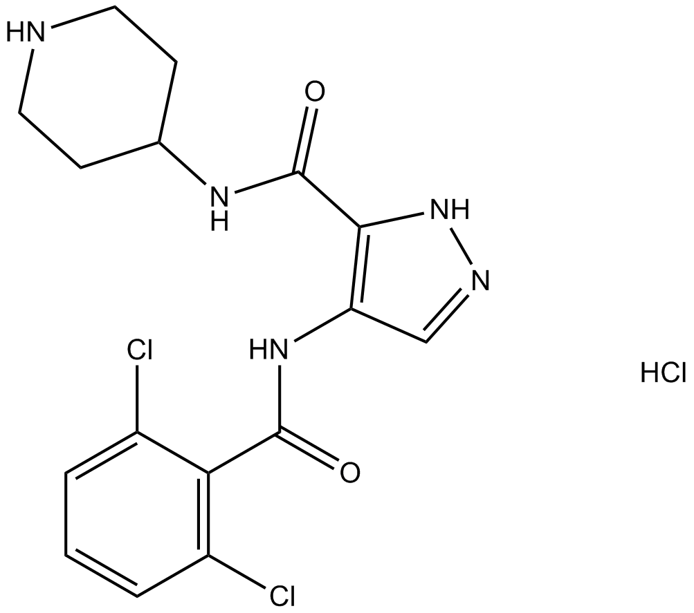
AT7519 Hydrochloride
A Cdk inhibitor
-
GC10638
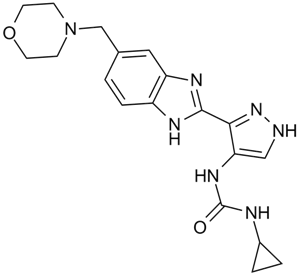
AT9283
A broad spectrum kinase inhibitor
-
GC18133
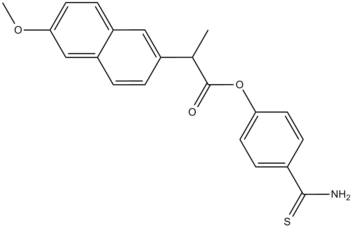
ATB-346
경구 활성 비스테로이드성 소염제(NSAID)인 ATB-346(ATB-346)은 사이클로옥시게나제-1 및 2(COX-1 및 2)를 억제합니다.
-
GC32704
Atezolizumab (MPDL3280A)
아테졸리주맙 (MPDL3280A)은 프로그램된 세포사멸 리간드 1 (PD-L1)에 대한 선택적 인간화 모노클로널 IgG1 항체로, 암 연구에 사용됩니다.

-
GC62499
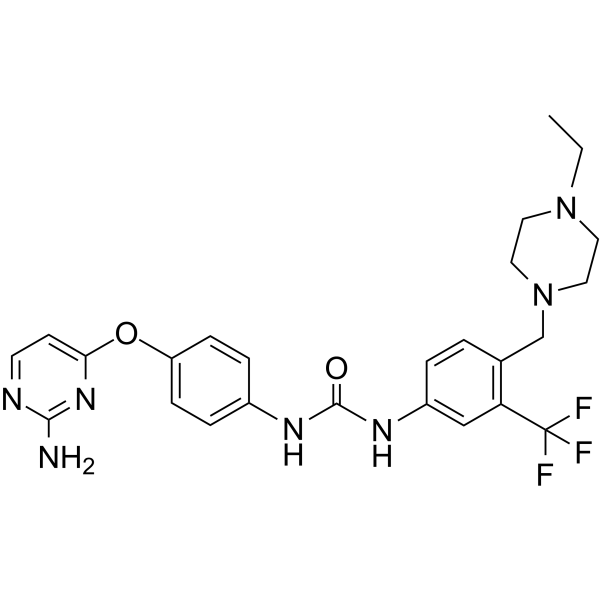
ATH686
ATH686은 강력하고 선택적이며 ATP 경쟁적인 FLT3 억제제입니다. ATH686은 돌연변이 FLT3 단백질 키나제 활성을 표적으로 하고 세포자살 유도 및 세포 주기 억제를 통해 FLT3 돌연변이를 보유하는 세포의 증식을 억제합니다. ATH686은 백혈병 효과가 있습니다.
-
GC46892
ATRA-BA Hybrid
A prodrug form of all-trans retinoic acid and butyric acid

-
GN10394
Atractylenolide III

-
GC15878
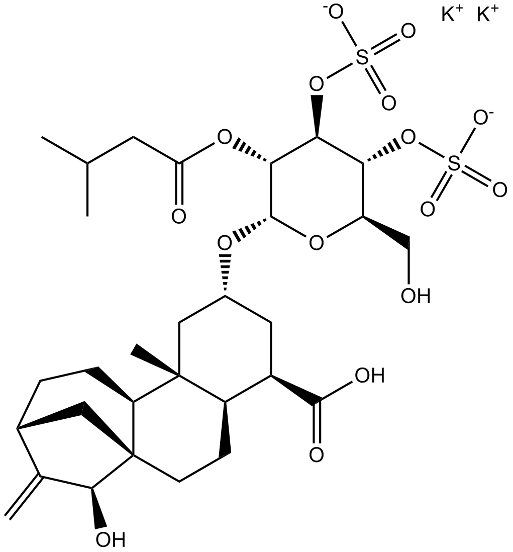
Atractyloside Dipotassium Salt
Inhibitor of ADP/ATP translocases
-
GC39699
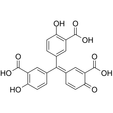
Aurintricarboxylic acid
아우린트리카르복실산은 αβ-메틸렌-ATP 민감성 P2X1R 및 P2X3R에 대한 선택성을 갖는 나노몰 효능의 알로스테릭 길항제이며, IC50은 rP2X1R 및 rP2X3R에 대해 각각 8.6 nM 및 72.9 nM입니다.
-
GC46895
Aurintricarboxylic Acid (ammonium salt)
A protein synthesis inhibitor with diverse biological activities

-
GC13332
Aurora A Inhibitor I
A potent and selective inhibitor of Aurora A kinase

-
GC15295
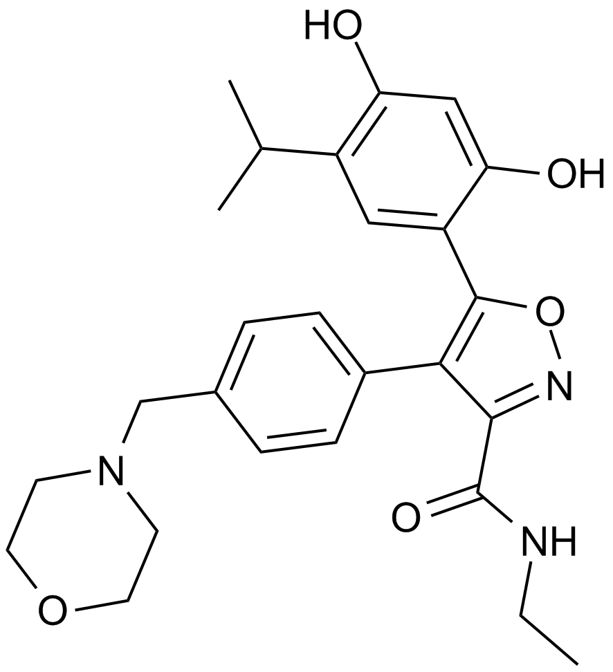
AUY922 (NVP-AUY922)
An Hsp90 inhibitor
-
GC31719
Avelumab (Anti-Human PD-L1, Human Antibody)
Avelumab(Anti-Human PD-L1, Human Antibody)은 잠재적인 항체 의존성 세포 매개 세포독성을 지닌 완전한 인간 IgG1 항-PD-L1 단일클론 항체입니다.

-
GC42880
Avenanthramide-C methyl ester
Avenanthramide-C methyl ester is an inhibitor of NF-κB activation that acts by blocking the phosphorylation of IKK and IκB (IC50 ~ 40 μM).

-
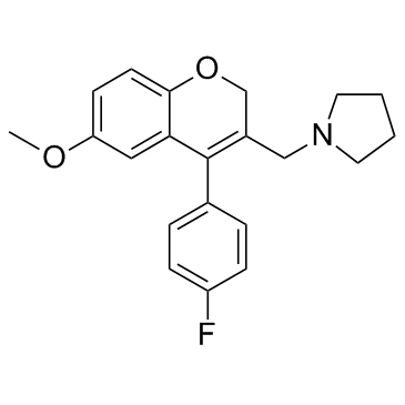
GC35440
AX-024
AX-024는 TCR에 의해 유발되는 T 세포 활성화를 IC50 ~1 nM으로 선택적으로 억제하는 TCR-Nck 상호작용의 경구 사용 가능한 동급 최초의 억제제입니다.
-
GC19046
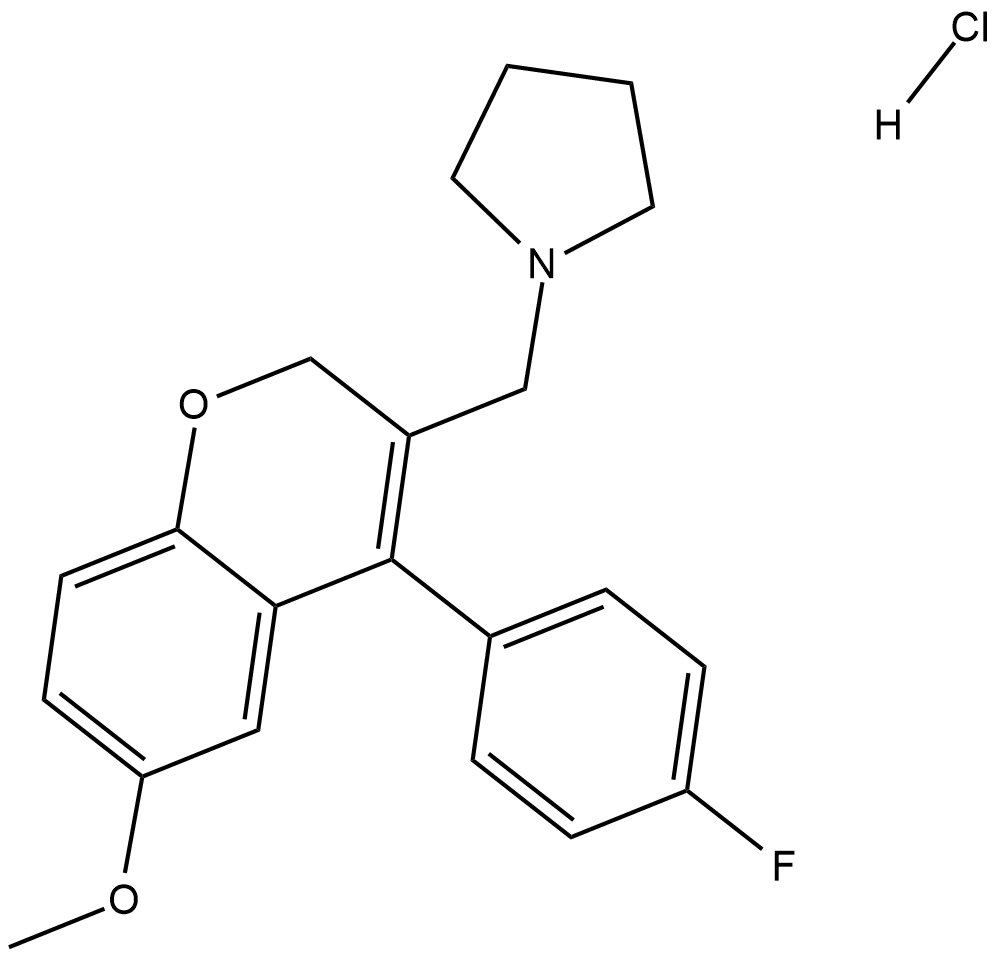
AX-024 hydrochloride
AX-024 염산염은 IC50 ~1 nM으로 TCR에 의해 유발되는 T 세포 활성화를 선택적으로 억제하는 TCR-Nck 상호작용의 구두로 이용 가능한 동급 최초의 억제제입니다.
-
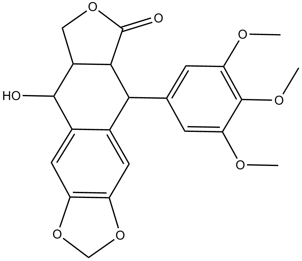
GC17045
AXL1717
A potent and selective inhibitor of IGF-1R
-
GC15055
AZ 628
AZ 628은 B-Raf, B-RafV600E 및 c-Raf-1에 대해 IC50이 각각 105, 34 및 29nM인 범-Raf 키나제 억제제입니다.

-
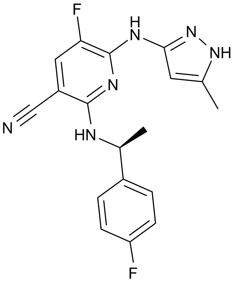
GC13433
AZ 960
A JAK2 inhibitor
-
GC46901
Azadirachtin
가장 유망한 식물성 살충제 중 하나인 Azadirachtin은 해충 방제에 널리 사용됩니다.

-
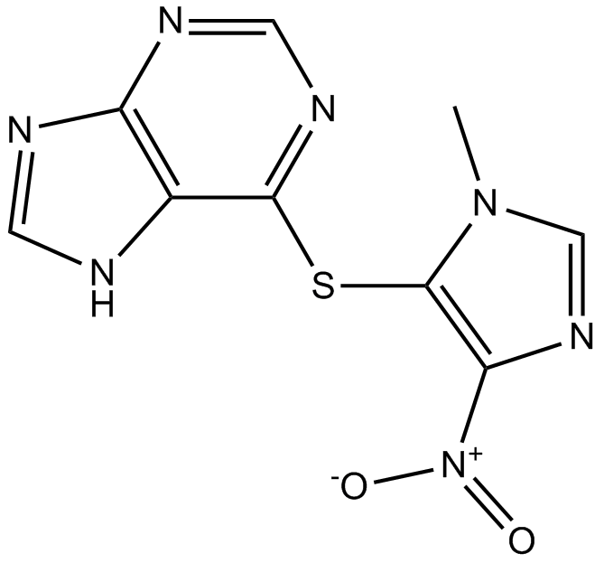
GC15033
Azathioprine
Azathioprine(BW 57-322)은 경구 활성 면역억제제입니다.
-
GC48971
AZD 1152 (hydrochloride)
A prodrug for a potent Aurora B inhibitor

-
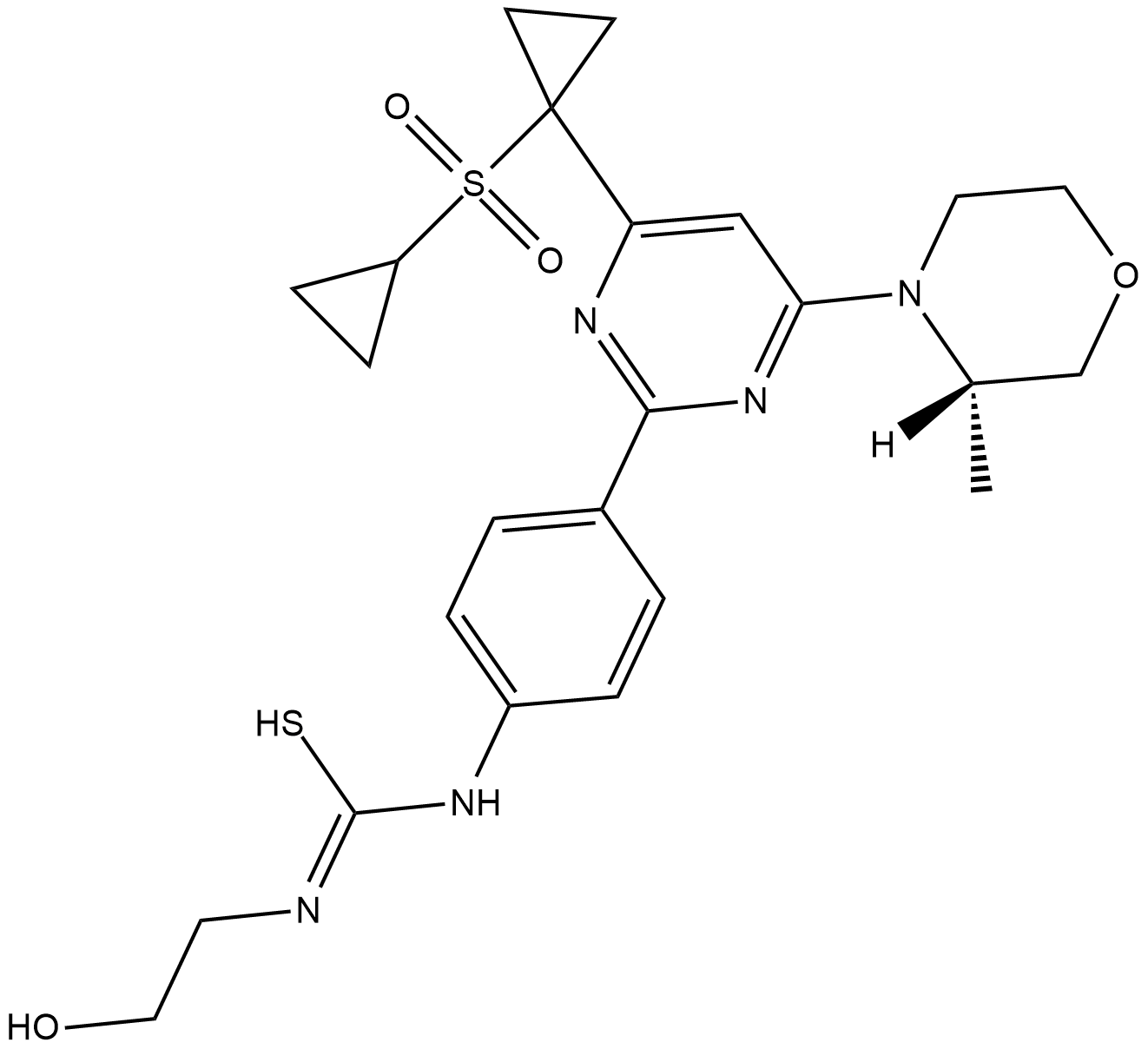
GC18566
AZD 3147
AZD 3147은 1.5nM의 IC50 값을 갖는 mTORC1 및 mTORC2의 강력한 경구 활성 선택적 이중 억제제입니다. AZD 3147은 또한 PI3K에 선택적 효과가 있습니다.
-
GC50109
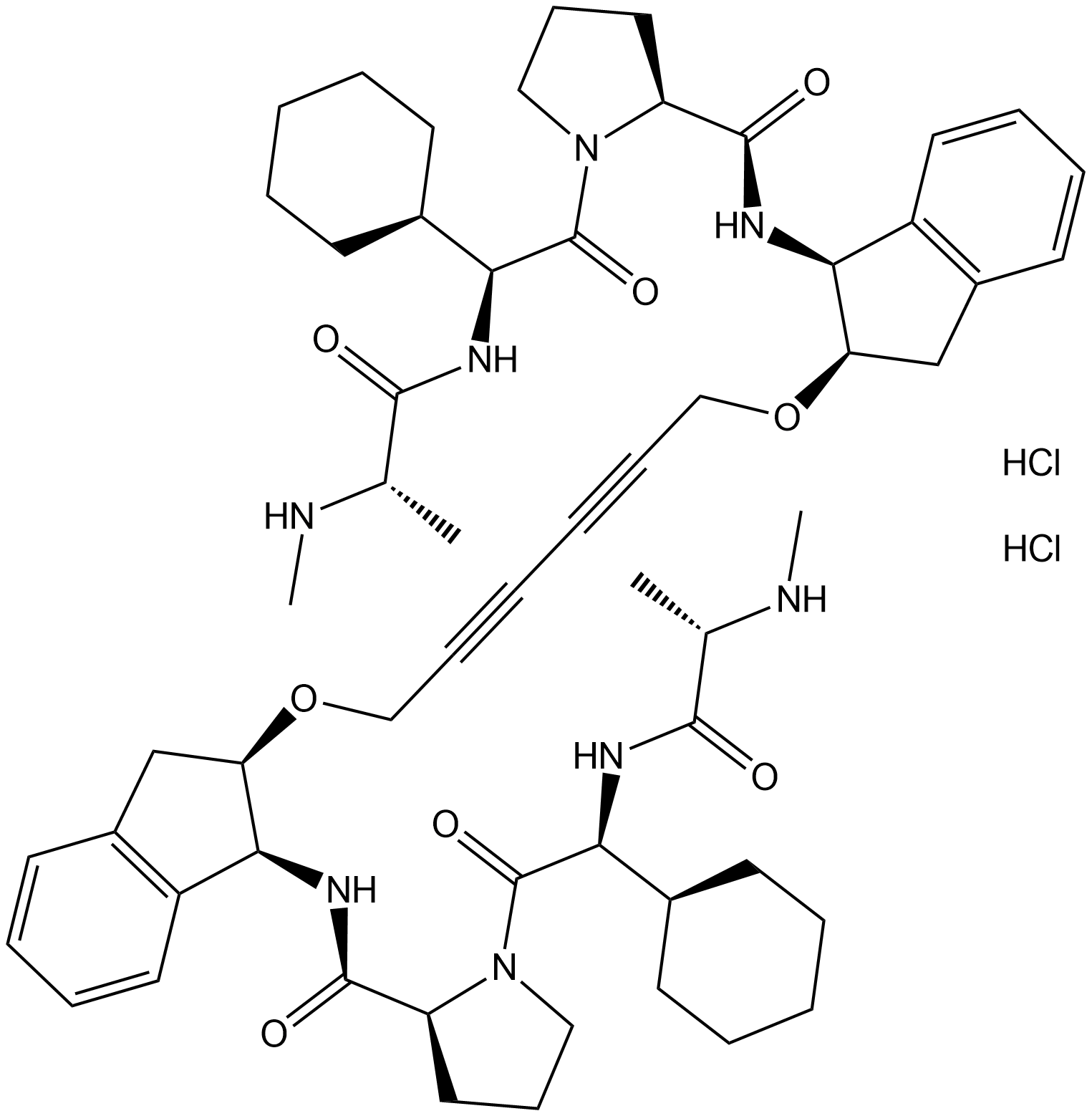
AZD 5582 dihydrochloride
AZD 5582 dihydrochloride는 각각 15, 21 및 15 nM의 IC50으로 BIR3 도메인 cIAP1, cIAP2 및 XIAP에 결합하는 세포자멸사 단백질 억제제(IAP)의 길항제입니다. AZD5582는 세포 사멸을 유도합니다.
-
GC33247
AZD-5991
AZD-5991은 FRET 분석에서 IC50이 0.7nM이고 표면 플라즈몬 공명(SPR) 분석에서 Kd가 0.17nM인 강력하고 선택적인 Mcl-1 억제제입니다.

-
GC33283
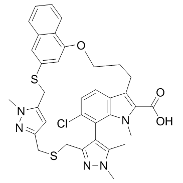
AZD-5991 Racemate
AZD-5991 Racemate는 AZD-5991의 racemate입니다. AZD-5991 Racemate는 FRET 분석에서 IC50이 3nM 미만인 Mcl-1 억제제입니다.
-
GC33239
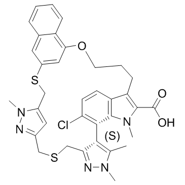
AZD-5991 S-enantiomer
AZD-5991 S-거울상 이성질체는 AZD-5991의 덜 활성인 거울상 이성질체입니다. AZD-5991 S-거울상 이성질체는 FRET 분석에서 IC50이 6.3μM이고 표면 플라즈몬 공명(SPR) 분석에서 Kd가 0.98μM인 Mcl-1 억제제입니다.
-
GC64938
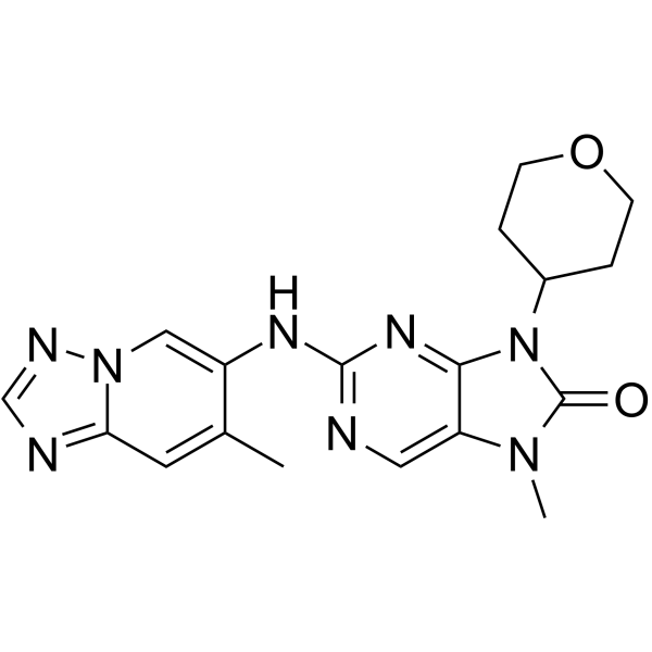
AZD-7648
AZD-7648은 IC50이 0.6nM인 강력한 경구 활성 선택적 DNA-PK 억제제입니다. AZD-7648은 세포사멸을 유도하고 항종양 활성을 나타낸다.
-
GC12660
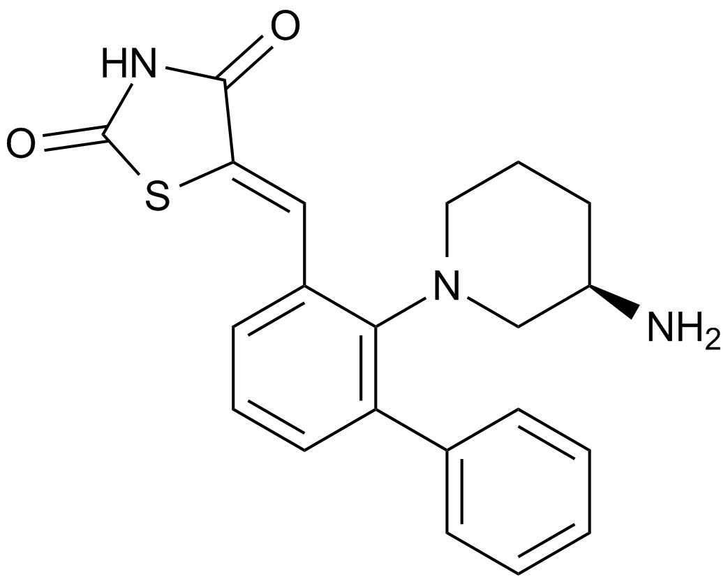
AZD1208
A pan-Pim kinase inhibitor
-
GC13029
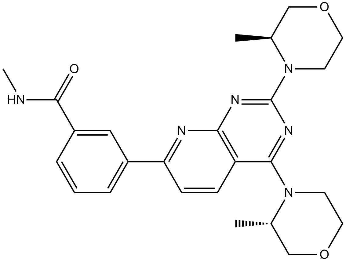
AZD2014
AZD2014(AZD2014)는 2.81nM의 IC50을 갖는 ATP 경쟁적 mTOR 억제제입니다. AZD2014는 mTORC1 및 mTORC2 복합체를 모두 억제합니다.
-
GC33255
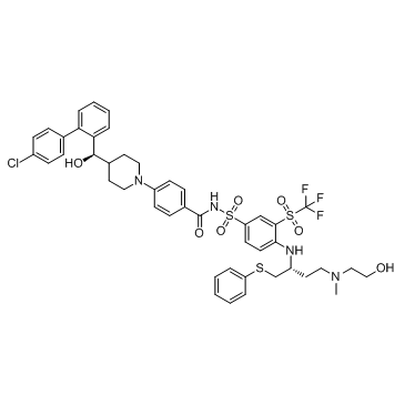
AZD4320
AZD4320은 KPUM-MS3, KPUM-UH1 및 STR-428 세포에 대해 IC50이 각각 26nM, 17nM 및 170nM인 새로운 BH3 모방 이중 BCL2/BCLxL 억제제입니다.
-
GC19050
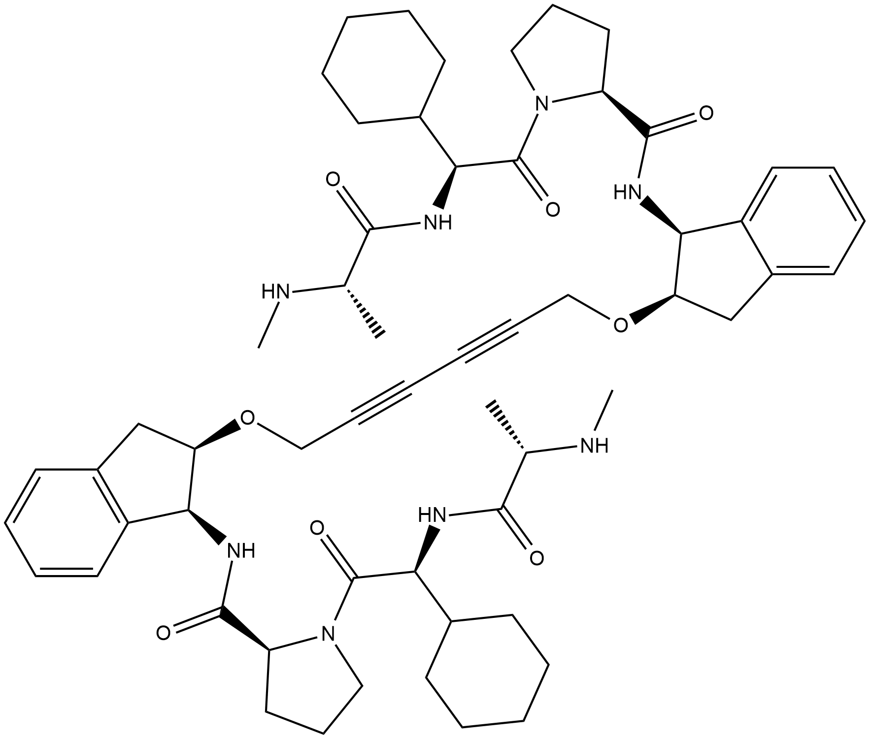
AZD5582
AZD5582는 BIR3 도메인 cIAP1, cIAP2 및 XIAP에 각각 15, 21 및 15nM의 IC50으로 결합하는 세포자멸사 단백질 억제제(IAP)의 길항제입니다. AZD5582는 세포 사멸을 유도합니다.
-
GC16380
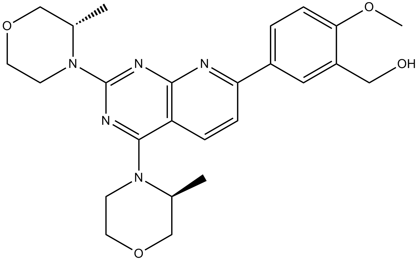
AZD8055
MTOR 억제제
-
GC19054
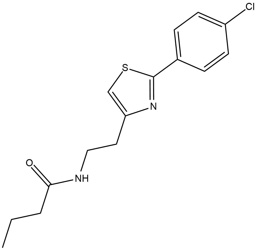
Azoramide
Azoramide는 UPR(unfolded protein response)의 강력한 경구 활성 소분자 조절제입니다.
-
GC46904
Azoxystrobin
아족시스트로빈은 광범위한 스펙트럼의 β-메톡시아크릴레이트 살균제입니다.

-
GC60616
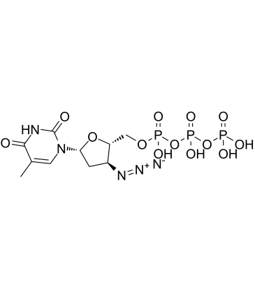
AZT triphosphate
AZT 삼인산(3'-Azido-3'-deoxythymidine-5'-triphosphate)은 지도부딘(AZT)의 활성 삼인산 대사 산물입니다.
-
GC60617
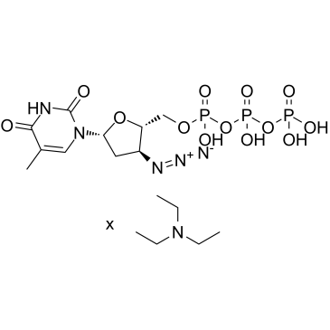
AZT triphosphate TEA
AZT 삼인산 TEA(3'-Azido-3'-deoxythymidine-5'-triphosphate TEA)는 지도부딘(AZT)의 활성 삼인산 대사 산물입니다.
-
GC35458
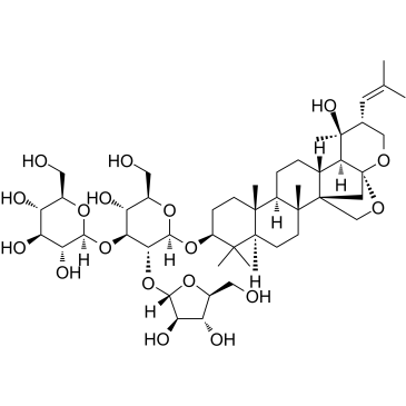
Bacopaside II
약초인 Bacopa monnieri의 추출물인 Bacopaside II는 Aquaporin-1(AQP1) 수로를 차단하고 AQP1을 발현하는 세포의 이동을 방해합니다. Bacopaside II는 세포 주기 정지와 세포 사멸을 유도합니다.
-
GC34263
Bak BH3
Bak BH3는 Bak의 BH3 도메인에서 파생되며 세포에서 Bcl-xL의 기능을 길항할 수 있습니다.

-
GC52344
Bak BH3 (72-87) (human) (trifluoroacetate salt)
A Bak-derived peptide

-
GC12053
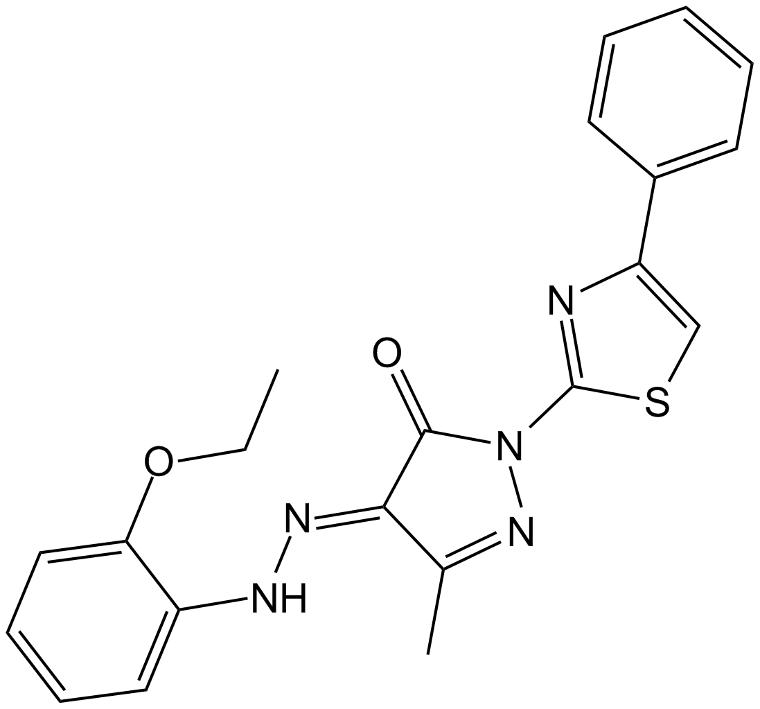
BAM7
A direct activator of Bax
-
GN10507
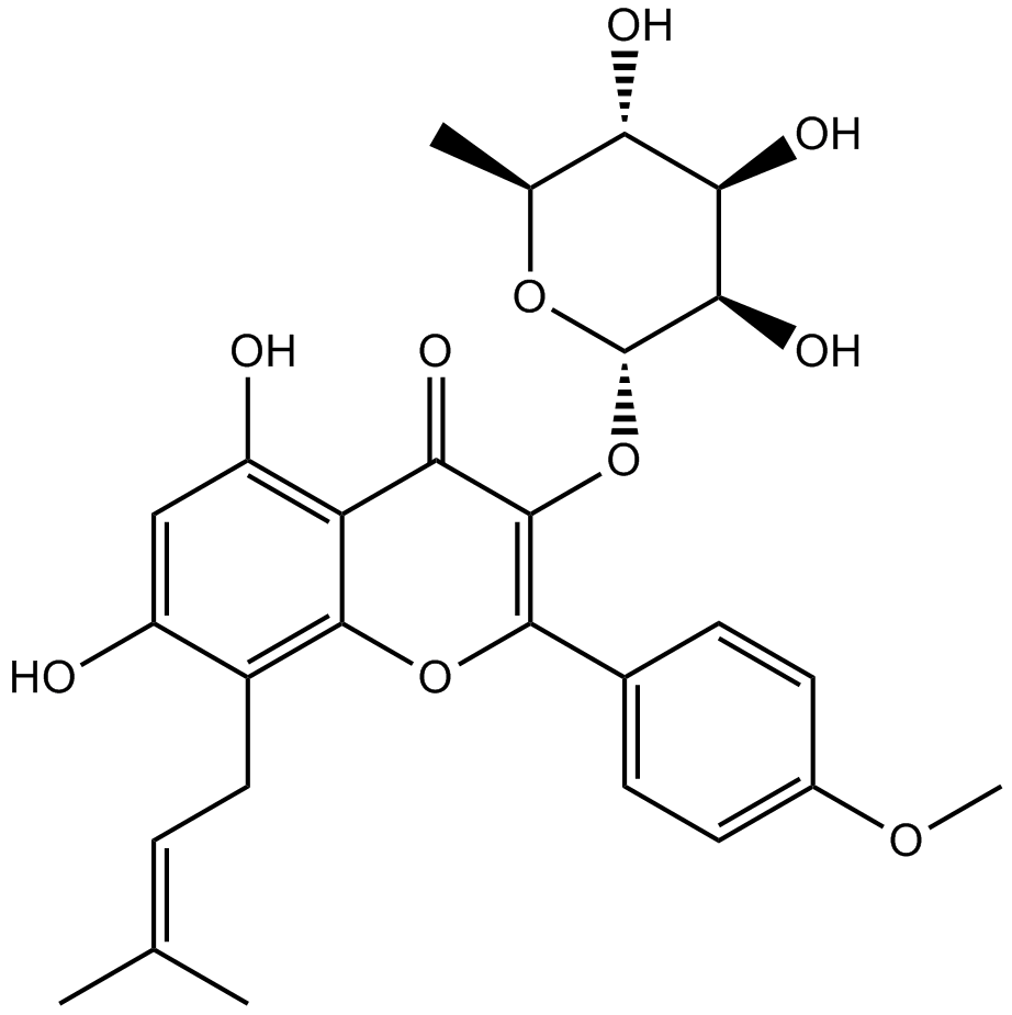
Baohuoside I
-
GC15371
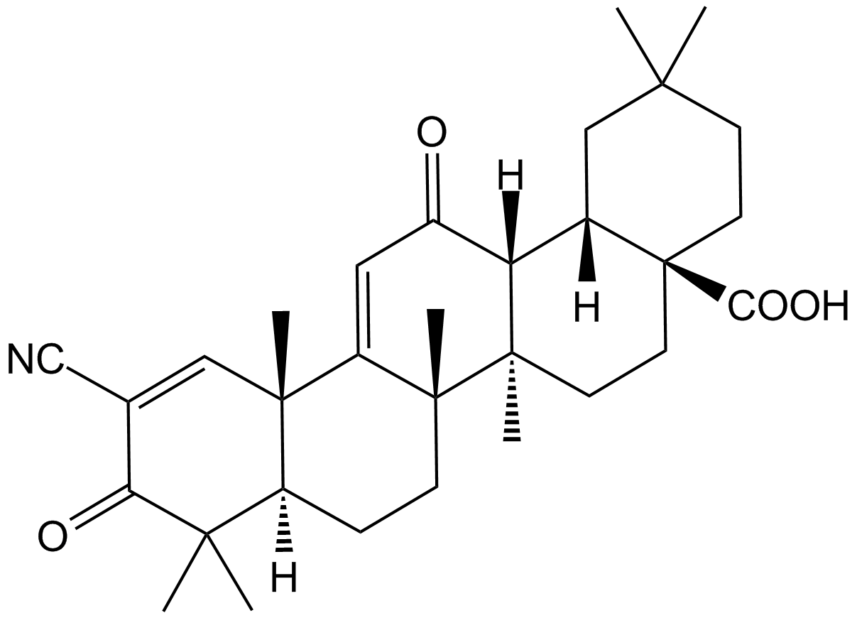
Bardoxolone
An anti-inflammatory compound that activates Nrf2/ARE signaling
-
GC11572
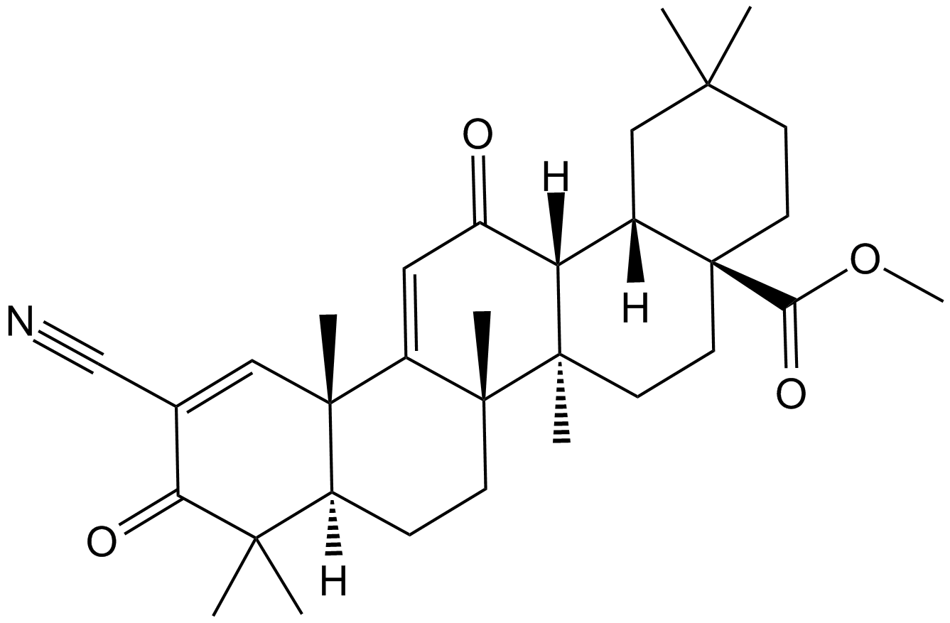
Bardoxolone methyl
A synthetic triterpenoid with potent anticancer and antidiabetic activity
-
GC60620
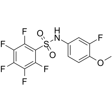
Batabulin
Batabulin(T138067)은 β-tubulin isotypes의 하위 집합에 공유 및 선택적으로 결합하여 미세소관 중합을 방해하는 항종양제입니다. Batabulin은 세포 형태에 영향을 미치고 세포 주기 정지를 유발하여 궁극적으로 세포 사멸을 유도합니다.
-
GC60621
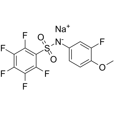
Batabulin sodium
바타불린 나트륨(T138067 나트륨)은 β-튜불린 이소타입의 하위 집합에 공유 및 선택적으로 결합하여 미세소관 중합을 방해하는 항종양제입니다. 바타불린 나트륨은 세포 형태에 영향을 미치고 세포 주기 정지를 유도하여 궁극적으로 세포 사멸을 유도합니다.
-
GC12763
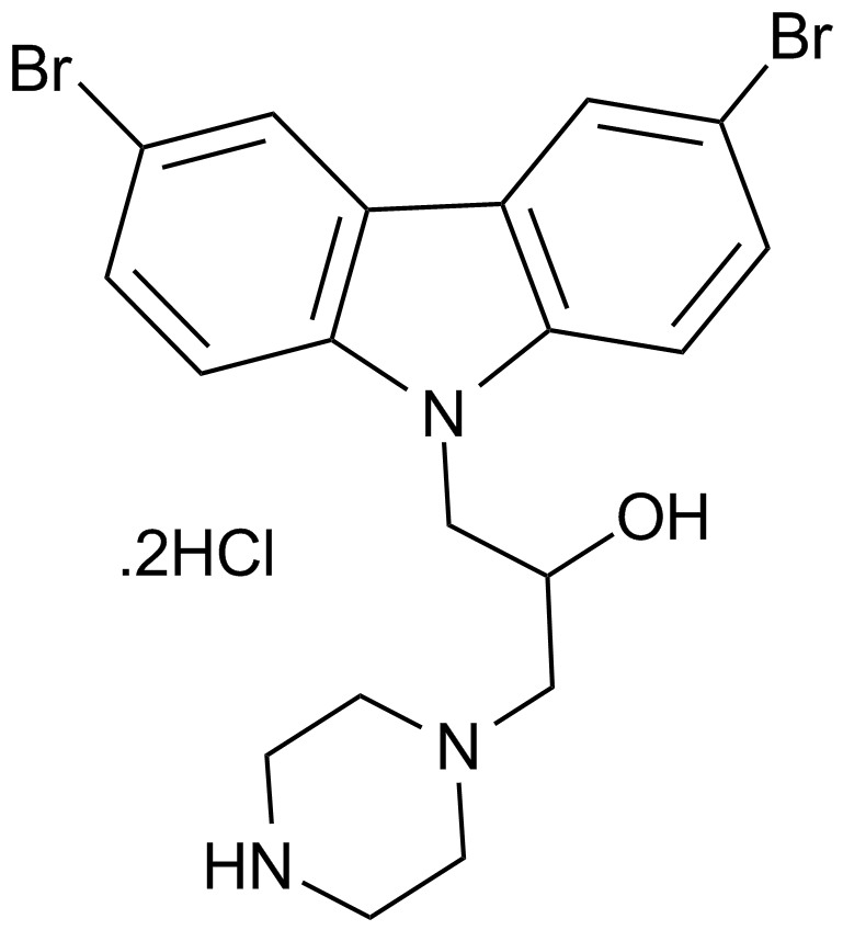
Bax channel blocker
-
GC16023
Bax inhibitor peptide P5
Bax inhibitor

-
GC17195
Bax inhibitor peptide V5
A Bax inhibitor

-
GC52476
Bax Inhibitor Peptide V5 (trifluoroacetate salt)
A Bax inhibitor

-
GC16695
Bax inhibitor peptide, negative control
Peptide inhibit Bax translocation to mitochondria

-
GC10345
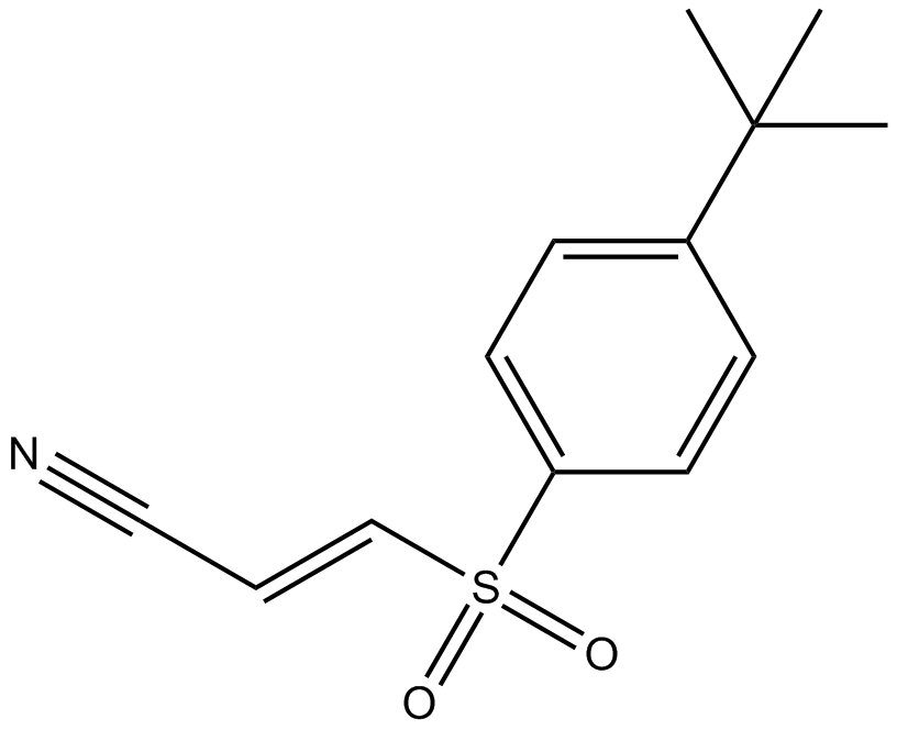
Bay 11-7085
BAY 11-7085(BAY 11-7083)는 NF-κB 활성화 및 IκBα의 인산화 억제제입니다. 10μM의 IC50으로 IκBα를 안정화합니다.
-
GC13035
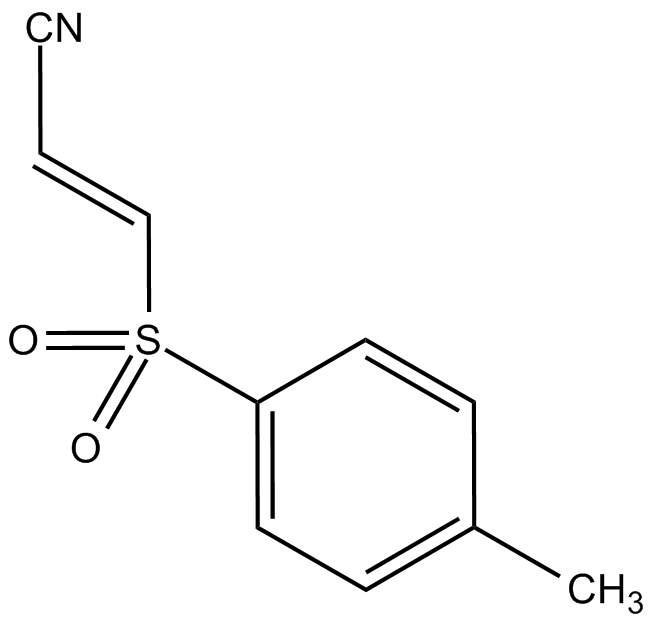
Bay 11-7821
선택적이고 불가역적인 NF-κB 억제제
-
GC16389
BAY 61-3606
A Syk inhibitor

-
GC42897
BAY 61-3606 (hydrochloride)
BAY 61-3606 is a cell-permeable, reversible inhibitor of spleen tyrosine kinase (Syk; Ki = 7.5 nM; IC50 = 10 nM).

-
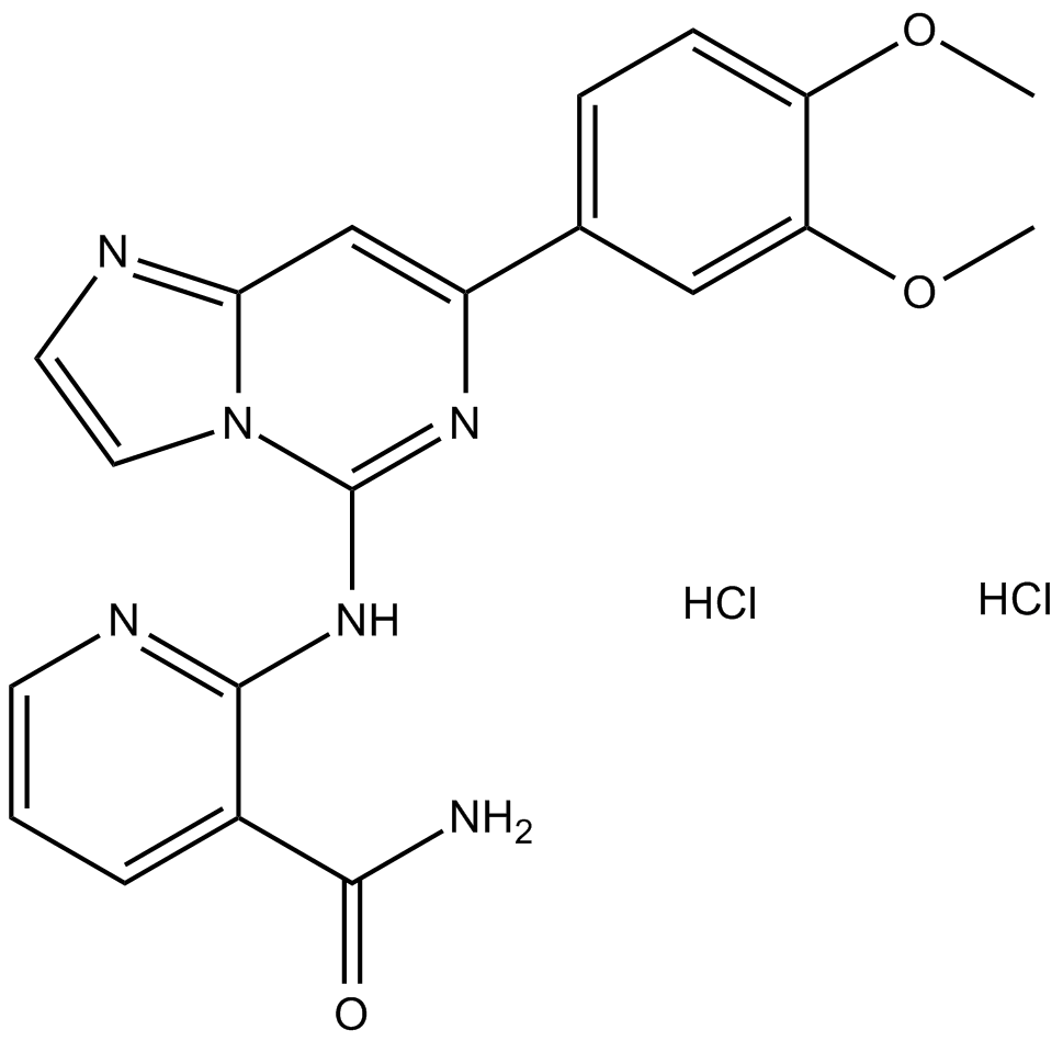
GC12136
BAY 61-3606 dihydrochloride
-
GC62164
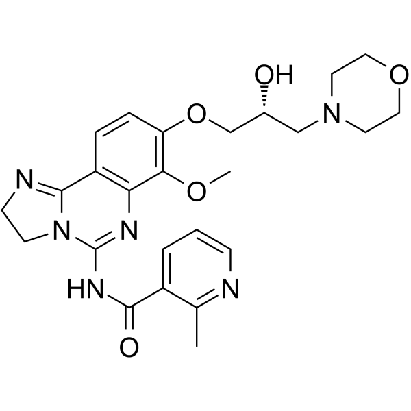
BAY1082439
BAY1082439는 경구 생체이용 가능한 선택적 PI3Kα/β/δ 억제제입니다. BAY1082439는 또한 PIK3CA의 돌연변이 형태를 억제합니다. BAY1082439는 Pten-null 전립선암 성장을 억제하는 데 매우 효과적입니다.
-
GC16516
BCH
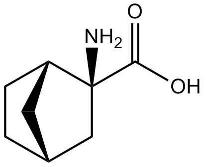
BCH(BCH)는 거대 중성 아미노산 수송체 1(LAT1)의 선택적이고 경쟁적인 억제제로 아미노산의 세포 흡수 및 mTOR 인산화를 유의하게 억제하여 암 성장 및 세포 사멸의 억제를 유도합니다.
-
GC63325
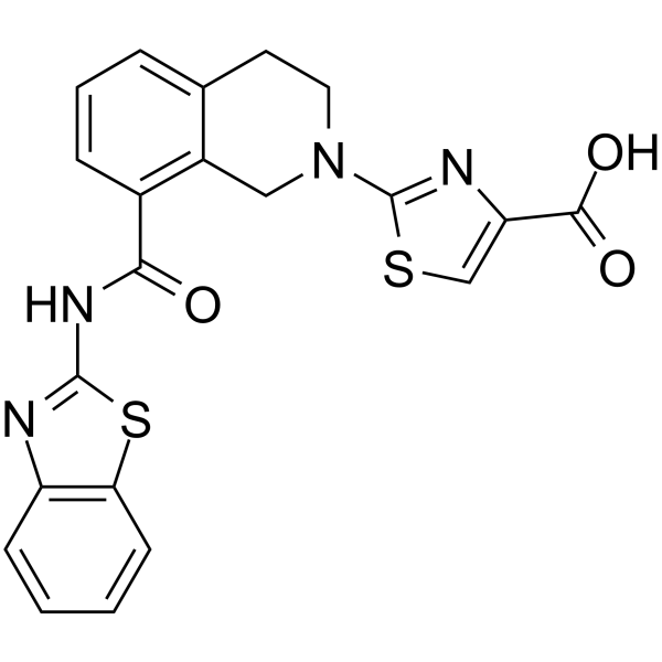
Bcl-xL antagonist 2
Bcl-xL 길항제 2는 IC50과 Ki가 각각 0.091μM 및 65nM인 BCL-XL의 강력하고 선택적인 경구 활성 길항제입니다. Bcl-xL 길항제 2는 암세포의 세포자멸사를 촉진합니다. Bcl-xL 길항제 2는 만성 림프구성 백혈병(CLL) 및 비호지킨 림프종(NHL) 연구에 대한 가능성이 있습니다.
-
GC62599
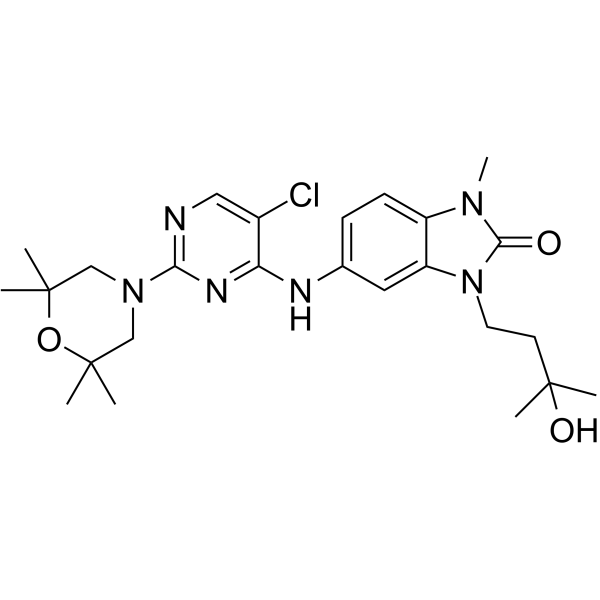
BCL6-IN-4
BCL6-IN-4는 IC50이 97nM인 강력한 B세포 림프종 6(BCL6) 억제제입니다. BCL6-IN-4는 항종양 활성이 있습니다.
-
GC68012
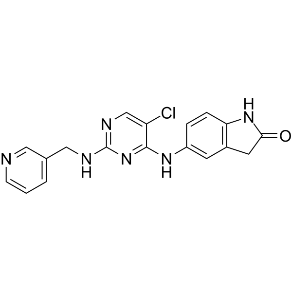
BCL6-IN-7
-
GC10721
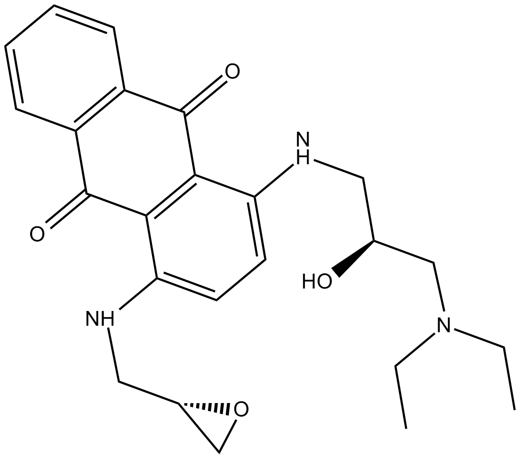
BDA-366
BDA-366은 강력한 Bcl2 길항제(Ki = 3.3nM)로 Bcl2-BH4 도메인에 높은 친화도와 선택도로 결합합니다. BDA-366은 Bcl2의 구조적 변화를 유도하여 항아폽토시스 기능을 없애고 이를 생존 분자에서 세포 사멸 유도제로 전환합니다. BDA-366은 폐암 세포의 성장을 억제합니다.
-
GC42912
Becatecarin
베카테카린은 항종양 효과가 있는 레베카마이신 유사체입니다. 베카테카린은 DNA에 삽입되어 토포이소머라제 I/II의 촉매 활성을 억제합니다.

-
GC68369
Belantamab

-
GC65031
Belimumab
벨리무맙(LymphoStat B)은 B 세포 활성화 인자(BAFF)를 억제하는 인간 IgG1Λ 단일클론 항체입니다.

-
GC49042
Benastatin A
A bacterial metabolite with diverse biological activities

-
GC64354
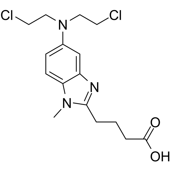
Bendamustine
퓨린 유사체인 벤다무스틴(SDX-105 유리 염기)은 DNA 가교제입니다. 벤다무스틴은 DNA 손상 스트레스 반응과 세포자멸사를 활성화합니다. 벤다무스틴은 강력한 알킬화, 항암 및 항대사 물질 특성을 가지고 있습니다.
-
GC10744
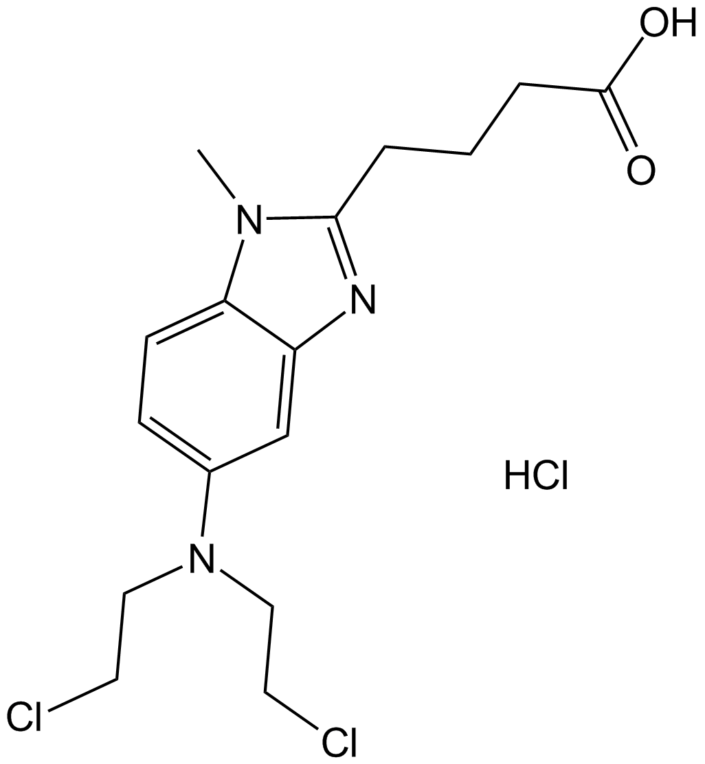
Bendamustine HCl
퓨린 유사체인 벤다무스틴 HCl(SDX-105)은 DNA 가교제입니다. Bendamustine HCl은 DNA 손상 스트레스 반응과 세포자멸사를 활성화합니다. Bendamustine HCl은 강력한 알킬화, 항암 및 항대사 물질 특성을 가지고 있습니다.
-
GC49781
Benomyl
A carbamate pesticide

-
GC62451
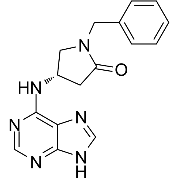
Benpyrine
Benpyrine은 82.1μM의 KD 값을 갖는 고도로 특이적이고 경구 활성인 TNF-α 억제제입니다.
-
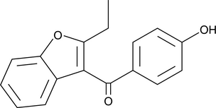
GC49403
Benzarone
Benzarone(Fragivix)은 난모세포에서 IC50이 2.8μM인 강력한 인간 요산 수송체 1(hURAT1) 억제제입니다.
-
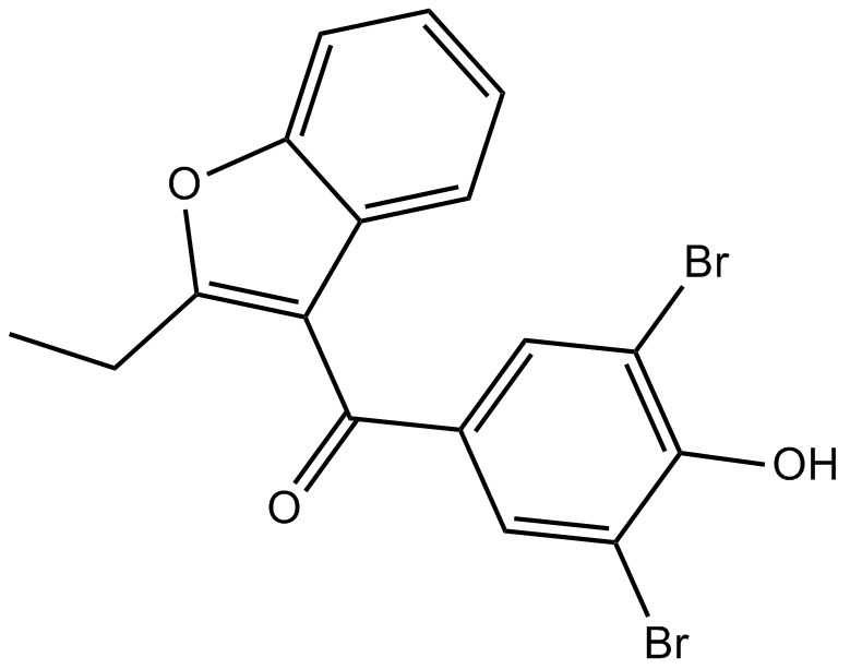
GC14930
Benzbromarone
Benzbromarone은 통풍 치료에 사용되는 요산 배출제로 사용되는 크산틴 산화효소의 매우 효과적이고 내약성이 뛰어난 비경쟁적 억제제입니다.
-
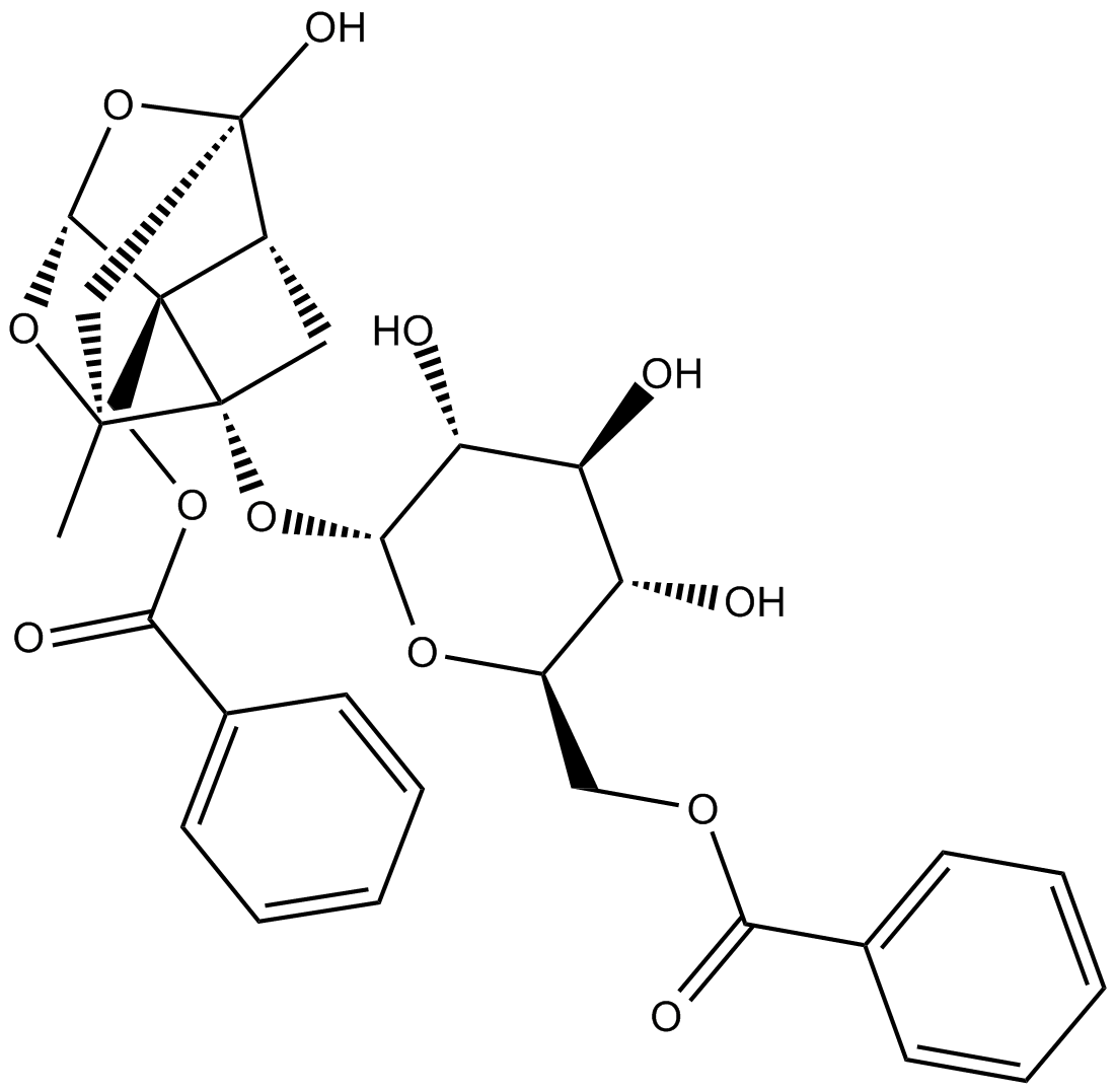
GN10520
Benzoylpaeoniflorin
-
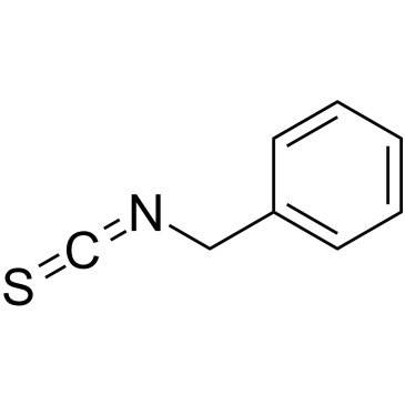
GC38683
Benzyl isothiocyanate
벤질 이소티오시아네이트는 항균 활성이 있는 천연 이소티오시아네이트의 구성원입니다.
-
GN10358
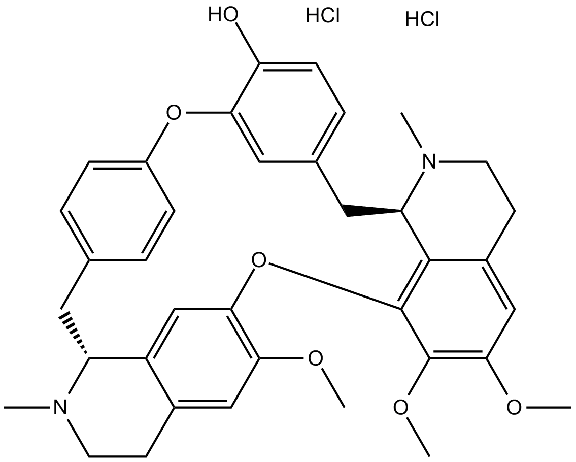
Berbamine hydrochloride
-
GN10539
Bergenin

-
GC42925
Berteroin
자연적으로 발생하는 설포라판 유사체인 베르테로인(Berteroin)은 항전이제입니다.

-
GC10734
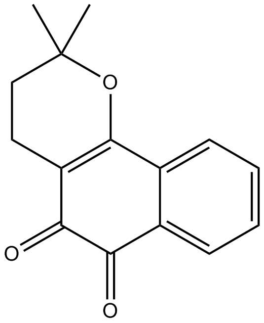
Beta-Lapachone
베타-라파콘(ARQ-501;NSC-26326)은 자연적으로 발생하는 O-나프토퀴논으로, 토포이소머라제 I 억제제로 작용하며, 세포 주기 진행을 억제하여 세포자멸사를 유도합니다.
-
GC35504
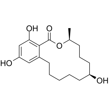
Beta-Zearalanol
Beta-Zearalenol은 Fusarium spp에 의해 생성되는 진균독으로 포유동물의 생식 세포에서 세포자멸사와 산화 스트레스를 유발합니다.
-
GN10632
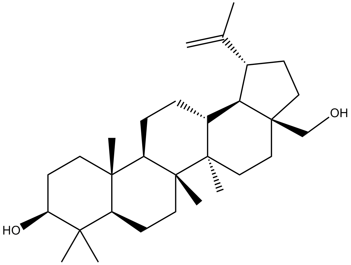
Betulin
-
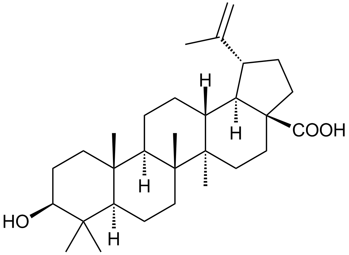
GC10480
Betulinic acid
A plant triterpenoid similar to bile acids
-
GC48477
Betulinic Acid propargyl ester
An alkyne derivative of betulinic acid

-
GC48504
Betulinic Aldehyde oxime
A derivative of betulin

-
GC48520
Betulonaldehyde
A pentacyclic triterpenoid

-
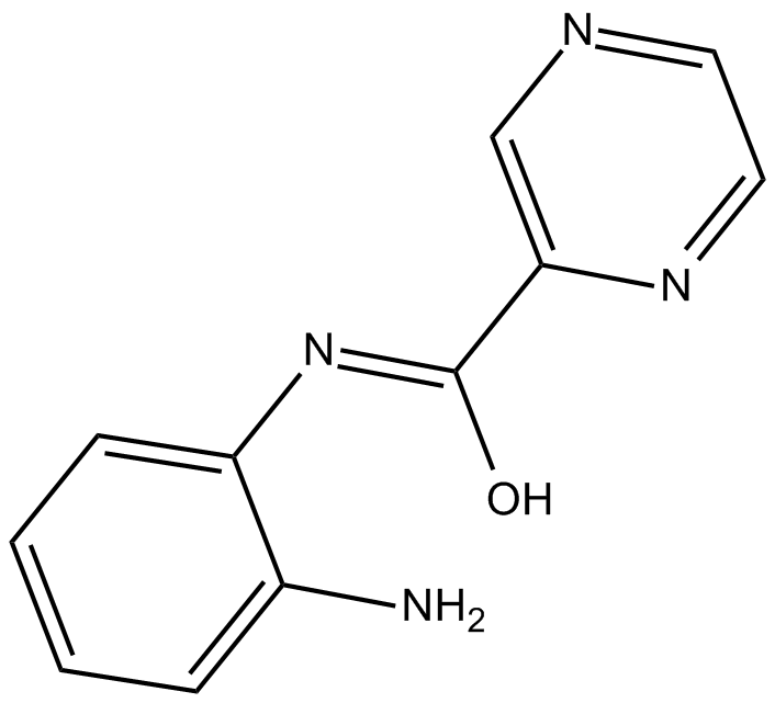
GC12074
BG45
BG45는 HDAC3에 대한 선택성이 있는 HDAC 클래스 I 억제제(IC50 = 289nM)입니다.
-
GC18136
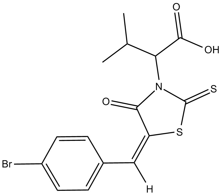
BH3I-1
BH3I-1은 Bcl-2 계열 길항제로 FP assay에서 2.4±0.2μM의 Ki로 Bcl-xL에 대한 Bak BH3 펩타이드의 결합을 억제합니다. BH3I-1은 p53/MDM2 쌍에 대해 5.3μM의 Kd를 가지고 있습니다.
-
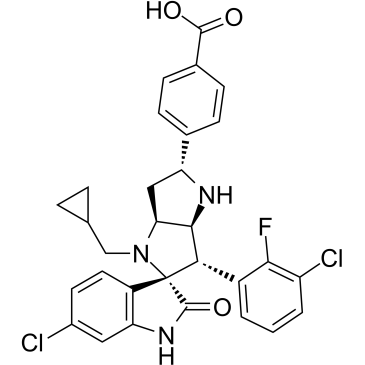
GC35511
BI-0252
BI-0252는 IC50이 4nM인 경구 활성 선택적 MDM2-p53 억제제입니다. BI-0252는 종양 단백질 p53(TP53) 표적 유전자 및 세포자멸사 마커의 동시 유도와 함께 마우스 SJSA-1 이종이식편의 모든 동물에서 종양 퇴행을 유도할 수 있습니다.
-
GC17828
BI-847325
BI-847325는 인간 MEK2 및 AK-C에 대해 IC50 값이 각각 4 및 15nM인 MEK 및 오로라 키나제(AK)의 ATP 경쟁적 이중 억제제입니다.

-
GC11224
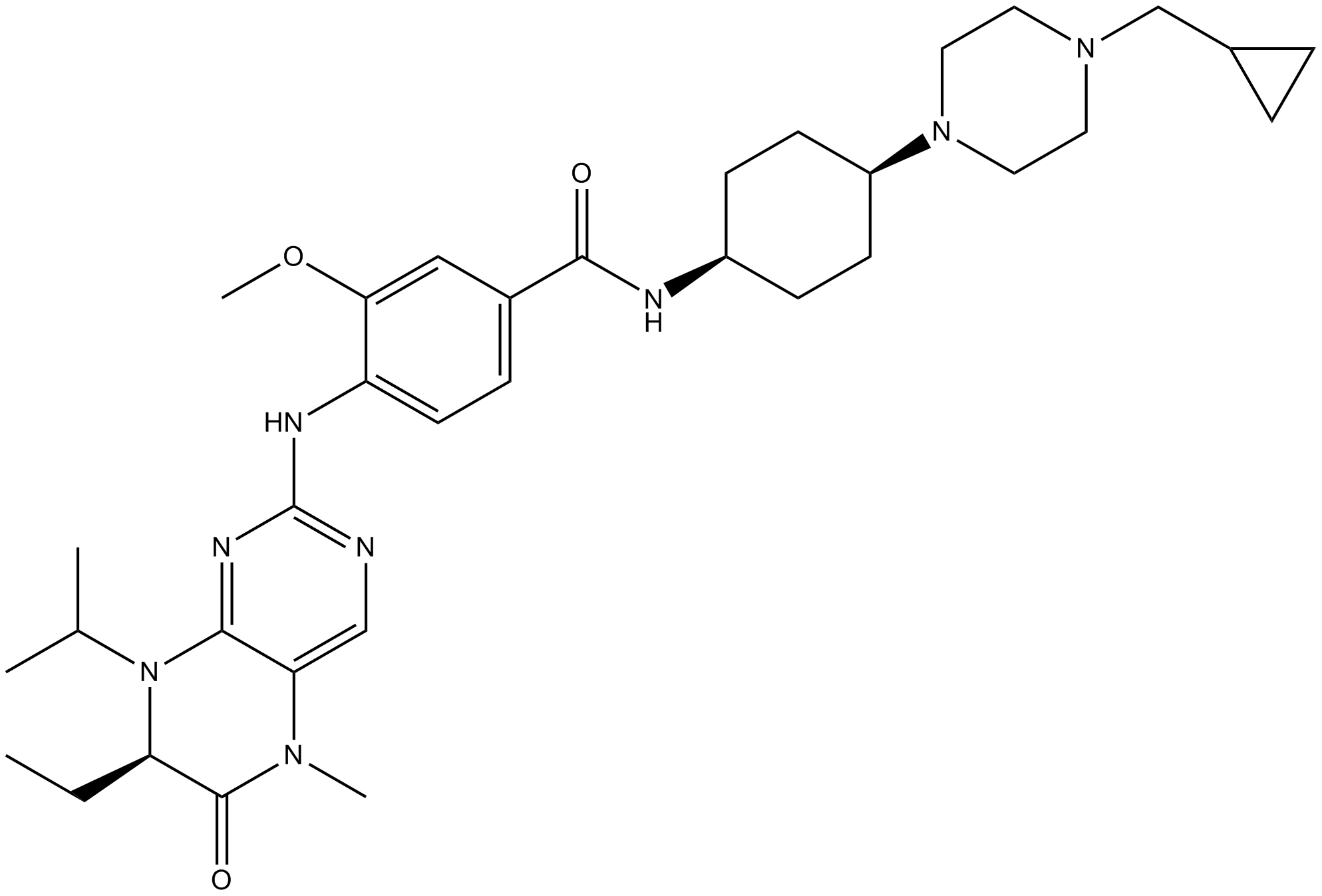
BI6727(Volasertib)
BI6727(Volasertib)(BI 6727)은 0.87 nM의 IC50을 갖는 경구 활성, 매우 강력하고 ATP-경쟁적인 Polo-like kinase 1(PLK1) 억제제입니다. BI6727(Volasertib)은 각각 5 및 56 nM의 IC50으로 PLK2 및 PLK3을 억제합니다. BI6727(Volasertib)은 유사분열 정지 및 세포자멸사를 유도합니다. 디히드로프테리디논 유도체인 BI6727(Volasertib)은 여러 암 모델에서 현저한 항종양 활성을 나타냅니다.
-
GC13636
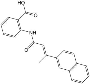
BIBR 1532
BIBR 1532는 무세포 분석에서 IC50이 100nM인 강력하고 선택적이며 비경쟁적인 텔로머라제 억제제입니다.
-
GC60076
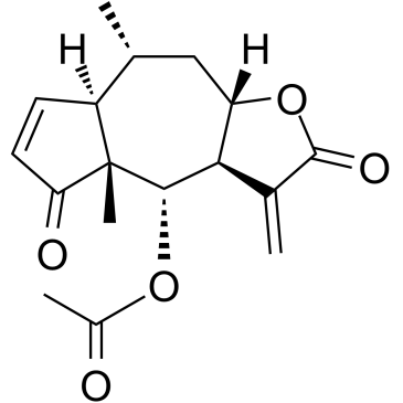
Bigelovin
Inula helianthus-aquatica에서 분리된 sesquiterpene lactone인 Bigelovin은 선택적 레티노이드 X 수용체 α 작용제입니다. Bigelovin은 ROS 생성에 의해 조절되는 mTOR 경로의 억제를 통해 apoptosis와 autophagy를 유도하여 종양 성장을 억제합니다.
-
GC15987
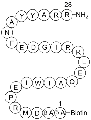
BIM, Biotinylated
Bim peptide fragment with a biotin moiety attached
-
GC49513
Bim/BOD (IN) Polyclonal Antibody
For immunodetection of Bim-
related proteins 
-
GC52355
BimS BH3 (51-76) (human) (trifluoroacetate salt)
A Bim-derived peptide

-
GC14233
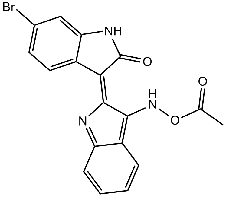
BIO-acetoxime
BIO-아세톡심(BIA)은 GSK-3α/β에 대해 IC50이 모두 10nM인 강력하고 선택적인 GSK-3 억제제입니다.
-
GC67680
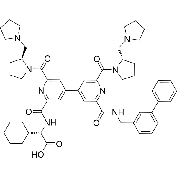
BIO8898
-
GC18476
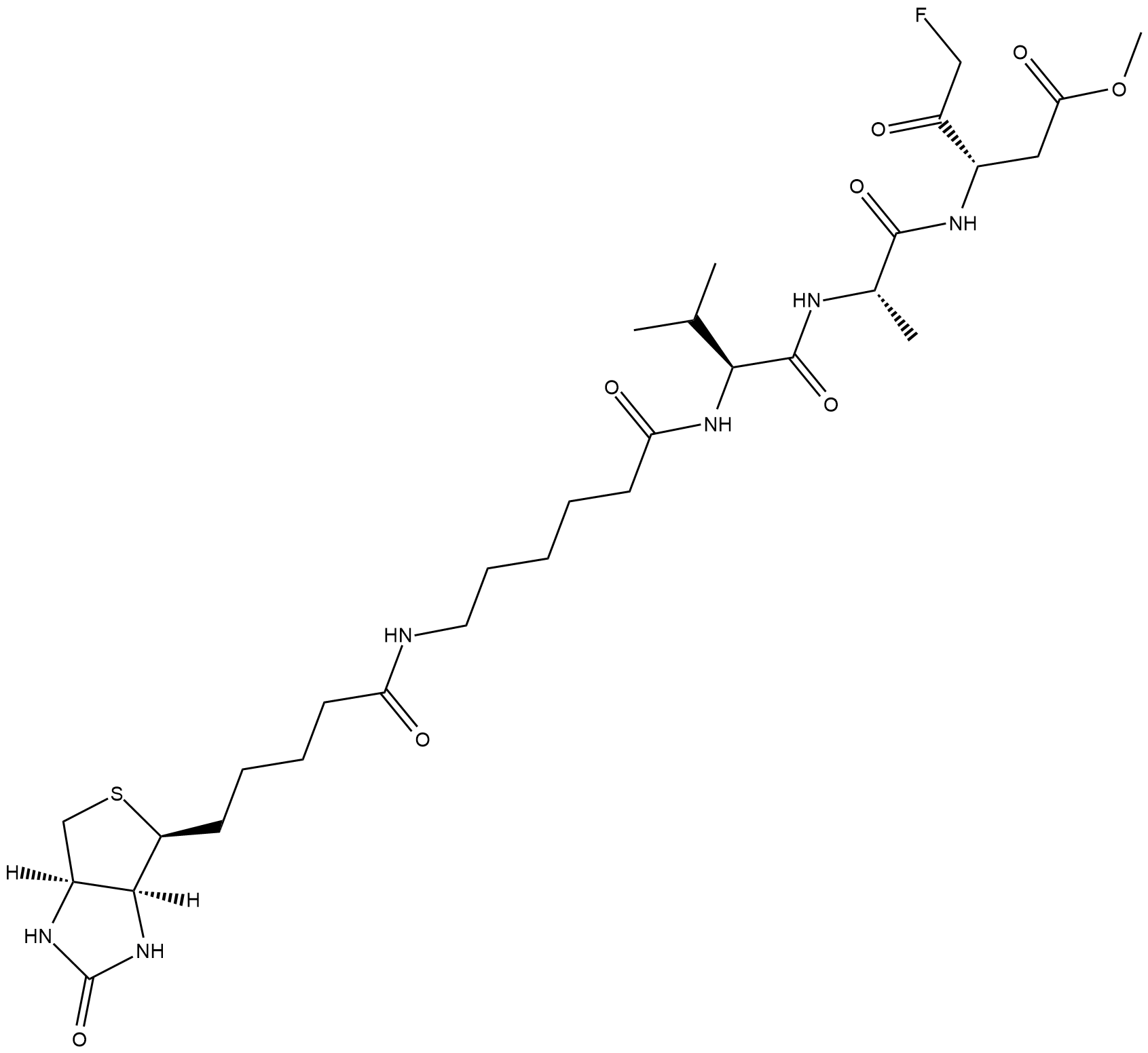
Biotin-VAD-FMK
Biotin-VAD-FMK는 세포 용해물에서 활성 카스파제를 식별하는 데 사용되는 세포 투과성, 비가역적 비오틴 표지 카스파제 억제제입니다.
-
GC35523
Bioymifi
강력한 TRAIL 수용체 DR5 활성화제인 Bioymifi(DR5 Activator)는 1.2μM의 Kd로 DR5의 세포외 도메인(ECD)에 결합합니다. Bioymif는 단일 에이전트로 작용하여 DR5 클러스터링 및 응집을 유도하여 세포자멸사를 유도할 수 있습니다.
