Oxidopamine hydrochloride (6-Hydroxydopamine hydrochloride) |
Catalog No.: GC30871 |
Oxidopamine (6-OHDA) hydrochloride is an antagonist of the neurotransmitter dopamine.
Products are for research use only. Not for human use. We do not sell to patients.
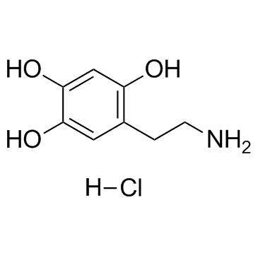
Cas No.:28094-15-7
Sample solution is provided at 25 µL, 10mM.
Quality Control & SDS
- View current batch:
-
Purity: >98.00%
- COA (Certificate Of Analysis)
- SDS (Safety Data Sheet)
- Datasheet
Oxidopamine hydrochloride is a neurotoxic synthetic organic compound, selectively destroys dopaminergic and noradrenergic neurons in the brain.
Cas No. | 28094-15-7 | SDF | |
Canonical SMILES | OC1=CC(CCN)=C(O)C=C1O.[H]Cl | ||
Formula | C8H12ClNO3 | M.Wt | 205.64 |
Solubility | DMSO : ≥ 32 mg/mL (155.61 mM) | Storage | Store at -20°C,unstable in solution, ready to use. |
General tips | For obtaining a higher solubility , please warm the tube at 37 ℃ and shake it in the ultrasonic bath for a while.Stock solution can be stored below -20℃ for several months. | ||
Shipping Condition | Evaluation sample solution : ship with blue ice All other available size: ship with RT , or blue ice upon request |
Step 1: Enter information below (Recommended: An additional animal making an allowance for loss during the experiment)
Step 2: Enter the in vivo formulation (This is only the calculator, not formulation. Please contact us first if there is no in vivo formulation at the solubility Section.)
Calculation results:
Working concentration: mg/ml;
Method for preparing DMSO master liquid: mg drug pre-dissolved in μL DMSO ( Master liquid concentration mg/mL, Please contact us first if the concentration exceeds the DMSO solubility of the batch of drug. )
Method for preparing in vivo formulation: Take μL DMSO master liquid, next addμL PEG300, mix and clarify, next addμL Tween 80, mix and clarify, next add μL saline, mix and clarify.
Method for preparing in vivo formulation: Take μL DMSO master liquid, next add μL Corn oil, mix and clarify.
Note: 1. Please make sure the liquid is clear before adding the next solvent.
2. Be sure to add the solvent(s) in order. You must ensure that the solution obtained, in the previous addition, is a clear solution before proceeding to add the next solvent. Physical methods such as vortex, ultrasound or hot water bath can be used to aid dissolving.
3. All of the above co-solvents are available for purchase on the GlpBio website.
6-Hydroxydopamine upregulates iron regulatory protein 1 by activating certain protein kinase C isoforms in the dopaminergic MES23.5 cell line
Iron-induced oxidative stress is thought to play a crucial role in the pathogenesis of Parkinson's disease. Our previous studies demonstrated that decreased expression of ferroportin 1 contributes to 6-hydroxydopamine induced intracellular iron accumulation and that decreased ferroportin 1 expression is caused by increased expression of iron regulatory protein 1. Iron regulatory protein 1 is a central regulator of iron homeostasis and is a likely target of extracellular agents to program changes in cellular iron metabolism. Therefore, the mechanism of iron regulatory protein 1 upregulation induced by 6-hydroxydopamine has become a significant focus of research. Iron regulatory protein 1 is regulated by protein kinase C, although this regulation is tissue specific. Therefore, in the present study, we aimed to determine whether alteration of protein kinase C activity modified iron regulatory protein 1 expression in the dopaminergic MES23.5 cell line, Furthermore, we investigated whether 6-hydroxydopamine induced iron regulatory protein 1 upregulation is mediated by protein kinase C, thus achieving regulation of cellular iron levels. The results showed that iron regulatory protein 1 was upregulated by phorbol 12-myristate-13-acetate, the PKC activator in dopaminergic MES23.5 cells, and ferroportin 1 expression and iron efflux were decreased as a result of iron regulatory protein 1 upregulation. The protein kinase C inhibitor bisindolylmaleimide I hydrochloride abolished the effect of phorbol 12-myristate-13-acetate. Protein kinase C-汛 and protein kinase C-汎, but not protein kinase C-? were activated by 6-hydroxydopamine. The protein kinase C-汛 inhibitor rottlerin inhibited protein kinase C-汛 phosphorylation and abolished iron regulatory protein 1 upregulation induced by 6-hydroxydopamine. The protein kinase C-汎 pseudo-substrate inhibitor inhibited protein kinase C-汎 phosphorylation and abolished iron regulatory protein 1 upregulation induced by 6-hydroxydopamine. These data indicate that iron regulatory protein 1 is regulated by protein kinase C in dopaminergic MES23.5 cells and that protein kinase C activated by 6-hydroxydopamine regulates iron regulatory protein 1 expression, thus achieving regulation of cellular iron levels.
Investigation of 6-hydroxydopamine-induced plasma extravasation in rat skin
Perfusion of 6-hydroxydopamine into the rat knee and trachea induces plasma extravasation, possibly by tissue-specific mechanisms involving sympathetic and sensory nerves respectively, and we aimed to identify the mediators which contribute to this response in skin. 6-Hydroxydopamine (both hydrobromide and hydrochloride salts), dose dependently increased plasma extravasation into rat dorsal skin, however, when compared to bradykinin or the tachykinin NK1 receptor agonist GR73632, high concentrations of 6-hydroxydopamine (1-10 mumol/site) were required. The response to 6-hydroxydopamine was not inhibited in chemically sympathectomised rats (6-hydroxydopamine, 300 mg/kg i.p. over 7 days) but was significantly reduced by co-administration with the histamine (H1) and the 5-HT receptor antagonists mepyramine and methysergide and in skin sites pre-injected with compound 48/80 (4 micrograms, -18 h) to degranulate dermal mast cells. The response was not inhibited by co-injection of the tachykinin NK1 receptor antagonist SRI40333 or by the cyclo-oxygenase inhibitor indomethacin (5 mg kg-1 i.p., -30 min) except at the lowest dose of 6-hydroxydopamine (1 mumol/site). We conclude that 6-hydroxydopamine is not a potent or selective mediator of increased vascular permeability in rat skin but, at high concentrations, may induce oedema formation via release of vasoactive amines from mast cells, augmented by generation of prostaglandins.
Effect of Roucongrong (Herba Cistanches Deserticolae) decoction on the substantia nigra through Wnt/汕-catenin signaling pathway in rats with Parkinson's disease induced by 6-hydroxydopamine hydrochloride
Objective: To investigate the effect of Roucongrong (Herba Cistanches Deserticolae) decoction on the substantia nigra in rats with Parkinson's disease (PD) induced by 6-hydroxydopamine hydrochloride (6-OHDA). To further determine whether the Wnt/汕-catenin signaling pathway is involved in the action.
Methods: A rat model of PD was established by intracranial injection of 6-OHDA. Subsequently, three concentrations of Roucongrong (Herba Cistanches Deserticolae) decoction were prepared and administered to rats by gavage therapy for 14 d. Behavioral changes were measured in PD rats. In vivo tyrosine hydroxylase (TH) levels in the substantia nigra were examined by immunohistochemistry. Additionally, gene and protein expression levels of members of the Wnt/汕-catenin signaling pathway were examined by Western blotting and polymerase chain reaction. Lastly, a Wnt/汕-catenin inhibitor was used to investigate the mechanism of action in 1-methyl-4-phenylpyridinium (MPP + )- treated MES23.5 cells in vitro.
Results: Roucongrong (Herba Cistanches Deserticolae) decoction improved performance in the stride and gait adjustment tests in PD rats. It also increased TH in the substantia nigra and altered the expression of genes and proteins in the Wnt/汕-catenin signaling pathway. Wnt/汕-catenin inhibitor reduced the effect of Roucongrong (Herba Cistanches Deserticolae) decoction in MPP +-treated MES23.5 cells.
Conclusion: Roucongrong (Herba Cistanches Deserticolae) decoction may promote neuronal survival in PD in vivo and in vitro by increasing TH content in the substantia nigra and by activating the Wnt/汕-catenin signaling pathway.
Nitroxide Radical-Containing Redox Nanoparticles Protect Neuroblastoma SH-SY5Y Cells against 6-Hydroxydopamine Toxicity
Parkinson's disease (PD) patients can benefit from antioxidant supplementation, and new efficient antioxidants are needed. The aim of this study was to evaluate the protective effect of selected nitroxide-containing redox nanoparticles (NRNPs) in a cellular model of PD. Antioxidant properties of NRNPs were studied in cell-free systems by protection of dihydrorhodamine 123 against oxidation by 3-morpholino-sydnonimine and protection of fluorescein against bleaching by 2,2-azobis(2-amidinopropane) hydrochloride and sodium hypochlorite. Model blood-brain barrier penetration was studied using hCMEC/D3 cells. Human neuroblastoma SH-SY5Y cells, exposed to 6-hydroxydopamine (6-OHDA), were used as an in vitro model of PD. Cells were preexposed to NRNPs or free nitroxides (TEMPO or 4-amino-TEMPO) for 2 h and treated with 6-OHDA for 1 h and 24 h. The reactive oxygen species (ROS) level was estimated with dihydroethidine 123 and Fluorimetric Mitochondrial Superoxide Activity Assay Kit. Glutathione level (GSH) was measured with ortho-phtalaldehyde, ATP by luminometry, changes in mitochondrial membrane potential with JC-1, and mitochondrial mass with 10-Nonyl-Acridine Orange. NRNP1, TEMPO, and 4-amino-TEMPO (25-150 米M) protected SH-SY5Y cells from 6-OHDA-induced viability loss; the protection was much higher for NRNP1 than for free nitroxides. NRNP1 were better antioxidants in vitro and permeated better the model BBB than free nitroxides. Exposure to 6-OHDA decreased the GSH level after 1 h and increased it considerably after 24 h (apparently a compensatory overresponse); NRNPs and free nitroxides prevented this increase. NRNP1 and free nitroxides prevented the decrease in ATP level after 1 h and increased it after 24 h. 6-OHDA increased the intracellular ROS level and mitochondrial superoxide level. Studied antioxidants mostly decreased ROS and superoxide levels. 6-OHDA decreased the mitochondrial potential and mitochondrial mass; both effects were prevented by NRNP1 and nitroxides. These results suggest that the mitochondria are the main site of 6-OHDA-induced cellular damage and demonstrate a protective effect of NRNP1 in a cellular model of PD.
Exercise attenuates levodopa-induced dyskinesia in 6-hydroxydopamine-lesioned mice
L-DOPA alleviates the motor symptoms of Parkinson's disease, but its long-term use is associated with undesirable dyskinesia. We now tested whether exercise can attenuate this L-DOPA-induced dyskinesia (LID). We tested the effects of exercise on LID in 6-hydroxydopamine hydrochloride-hemiparkinsonian mice. Animals were treated with L-DOPA/benserazide (25/12.5 mg/kg, i.p.) without and with possibility to exercise (running wheel) during 2 weeks. Exercise drastically prevented the development of LID, and its associated aberrant striatal signaling, namely the hyperphosphorylation of dopamine and cAMP-regulated phosphoprotein 32 kDa protein and c-Fos expression. Our results indicate that exercise can partially prevent the development of LID through the normalization of striatopallidal dopaminergic signaling.
Keywords:
Average Rating: 5
(Based on Reviews and 10 reference(s) in Google Scholar.)GLPBIO products are for RESEARCH USE ONLY. Please make sure your review or question is research based.
Required fields are marked with *