Home >> Signaling Pathways >> Others >> DNA Stain

DNA Stain

DNA stain is a technique using dye for visualization and detection of DNA after separation by gel electrophoresis.

Products for  DNA Stain

  1. Cat.No. Product Name Information
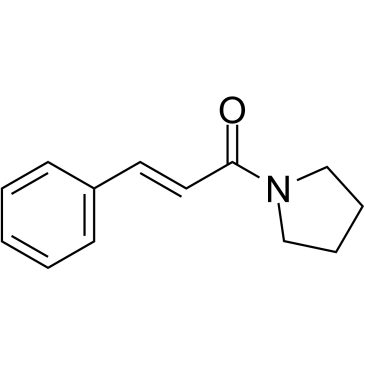
  2. GC35064 1-Cinnamoylpyrrolidine 1-Cinnamoylpyrrolidine (Compound 3), a crude extract prepared from Piper caninum, is a DNA strand scission agent, induces the relaxation of supercoiled pBR322 plasmid DNA. 1-Cinnamoylpyrrolidine  Chemical Structure
  3. GC71740 Biotin-16-dUTP trisodium Biotin-16- dUTP (Biotin-16-deoxyuridine-5'-triphosphate) trisodium can be used to replace its natural counterpart dTTP by enzymatically incorporating it into DNA/cDNA. Biotin-16-dUTP trisodium  Chemical Structure
  4. GC71772 Biotin-16-UTP tetrasodium Biotin-16-UTP tetrasodium is an active substrate for RNA polymerase. Biotin-16-UTP tetrasodium  Chemical Structure
  5. GC68850 CellTracker Blue CMF2HC Dye

    CellTracker Blue CMF2HC Dye is a blue fluorescent dye that can be used for dual-channel nucleic acid sequencing, using blue and purple excitation light (450-460 nm/400-405nm or 415-450 nm/480-525nm). CellTracker Blue CMF2HC Dye can also be used for rapid determination of microbial antibiotic sensitivity.

    CellTracker Blue CMF2HC Dye  Chemical Structure
  6. GC68919 Cyanine 5 Tyramide methyl indole

    Cyanine 5 Tyramide (Tyramide-Cy5) methyl indole is a red fluorescent dye used as a reporting fluorescence substrate for horseradish peroxidase (HRP) catalyzed deposition. It is a signal amplification technology in immunological assays and nucleic acid in situ hybridization. Storage method: avoid light.

    Cyanine 5 Tyramide methyl indole  Chemical Structure
  7. GC67839 DMT-5Me-dC(Bz)-CE Phosphoramidite DMT-5Me-dC(Bz)-CE Phosphoramidite  Chemical Structure
  8. GC71783 DRAQ5 DRAQ5 is a novel cell permeant and far red-fluorescing DNA probe. DRAQ5  Chemical Structure
  9. GC71779 EthD-III EthD-III is a nucleic acid probe. EthD-III  Chemical Structure
  10. GC71005 G-quadruplex DNA fluorescence probe 1 G-quadruplex DNA fluorescence probe 1 (Compound E1) is a selective G-quadruplex DNA targeting fluorescent probe. G-quadruplex DNA fluorescence probe 1  Chemical Structure
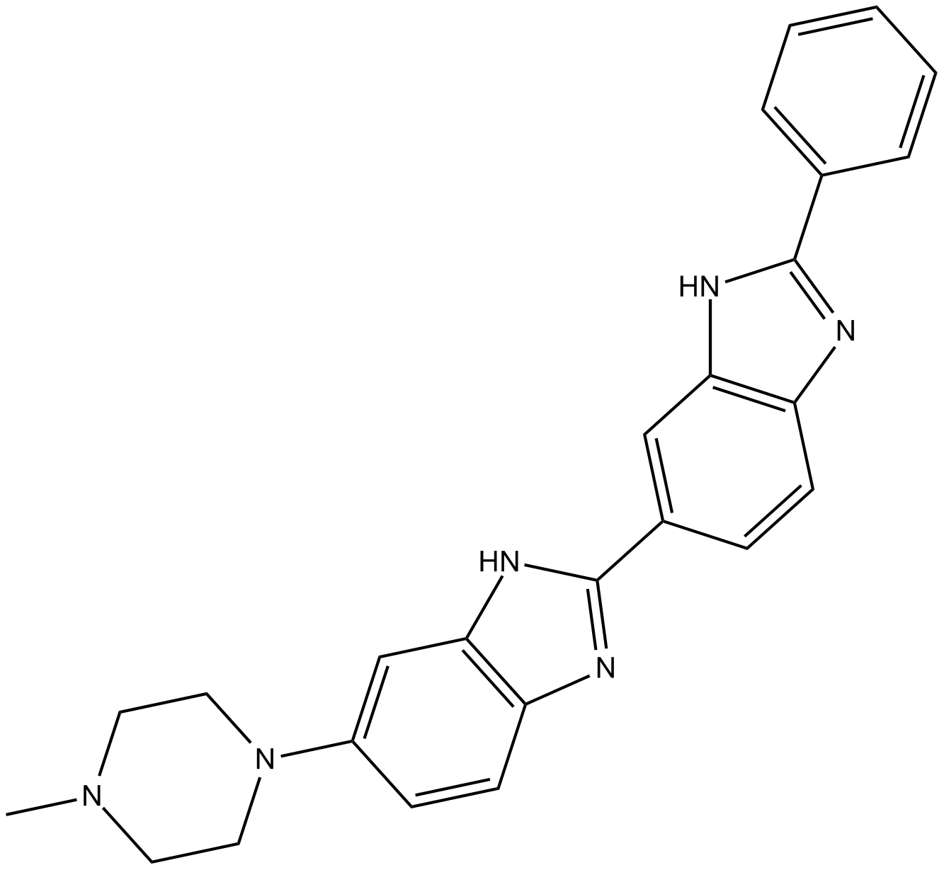
  11. GC11241 HOE 33187

    HOE33187;HOE-33187

    HOE 33187  Chemical Structure
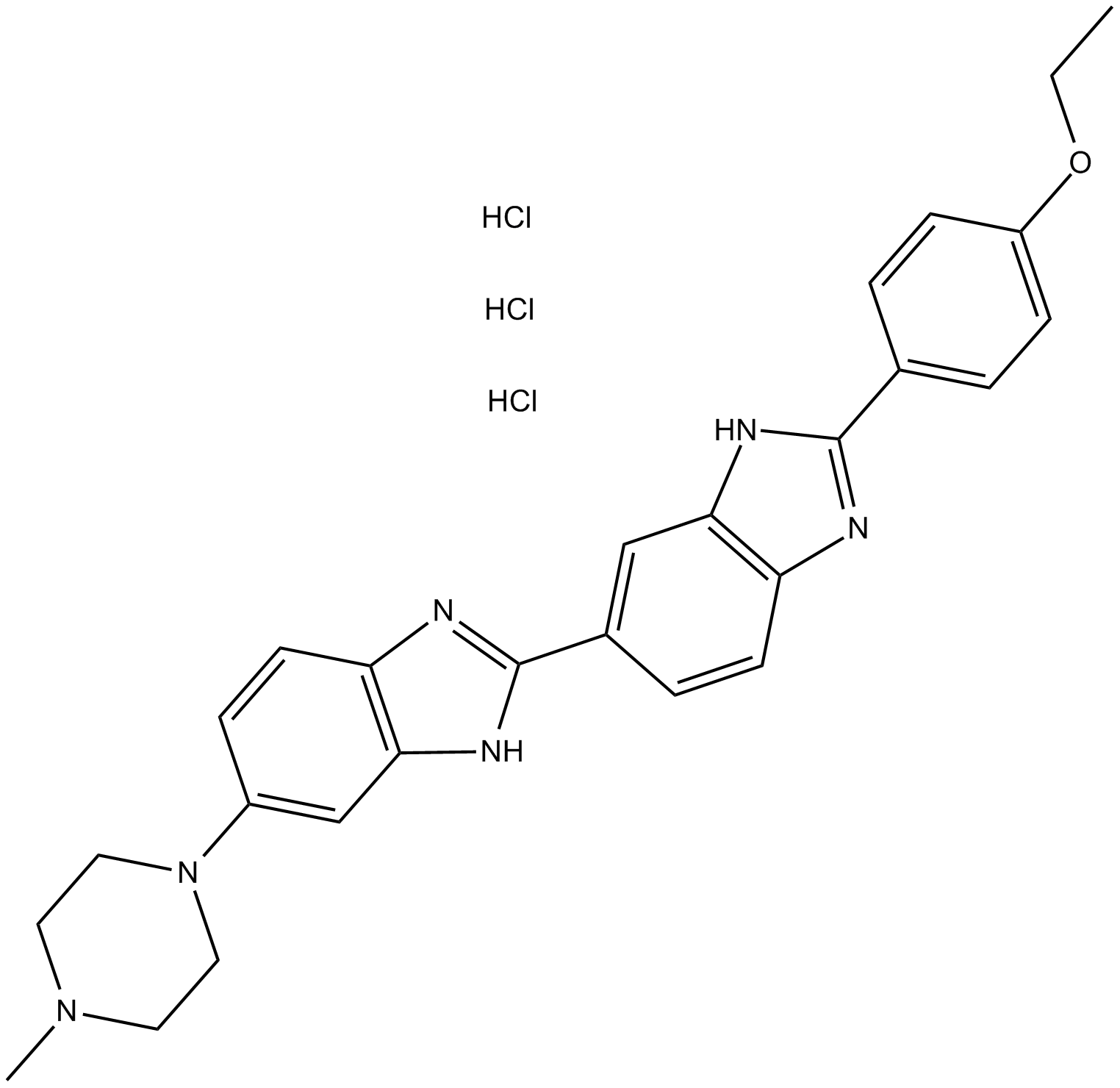
  12. GC12187 Hoechst 33258 trihydrochloride

    Bisbenzimide, HOE 33258, NSC 322921, Pibenzimol

    Hoechst 33258 trihydrochloride is an analog of the nucleic acid stain Hoechst 33258. It emits blue fluorescence after binding to dsDNA, with a maximum excitation/emission light of 350/461nm.

    Hoechst 33258 trihydrochloride  Chemical Structure
  13. GC10939 Hoechst 33342 trihydrochloride

    Bisbenzimide, HOE 33342, NSC 334072

    Hoechst 33342 trihydrochloride is a DNA fluorescent dye with excitation/emission maxima at 350/460 nm, respectively.

    Hoechst 33342 trihydrochloride  Chemical Structure
  14. GC17657 Hoechst 34580

    HOE 34580;Proamine;Hoechst-34580;Hoechst34580

    The nucleic acid stain Hoechst 34580 (Ex/Em: 392/440 nm) is frequently utilized as a cell-permeable nuclear counterstain that emits a blue fluorescence upon binding to dsDNA.
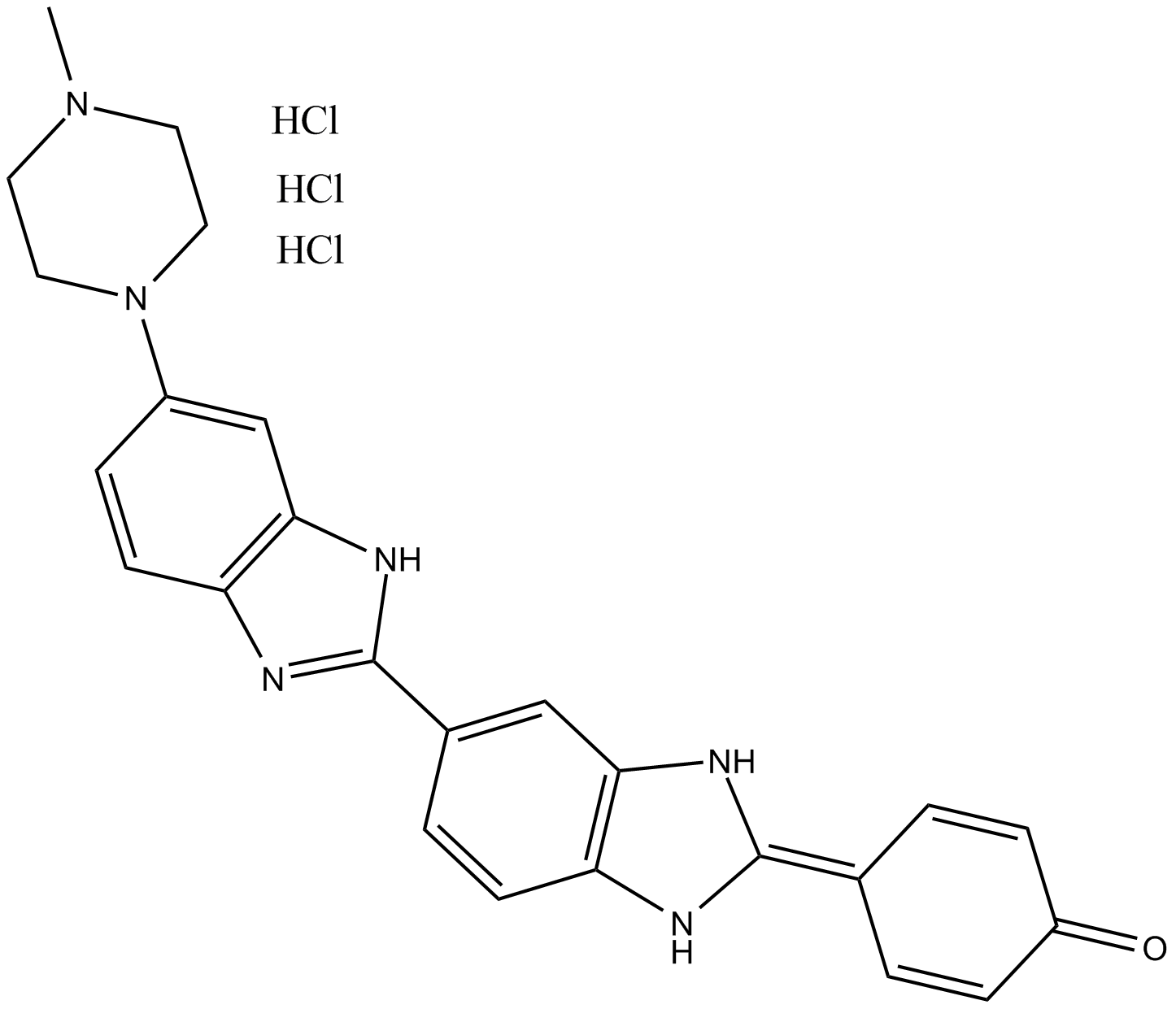
    Hoechst 34580  Chemical Structure
  15. GC69446 MeCY5-NHS ester triethylamine

    Sulfo-Cyanine5 NHS ester triethylamine

    MeCY5-NHS ester (potassium) is an active dye that can be used to label proteins and nucleic acids.

    MeCY5-NHS ester triethylamine  Chemical Structure
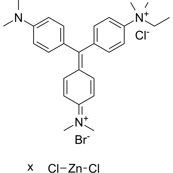
  16. GC67838 Methyl Green zinc chloride Methyl Green zinc chloride  Chemical Structure
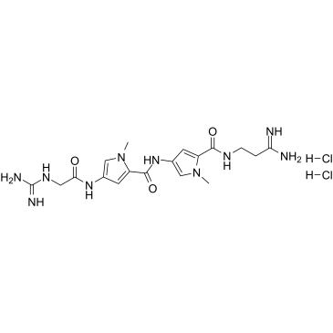
  17. GC36724 Netropsin dihydrochloride

    Congocidine, Sinanomycin

    A DNA minor groove binder Netropsin dihydrochloride  Chemical Structure

16 Item(s)

per page

Set Descending Direction