


ZnAF-2 DA is a membrane-permeable derivative of the zinc ion fluorescent probe ZnAF-2. Once it enters the cell, it is hydrolyzed by esterase in the cytoplasm to generate ZnAF-2F, which is retained in the cell. ZnAF-2 DA is suitable for bioimaging analysis of living cells or tissues (such as hippocampal slices). ZnAF-2 has a fluorescein structure linked to a TPEN analog and highly specifically binds Zn2+ in buffer. High affinity allows detection of low concentrations of Zn2+ (dissociation constant: 2.7nM). Low fluorescence background enables highly sensitive determination of Zn2+ in living samples. ZnAF-2 emits green fluorescence (Ex/Em=492/515nm) after combining with Zn2+ [1-3].
References:
[1] Highly Zinc-Selective Fluorescent Sensor Molecules Suitable for Biological Applications: T. Hirano, et al; JACS 122,12399 (2000)
[2] Mossy fiber Zn2+ spilover modulates heterosynaptic N-methy-D-aspartate receptor activity in hippocampal CA3 circuits: S.Ueno, et al; J. Cell Biol, 158, 215 (2002)
[3] lmprovement and biological applications of fluorescent probes for zinc, ZnAFs: T. Hirano, et al,; JACS 124, 6555 (2002)
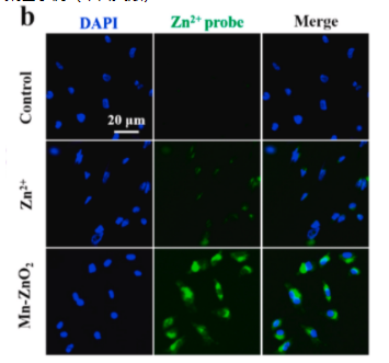 Fig 1. ntracelular elevation of dual Zn-Mn ions and ROS by Mn-Zn02 NPs. (a) Schematic ilustration of intracellular release of dualZn-Mn ions and H202 and subsequent 0H generation from Mn-Zn02 NPs. (b) Fluorescence images of MDA-MB-231 cells stainedwith Zn2+ dye- ZnAF-2 DA (green, 5 uM ) after incubation with ZnCl2 or Mn-Zn02 NPs for 4 h.
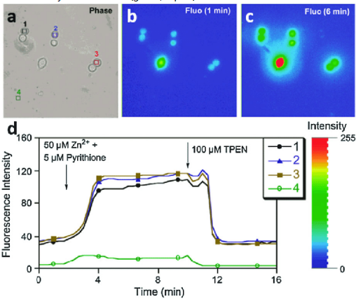 a)Fig Fig 2 (a) Bright-field and (b, c) fluorescence images of RAW 2647 cells incubated with 10 mlM ZnAF-2F DA. (d) Changes in the average fluorescence intensity of the coresponding areas (1-3, intrace ular redion: 4. extrace ular redion) as a function of time, The fluorescence excited at 470-490 nm was measured at the intervals of 20 s. At 2 min, Zn 2+-pyrithione were added to the medium, and at 10 min 100 mM TPEN was added.