
EdU-488 Cell Proliferation Detection Kit
Catalog No.: GK10046
The EdU-488 Cell Proliferation Detection Kit can directly detect DNA synthesis in cells.
Les produits sont exclusivement destinés à la recherche. Ils ne doivent pas être utilisés chez l’être humain. Nous ne vendons pas aux patients.
50-500TIn Stock
$55.00
1
200-2000TIn Stock
$175.00
1
| Pack Size | Price | Availability | Quantity | Action |
|---|---|---|---|---|
| 50-500T | $55.00 | In Stock | 1 | |
| 200-2000T | $175.00 | In Stock | 1 |
Tel: (909) 407-4943 / (909) 689-7124Email: sales@glpbio.com
Citations
1 publicationsPublications de chercheurs utilisant ce produitDescription
The EdU-488 Cell Proliferation Detection Kit can directly detect DNA synthesis in cells. The thymidine analogue EdU (5-ethynyl-2 '- deoxyuridine) in this kit can replace thymidine and incorporate into DNA during active DNA synthesis. Subsequently, EdU conjugates with fluorescently labeled small molecule azides (such as AF 488 azide, AF 647 azide, etc.) via the Click reaction, thereby labeling the newly synthesized DNA.
The EdU-488 Cell Proliferation Detection Kit can be used to detect the proliferation of individual cells in adherent cells, suspended cells, or tissue samples, as well as to quantitatively detect the total cell proliferation of cell or tissue samples.
The samples processed by this kit can be detected by fluorescence microscopy, flow cytometer, or fluorescent microplate reader, providing broad applications in cell proliferation studies.
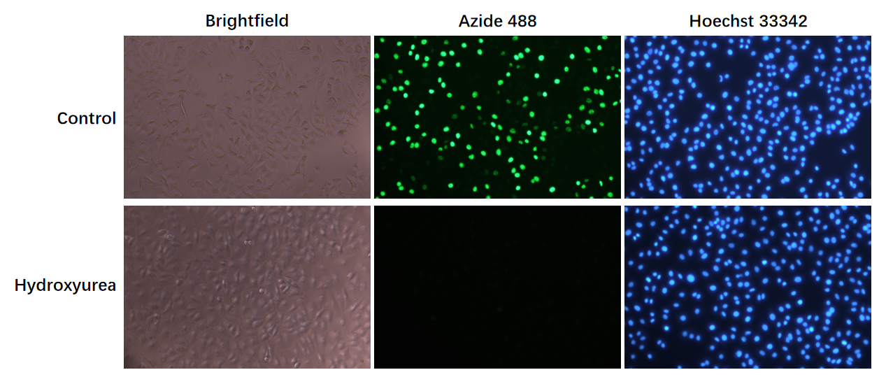
Figure 1: A549 cell proliferation detection (fluorescence microscopy). The group incubated with EdU (10μM) for 2 hours showed bright green fluorescence (first row). After pretreatment with 10mM Hydroxyurea (a DNA synthesis inhibitor) for 0.5 hours, followed by incubation with EdU (10μM) for 2 hours, the green fluorescence significantly decreased, indicating that DNA synthesis was inhibited (second row).
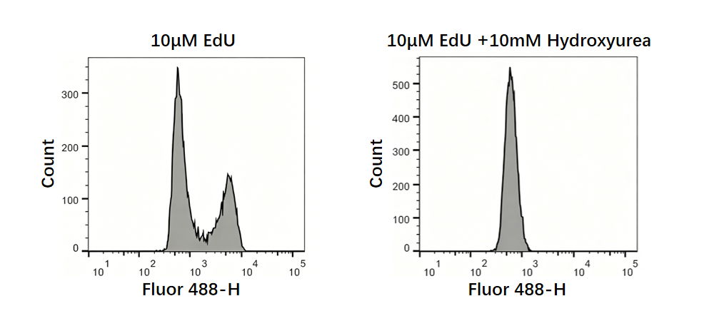
Figure 2: A549 cell proliferation assay (flow cytometry). After incubation with EdU (10μM) for 2 hours, the proliferating cells (Fluor 488-H positive cells) incorporating EdU showed clear separation from the weakly stained non-proliferating cell group. After treatment with the inhibitor Hydroxyurea (10mM), the Fluor 488-H positive cell population almost completely disappeared, leaving only the negative cell group (right figure). The data in the figure are for reference only; actual data may vary due to factors such as cell types, cell proliferation status, and assay instrument.
Protocole (Protocol)
I Reagents to be prepared separately:
1. PBS (pH 7.2-7.6).
2. Fixative: 4% paraformaldehyde (freshly prepared, PBS solution).
3. Transparent solution: PBS containing 0.3% Triton X-100.
4. Washing solution: PBS containing 3% BSA.
II Reagent Preparation
Before the initial use, completely transfer 1.3ml of Click Additive Buffer into the tube containing Click Additive, mix well until completely dissolved to prepare the Click Additive Solution. This reagent is prone to oxidation. it is recommended to aliquot appropriately and store at -20°C.
Note: It is recommended to prepare the next tube only after using up the previous one.
III Selection of the Detection System
The following experimental methods are based on the 6-well plate or conventional slice detection system. For 12-well, 96-well or 384-well plates, proportionally reduce the reaction volume, and ensure complete coverage of samples.
IV. Experimental Procedure
1. EdU labeling
1.1. Cell EdU labeling
1.1.1. Prepare 2× EdU working solution: It is recommended to prepare a 2× working solution, and then add an equal volume of EdU working solution and culture medium to the well plate. The recommended final concentration of EdU is 10μM (1×). Dilute EdU (10mM) with cell culture medium 1:500 to obtain 2× EdU working solution (20μM).
Note:
a. To minimize the potential effects on cell proliferation, complete replacement of the culture medium is not recommended.
b. The working concentration of EdU should be optimized according to the cell type used. It is recommended to conduct preliminary experiments and set EdU working solutions determine the optimal concentration.
1.1.2. Retain an appropriate volume of culture medium in the well plate, add an equal volume of 2× EdU working solution preheated at 37℃ to the well plate, and continue incubating for 2 hours.
Attention: Incubation duration is determined by cell growth rate. Generally set it to approximately 10% of the total cell cycle duration. Common mammalian cells such as A549, HCT116, HEK293, etc. have a cell cycle of about 18-25 hours, and it is recommended to control the incubation time at around 2 hours. The cell cycle of human embryonic cells is about 30 minutes, and the recommended incubation time is 5 minutes. The cell cycle of yeast cells is about 3 hours, and the recommended incubation time is 20 minutes. The cell cycle of proliferating nerve cells is about 5 days, and the recommended incubation time is 1 day. When the incubation time is less than 45 minutes, it is recommended to increase the concentration of EdU. When the incubation time is greater than 20 hours, it is recommended to reduce the concentration of EdU appropriately.
1.1.3. After EdU labeling of cells is completed, discard the culture medium, add 1ml fixative, and fix at room temperature for 15 minutes.
Attention: If flow cytometer is required for detection, the adherent cells should be digested with trypsin before fixation.
1.1.4. Remove the fixative and wash the samples three times with 1ml washing solution,3-5 minutes each time.
1.1.5. Remove the washing solution, add 1ml transparent solution to each well, and incubate at room temperature for 10-15 minutes.
1.1.6. Remove the transparent solution and wash the samples three times with 1ml washing solution,3-5 minutes each time.
1.1.7. Go to step 2 EdU detection.
1.2. In vivo EdU labeling
EdU can be used for in vivo labeling of animals through appropriate methods such as injection or oral administration. The experimental protocol below takes mice as an example, please refer to relevant literature for EdU labeling in other animals.
1.2.1. Reference dosage: 10-200mg EdU/kg body weight. Prepare EdU at an appropriate concentration in PBS/Normal Saline, filter and sterilize using a 0.22μm filter. It is recommended to use the freshly prepared EdU on the day of the experiment. It can be injected into mice via intraperitoneal injection, local injection into specific tissues or organs, or addition to drinking water.
Note:
a. If the EdU usage is large and the product specifications provided by this kit do not meet the experimental requirements, it is recommended to purchase an additional item (Catalog No.: GC42504).
b. The specific dosage depends on the type, weight, and EdU injection method of the experimental animals. It is recommended to refer to relevant literature.
c. For the first experiment, it is recommended to explore the dosage of EdU or test it at 50mg EdU/kg body weight.
d. If BrdU has been used for experiments before, you can refer to the dosage of BrdU for experimentation or exploration.
1.2.2. After 4 hours of EdU treatment, mice were quickly euthanized, and the required tissues were removed. Frozen or paraffin sections were prepared according to conventional procedures.
Note: The duration can be adjusted according to specific experiments or relevant literature.
1.2.3. Frozen section processing
1.2.3.1. Add an appropriate amount of fixative to completely immerse the samples and fix at room temperature for 15 minutes.
1.2.3.2. Remove the fixative and wash the samples three times with appropriate amount of washing solution,3-5 minutes each time.
1.2.3.3. Remove the washing solution, add an appropriate amount of transparent solution, and incubate at room temperature for 10-15 minutes.
1.2.3.4. Remove the transparent solution and wash the samples three times with appropriate amount of washing solution,3-5 minutes each time.
1.2.3.5. Antigen retrieval (optional): If immunofluorescence staining of proteins is required and antigen retrieval is necessary, it is recommended to use an appropriate antigen retrieval solution.
Note: If proteinase K or trypsin is used for antigen retrieval, repeated washing is essential, as residual enzymes can severely interfere with subsequent labeling reactions.
1.2.3.6. Go to step 2 EdU detection.
1.2.4. Paraffin section processing
1.2.4.1 Dewaxing: Soak the paraffin sections in xylene for 10 minutes. Soak in fresh xylene for another 10 minutes. Soak in 50% xylene for 5 minutes. Soak in anhydrous ethanol for 5 minutes. Soak in new anhydrous ethanol for another 5 minutes. Soak in 95% ethanol for 5 minutes. Soak in 85% ethanol for 5 minutes. Soak in 75% ethanol for 5 minutes. Soak in 50% ethanol for 5 minutes. Soak in 30% ethanol for 5 minutes. Soak in PBS for 5 minutes.
1.2.4.2 Antigen retrieval (optional): If immunofluorescence staining of proteins is required and antigen retrieval is necessary, it is recommended to use an appropriate antigen retrieval solution.
Note: If proteinase K or trypsin is used for antigen retrieval, repeated washing is essential, as residual enzymes can severely interfere with subsequent labeling reactions.
1.2.4.3 Go to Step 2 EdU detection.
2. EdU detection
2. 1. Refer to the table below to prepare Click reaction cocktail:
|
Component |
Number of 6-well plate samples |
||||||
|
1 |
2 |
4 |
5 |
10 |
25 |
50 |
|
|
Click Reaction Buffer |
430μl |
860μl |
1.72ml |
2.15ml |
4.3ml |
10.75ml |
21.5ml |
|
CuSO4 |
20μl |
40μl |
80μl |
100μl |
200μl |
500μl |
1ml |
|
Azide 488 |
1μl |
2μl |
4μl |
5μl |
10μl |
25μl |
50μl |
|
Click Additive Solution |
50μl |
100μl |
200μl |
250μl |
500μl |
1.25ml |
2.5ml |
|
Total Volume |
500μl |
1ml |
2ml |
2.5ml |
5ml |
12.5ml |
25ml |
Note:
a. Click reaction cocktail should be prepared strictly according to the order and volume of the components in the table, otherwise, the reaction will not proceed optimally.
b. Click reaction cocktail should be used within 15 minutes of preparation.
c. For 6-well plates, it is recommended to add 500μl of Click reaction cocktail per well. For 12, 24, 48, 96, and 384-well plates, it is recommended to add 200μl, 100μl, 70μl, 50μl, and 20μl of Click reaction cocktail per well, respectively. The dosage can be adjusted according to the actual situation.
d. For sliced samples, use 100-200μl of Click reaction cocktail per slice, depending on its size, ensuring complete coverage of the sample.
2.2. Remove the washing solution from the previous step, add 0.5 ml of Click reaction cocktail to each well, and gently shake to ensure the sample is evenly covered.
2. 3. Incubate at room temperature in the dark for 30 minutes, or extend the incubation time appropriately.
2. 4. Remove Click reaction cocktail and wash 3 times with washing solution, each time for 3-5 minutes.
2. 5. Resuspension the cells in washing solution and use fluorescence microscopy, flow cytometer, or fluorescence microplate reader for detection. The maximum excitation/emission wavelengths of Azide 488 are 495/519nm.
Note:
a. If staining of the cell nucleus is required, proceed to step 3. Nuclear staining.
b. If traditional hydrodynamic focusing Flow cytometer is used to detect total DNA content, it is recommended to use low flow rate during the detection process, and the same flow rate and cell concentration should be used for each sample in the experiment.
3. Nuclear staining
3.1. Configure 1× Hoechst 33342 solution: Dilute Hoechst 33342 (1000×) with PBS at a ratio of 1:1000.
3.2. Remove the washing solution, add 1ml 1× Hoechst 33342 per well, and incubate at room temperature in the dark for 10 minutes.
3.3. Remove Hoechst 33342 solution and wash the samples three times with an appropriate amount of washing solution,3-5 minutes each time.
3.4. Fluorescence detection. The maximum excitation/emission wavelengths of Hoechst 33342 are 346/460nm.
Notes:
1. For flow cytometer, it is recommended to use cell samples without EdU labeling as a negative control and select an appropriate voltage.
2. Due to the sensitivity in flow cytometer detection, it is recommended to adjust the concentration of Azide 488 appropriately based on cell type and actual staining conditions.
3. After preparing the Click Additive solution, it is recommended to pack it appropriately. If white substances precipitate after reheating, they can be completely dissolved by vortexing or flipping up and down, and then used normally. If the color of the solution turns brown, it has expired and should be discarded.
4. If your samples require staining with other fluorescent antibodies or organelle dyes, this kit is fully compatible with various organic dyes, such as Alexa Fluor series dyes, FITC (overlaps with Azide 488 fluorescence, it is not recommended to use with this kit), APC, APCE-tandems dyes. If Qdot probes, HRP, R-PE, and R-PE-tandems are needed, the reaction and detection should be performed after the click reaction.
5. The fluorescence of fluorescent proteins such as GFP, RFP, and mCherry can be affected by copper ions in this kit, should be reacted or detected before the click reaction.
6. Phalloidin is not compatible with click reactions. It is recommended to use Tubulin for microtubule detection.
7. All fluorescent dyes all have quenching problems. Please try to avoid light exposure during the experiment to mitigate fluorescence quenching.
8. For your safety and health, please wear lab coat and disposable gloves when handling these materials.
Documents produit (Product Documents)
Automatic documents are temporarily unavailable. Please refresh this page to try again.
Composants du kit (Kit Components)
| Components | 50-500T | 200-2000T |
| EDU (10mM) | 200μL | 800μL |
| Azide 488 | 55μL | 220μL |
| CuSO4 | 1.1mL | 4.4mL |
| Click Additive | 1 tube | 1 tube x4 |
| Click Additive Buffer | 1.4mL | 6mL |
| Click Reaction Buffer | 30mL | 120mL |
| Hoechst 33342(1000X) | 50μL | 200μL |
Attributs du produit
Applications et caractéristiques
Good safety: No use of 3HTdR, no radioactive contamination.
Simple operation, quick response, and short experimental time.
High sensitivity and good labeling rate, only a small amount of small molecule azide probes are needed to effectively label the doped EdU, and single proliferating cells can also be accurately detected.
Good compatibility, Antibody-based immunofluorescence and immunohistochemistry testing can be performed after EdU detection is completed.
Widely applicable: suitable for detecting adherent cells, suspended cells, and cultured tissue samples.
There are various detection methods available: compatible with fluorescence microscopy, laser confocal microscopy, flow cytometer, and fluorescence enzyme-linked immunosorbent assay, and can also be used for high content screening.
Expédition
Ship with blue ice.
Conditions de conservation
Store kit at -20℃ for up to 1 year.
Utilisation prévue
Réservé à la recherche. Ne pas utiliser chez l’être humain.
| Applications et caractéristiques | Good safety: No use of 3HTdR, no radioactive contamination. Simple operation, quick response, and short experimental time. High sensitivity and good labeling rate, only a small amount of small molecule azide probes are needed to effectively label the doped EdU, and single proliferating cells can also be accurately detected. Good compatibility, Antibody-based immunofluorescence and immunohistochemistry testing can be performed after EdU detection is completed. Widely applicable: suitable for detecting adherent cells, suspended cells, and cultured tissue samples. There are various detection methods available: compatible with fluorescence microscopy, laser confocal microscopy, flow cytometer, and fluorescence enzyme-linked immunosorbent assay, and can also be used for high content screening. |
| Expédition | Ship with blue ice. |
| Conditions de conservation | Store kit at -20℃ for up to 1 year. |
| Utilisation prévue | Réservé à la recherche. Ne pas utiliser chez l’être humain. |
Reviews
Review for EdU-488 Cell Proliferation Detection Kit
Rating: 5
- 5 Star100%
- 4 Star0%
- 3 Star0%
- 2 Star0%
- 1 Star0%