
IF 16
PMID: 39846120 ↗Hoechst 33342
Catalog No.: GC63515Purity:>98.00%
يتم استخدام صبغة حمض النووي هوكست 33342 (Ex/Em: 350/461 نانومتر) كصبغة مضادة للنفاذية تستخدم بشكل شائع كصبغة عد الأنوية التي يسهل اختراقها والتي تطلق ضوءًا أزرق عند الارتباط بـ dsDNA.
المنتجات مخصصة للاستخدام البحثي فقط، وليست للاستخدام البشري. نحن لا نبيع للمرضى.
Cas No.: 23491-52-3
25mgIn Stock
$45.00
1
50 mgIn Stock
$72.00
1
100 mgIn Stock
$108.00
1
| Pack Size | Price | Availability | Quantity | Action |
|---|---|---|---|---|
| 25mg | $45.00 | In Stock | 1 | |
| 50 mg | $72.00 | In Stock | 1 | |
| 100 mg | $108.00 | In Stock | 1 |
Tel: (909) 407-4943 / (909) 689-7124Email: sales@glpbio.com
المراجع (Citations)
6 publicationsمنشورات الباحثين الذين استخدموا هذا المنتجالوصف (Description)
The nucleic acid stain Hoechst 33342 (Ex/Em: 350/461 nm) is frequently utilized as a cell-permeable nuclear counterstain that emits a blue fluorescence upon binding to dsDNA. Hoechst 33342 is commonly employed in various studies related to cell counting, cell cycle analysis, and cell replication. It is particularly useful in identifying condensed nuclei in apoptotic cells, as well as in combination with BrdU staining for cell-cycle studies.
Hoechst dyes are also useful for monitoring cell viability by tracking changes in their emission spectra. As minor groove-binding DNA stains with AT selectivity, the Hoechst dyes are able to bind to all nucleic acids, but they show a greater fluorescence enhancement for AT-rich double-stranded DNA strands compared to GC-rich strands [1]. This property has been exploited to identify Q-bands in chromosomes, which are regions rich in AT base pairs that fluoresce brightly when stained with the quinacrine dye [2].
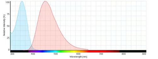
Fig. Fluorescence excitation and emission spectra of Hoechst 33342 bound to DNA
References:
[1]. Portugal J, Waring MJ. Assignment of DNA binding sites for 4′, 6-diamidine-2-phenylindole and bisbenzimide (Hoechst 33258). A comparative footprinting study. Biochimica et Biophysica Acta (BBA)-Gene Structure and Expression. 1988 Feb 28;949(2):158-68.
[2]. Weisblum B, Haenssler E. Fluorometric properties of the bibenzimidazole derivative Hoechst 33258, a fluorescent probe specific for AT concentration in chromosomal DNA. Chromosoma. 1974 Sep;46(3):255-60.
البروتوكول (Protocol)
This plan only provides a guide, please modify it to meet your specific needs.
1. Prepare Hoechst 33342 staining solution
(1) Prepare Hoechst 33342 dye stock solution: Use DMSO to dissolve solid Hoechst 33342 and prepare a 10 mg/mL Hoechst 33342 dye stock solution.
Note: Hoechst stock solution is recommended to be aliquoted and stored in the dark at -4°C or -20°C to avoid repeated freezing and thawing.
(2) Working solution preparation: Use preheated serum-free medium or buffer (such as HBSS or PBS) to dilute the stock solution and prepare a Hoechst 33342 working solution with a concentration of 10 μg/mL.
Note: Please adjust the concentration of Hoechst working fluid according to the actual situation and prepare it immediately.
2. Cell staining
2.1 Suspension cells (taking 6-well plate as an example)
(1) Centrifuge suspended cells at 1000g for 3-5 minutes. Discard the supernatant and wash twice with PBS for 5 minutes each time.
(2) Add 1 mL of Hoechst 33342 dye working solution and incubate at room temperature in the dark for 5-10 minutes.
(3) After the incubation, centrifuge at 1000g for 5 minutes, remove the supernatant, add PBS and wash 2-3 times, 5 minutes each time.
(4) Use serum-free cell culture medium or PBS to resuspend the cells and observe them through fluorescence microscopy or flow cytometry.
2.2 Adherent cells
(1) Culture adherent cells on sterile coverslips.
(2) Remove the coverslip from the culture medium, suck out the excess culture medium, and place the coverslip in a humid environment.
(3) Add 100uL of Hoechst 33342 dye working solution from one corner of the coverslip, shake gently to evenly cover all cells with the dye, and incubate at room temperature in the dark for 5-15 minutes.
(4) Aspirate away the dye working solution, wash the coverslip 2 to 3 times with culture solution, and observe with a fluorescence microscope.
3. Microscope detection: The excitation/emission light of Hoechst 33342 is 350/461nm respectively.
Precautions:
① For staining of fixed cells or tissue samples, the fixative needs to be removed by rinsing after fixation;
② Hoechst 33342 staining is usually performed after other staining. If no other staining is required, Hoechst 33342 staining is performed directly;
③ To slow down fluorescence quenching, it is recommended to use anti-fluorescence quenching mounting agent;
④ Fluorescent dyes all have quenching problems, so please try to avoid light;
⑤ Hoechst 33342 is irritating to the human body. For your safety and health, please wear a lab coat and disposable gloves when operating.
References:
[1]. Lisa C Crowley, Brooke J Marfell , Nigel J Waterhouse. Analyzing Cell Death by Nuclear Staining with Hoechst 33342. 2016 Sep 1;2016(9). doi: 10.1101/pdb.prot087205.
وثائق المنتج (Product Documents)
Purity:>98.00%Appearance:A solid
البيانات البيولوجية (Biological Data)
1 / 2
الخصائص الكيميائية (Chemical Properties)
رقم CAS
23491-52-3
الصيغة الجزيئية
C27H28N6O
الوزن الجزيئي
452.55
الذوبانية
DMSO : 10 mg/mL (22.10 mM; Need ultrasonic)|Water : 1 mg/mL (2.21 mM; ultrasonic and warming and heat to 80°C)
التخزين
4°C, protect from light
إرشادات عامة
اختر مذيبًا مناسبًا وفقًا لبيانات ذوبانية المنتج. بعد التحضير، قسّم المحلول المخزون إلى أجزاء لتجنب دورات التجميد والذوبان المتكررة.
استخدم المحاليل المخزونة خلال 6 أشهر عند حفظها في -80°C، أو خلال شهر واحد عند حفظها في -20°C.
لتحسين الذوبانية، سخّن الأنبوب إلى 37°C وعرّضه للموجات فوق الصوتية لمدة مناسبة.
استخدم المحاليل المخزونة خلال 6 أشهر عند حفظها في -80°C، أو خلال شهر واحد عند حفظها في -20°C.
لتحسين الذوبانية، سخّن الأنبوب إلى 37°C وعرّضه للموجات فوق الصوتية لمدة مناسبة.
شروط الشحن
تُشحن عينات التقييم مع عبوات تبريد. وتُشحن الأحجام الأخرى المتاحة في درجة حرارة الغرفة، أو مع عبوات تبريد عند الطلب.
الحاسبات (Calculators)المولارية / التخفيف / الوزن الجزيئي / تحويل الوحدات / تركيبة داخل الجسم الحي / الذوبانية
g/mol
Reviews
Review for Hoechst 33342
Rating: 5
- 5 Star100%
- 4 Star0%
- 3 Star0%
- 2 Star0%
- 1 Star0%