GLPBio

Mito-Tracker Deep Red 633

Catalog No.: GC20140

Mito-Tracker Deep Red 633 is a novel deep red fluorescent probe that detects mitochondrial membrane potential and specifically stains mitochondria in living cells.

제품은 연구용으로만 사용됩니다. 인체에 사용할 수 없으며 환자에게 판매하지 않습니다.


Mito-Tracker Deep Red 633
50μgIn Stock
$60.00
1
250μgIn Stock
$200.00
1

인용 문헌 (Citations)

현재 이 제품에 대한 인용 문헌이 없습니다. 아래는 GlpBio 제품을 인용한 주요 논문입니다.
  • Nature cover
    Nature
    641, 529–536 (2025)
  • Nature cover
    Nature
    628, 630–638 (2024)
  • Nature cover
    Nature
    632, 686–694 (2024)
  • Nature cover
    Nature
    618, 1017–1023 (2023)
  • Nature cover
    Nature
    610, 366–372 (2022)
  • Cell cover
    Cell
    187(9):2288-2304 (2024)
  • Cell cover
    Cell
    183(7):1867-1883 (2020)
  • Science cover
    Science
    388(6745) (2025)
  • Science cover
    Science
    387(6739) (2025)
  • Science cover
    Science
    387(6734) (2025)
  • Cell Research cover
    Cell Research
    35, 97–116 (2025)
  • Cell Research cover
    Cell Research
    34, 683–706 (2024)
  • Cell Research cover
    Cell Research
    33, 273–287 (2023)
  • Cell Research cover
    Cell Research
    33, 546–561 (2023)
  • Cell Research cover
    Cell Research
    33, 904–922 (2023)
  • Cell Research cover
    Cell Research
    31, 1291–1307 (2021)

제품 설명 (Description)

Mito-Tracker Deep Red 633 is a novel deep red fluorescent probe that detects mitochondrial membrane potential and specifically stains mitochondria in living cells. By simply incubating with cells, it can be transported passively across the cell membrane and accumulate in biologically active mitochondria, resulting in bright deep red fluorescence.

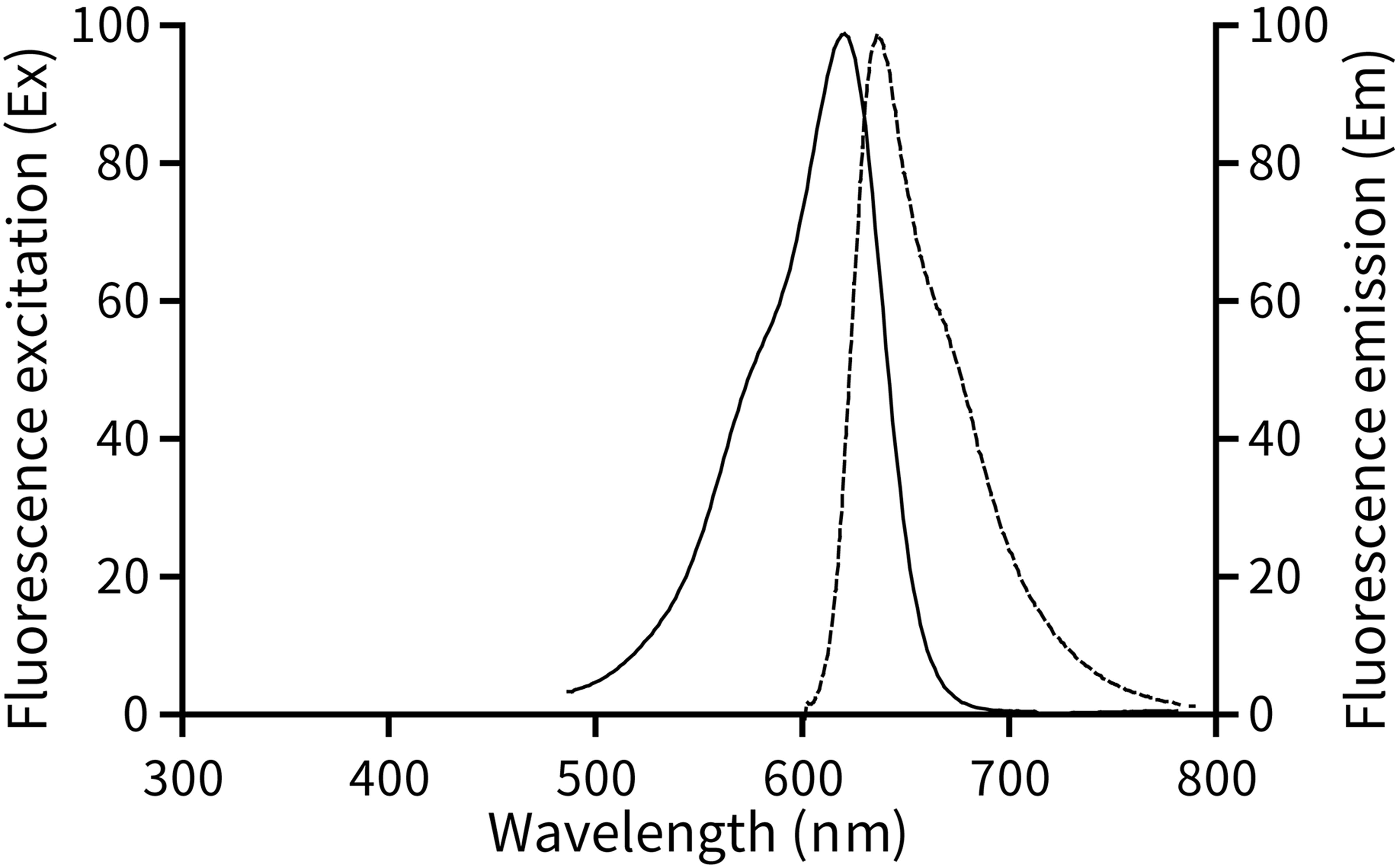
Mito-Tracker Deep Red 633, with a molecular weight of 586.23, has a maximum excitation wavelength of 622nm and a maximum emission wavelength of 648nm. Please refer to Figure 1 for the excitation and emission spectra and structural formula of Mito-Tracker Deep Red 633.
Mito-Tracker Deep Red 633

Figure 1. Excitation and emission spectra of Mito-Tracker Deep Red 633.

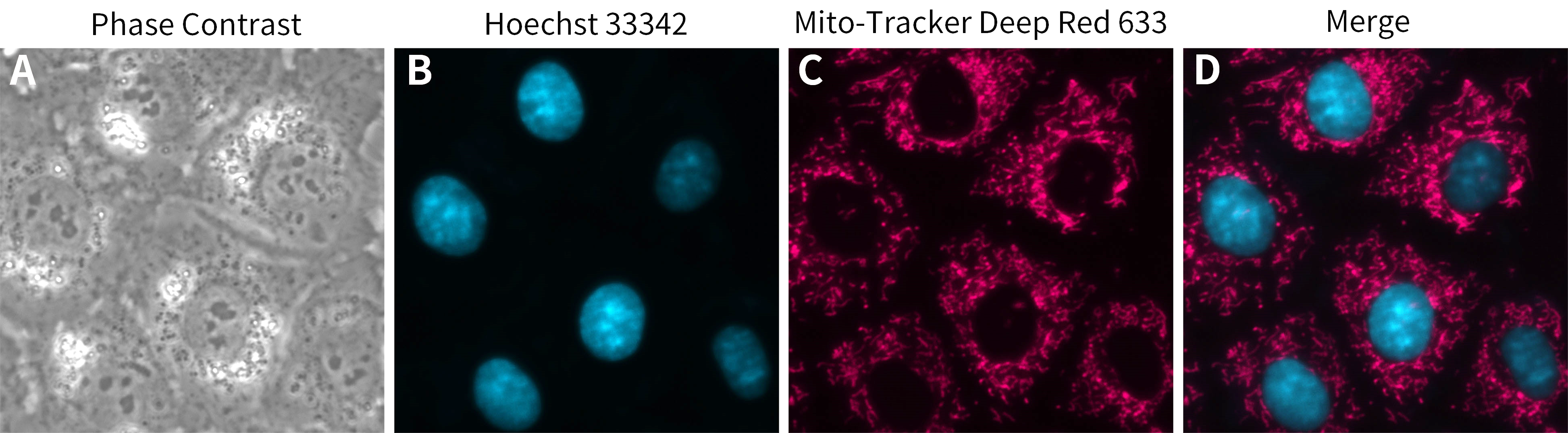
Mito-Tracker Deep Red 633 can be used as a mitochondria-specific fluorescent probe. Similar to Rhodamine 123 or JC-1, Mito-Tracker Deep Red 633 staining of mitochondria is dependent on the mitochondrial membrane potential, so the probe can only stain live cells or tissues, not fixed or permeabilized cells or tissues. Please refer to Figure 2 for the staining effect of Mito-Tracker Deep Red 633 on live cells.
Mito-Tracker Deep Red 633

Figure 2. The staining effect of Mito-Tracker Deep Red 633 on mitochondria in NRK-52E cells (rat renal tubular epithelial cells). The mitochondria of Mito-Tracker Deep Red 633-stained NRK-52E cells showed magenta (pseudo-color) fluorescence (C). The nuclei were stained with Hoechst 33342. This figure is for reference only, which may vary due to different experimental conditions.

Since the accumulation of Mito-Tracker Deep Red 633 in mitochondria is dependent on the mitochondrial membrane potential, this product can also be used as an indicator probe to detect apoptosis by detecting changes in the mitochondrial membrane potential.

Based on a final working concentration of 50-200nM, each 50µg of this product can be used to prepare approximately 400-1700ml of Mito-Tracker Deep Red 633 staining solution.

Storage Conditions:
Store at -20℃ in the dark, valid for at least one year.

Precautions:
Mito-Tracker Deep Red 633 can only be used for mitochondrial fluorescent labeling of living cells, it cannot be used on fixed cells or tissues, nor can fixation and permeabilization be performed after staining.

For trace amounts of liquid, centrifuge for a few seconds before each use to collect the liquid at the bottom of the tube.

Fluorescence intensity decays. Avoid light to minimize the quenching of fluorescence.

This product is for R&D only. Not for drug, household, or other uses.

For your safety and health, please wear a lab coat and disposable gloves during the operation.

실험 프로토콜 (Protocol)

Instructions for Use:

1. Preparation of Mito-Tracker Deep Red 633 stock solution.

After equilibrating a tube of 50μg Mito-Tracker Deep Red 633 powder to room temperature, add 426μl of anhydrous DMSO (anhydrous dimethylsulfoxide), and dissolve thoroughly to obtain 0.2mM Mito-Tracker Deep Red 633 stock solution. Store at -20℃ or lower temperatures after proper dispensing and protected from light.

2. Preparation of Mito-Tracker Deep Red 633 staining solution.

a. Add 0.2mM Mito-Tracker Deep Red 633 stock solution to cell culture medium or an appropriate solution (e.g., HBSS with calcium and magnesium ions) at a ratio of 1:4000-1:1000 to a final concentration of 50nM-200nM. For example, take 1µl of 0.2mM Mito-Tracker Deep Red 633 and add it to 4ml or 1ml of cell culture medium, mix well, and then it will be the Mito-Tracker Deep Red 633 staining solution, with a final concentration of 50nM or 200nM, respectively.

b. Mito-Tracker Deep Red 633 staining solution should be pre-warmed at 37℃ before use.

Note: The concentration of staining solution can be adjusted appropriately according to the actual situation. To reduce non-specific fluorescent staining and mitochondrial toxicity, we recommend using a low concentration.

3. Mitochondrial staining of adherent cells.

a. When the cells are incubated to a certain density in cell culture plates or Petri dishes, remove the cell culture medium, add the Mito-Tracker Deep Red 633 staining solution prepared in step 2, and incubate at 37℃ for 15-30 minutes.

Note: The optimal incubation time should be optimized appropriately according to the cell type.

b. Remove the Mito-Tracker Deep Red 633 staining solution and add fresh cell culture medium pre-incubated at 37℃.

c. Observe with a fluorescence microscope, laser confocal microscope, or fluorescence microplate reader. If the staining is not satisfactory, the concentration of Mito-Tracker Deep Red 633 staining solution can be increased or the staining time can be extended appropriately within the recommended range.

Note: Mito-Tracker Deep Red 633 is prone to quenching, so please perform fluorescence assays or take pictures as soon as possible. Quenching can also be delayed by reducing the excitation intensity of the fluorescence microscope (i.e. mercury lamp or LED light source) or by appropriately reducing the concentration of Mito-Tracker Deep Red 633 staining solution.

4. Mitochondrial staining of suspension cells.

a. Centrifuge at 1000g for 5 min, discard the supernatant, gently resuspend cells with 37℃ pre-warmed Mito-Tracker Deep Red 633 staining solution and incubate at 37℃ for 15-30 min.

Note: The optimal incubation time should be optimized appropriately according to cell type.

b. After incubation, centrifuge at 1000g for 5 minutes, discard the supernatant and resuspend the cells by adding fresh cell culture medium pre-incubated at 37℃.

c. Observe with a fluorescence microscope, laser confocal microscope, flow cytometer, or fluorescence microplate reader.

Note: If suspension cells need to be fixed on a slide or coverslip, the slide or coverslip can first be treated with Poly-D-lysine.

제품 문서 (Product Documents)

Appearance:A solid

화학적 특성 (Chemical Properties)

보관
Store at -20°C, protect from light
일반 주의사항
제품의 용해도 데이터에 따라 적절한 용매를 선택하십시오. 조제 후에는 반복적인 동결·해동을 피할 수 있도록 저장 용액을 소분하십시오.
저장 용액은 -80°C 보관 시 6개월 이내, -20°C 보관 시 1개월 이내에 사용하십시오.
용해도를 높이려면 튜브를 37°C로 데운 뒤 적절한 시간 동안 초음파 처리하십시오.
배송 조건
평가용 샘플은 아이스팩과 함께 배송됩니다. 그 외 용량은 실온 배송되며, 요청 시 아이스팩과 함께 배송할 수 있습니다.

계산 도구 (Calculators)몰 농도 / 희석 / 분자량 / 단위 변환

g/mol

Reviews

Review for Mito-Tracker Deep Red 633

Rating: 5
  • 5 Star100%
  • 4 Star0%
  • 3 Star0%
  • 2 Star0%
  • 1 Star0%