Acridine Orange hydrochloride |
رقم الكتالوجGC12913 |
أكريدين أورانج هيدروكلوريد هو صبغة فلورية قابلة للنفاذ للخلايا وترتبط بالأحماض النووية ، مما يؤدي إلى انبعاث طيفي متغير
Products are for research use only. Not for human use. We do not sell to patients.
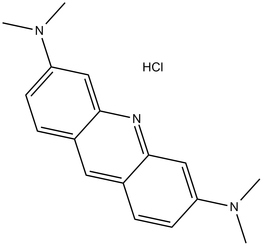
Cas No.: 65-61-2
Sample solution is provided at 25 µL, 10mM.
Acridine Orange hydrochloride is a cell-permeable fluorescent dye that binds to nucleic acids, resulting in an altered spectral emission.
Acridine Orange has been employed extensively as a cytochemical stain and has shown to stain differentially DNA and RNA, and double-restranded nucleic acids in situ. Acridine Orange can either intercalate into double helical nucleic acids (green fluorescence at 530 nm), or bind electrostatically to phosphate groups of single-stranded molecules (red fluorescence at 640 nm). This unique characteristic makes acridine orange useful for cell-cycle studies[1]. Acridine Orange staining of unfixed cells may be used as a simple, fast means of obtaining information on cell ploidy levels and cell cycle status from DNA measurements (green fluorescence), and cell transcriptional activity from RNA staining (red fluorescence), in human and murine cells lines, peripheral blood and bone marrow specimens from patients with leukemia and mitogenically (phytohemagglutinin) or antigenically (mixed lymphocyte culture) stimulated human peripheral blood cultures[2].
References:
[1]. McMaster GK, et al. Analysis of single- and double-stranded nucleic acids on polyacrylamide and agarosegels by using glyoxal and acridine orange. Proc Natl Acad Sci U S A. 1977 Nov;74(11):4835-8.
[2]. Traganos F, et al. Simultaneous staining of ribonucleic and deoxyribonucleic acids in unfixed cells using acridine orange in a flow cytofluorometric system. J Histochem Cytochem. 1977 Jan;25(1):46-56.
This plan only provides a guide, please modify it to meet your specific needs.
1. Prepare Acridine Orange staining solution
(1) Acridine Orange dye stock solution: Use DMSO to dissolve Acridine Orange hydrochloride into a 1mg/ml stock solution.
Note: It is recommended that unused stock solutions be aliquoted and stored in the dark at -4°C or -20°C (stored under nitrogen), and avoid repeated freezing and thawing.
(2) Preparation of Acridine Orange dye working solution: Dilute the stock solution with a suitable buffer (such as serum-free medium, HBSS or PBS) to prepare an Acridine Orange working solution with a concentration of 0.5-20 μg/ml.
Note: Please adjust the concentration of the working fluid according to the actual situation, and use it now.
2. DNA and RNA staining of cells:
(1) Collect the cell suspension, transfer about 0.2ml/portion of the original cell suspension into a small glass or plastic tube with a volume of 2/5 mL, and place it on ice to cool;
(2) Slowly add 0.4 mL of pre-cooled cell permeabilization solution, and place the cells on ice for about 15 seconds;
(3) Slowly add 1.2 mL of pre-cooled Acridine Orange working solution, and incubate on ice in the dark for 2-15 minutes;
(4) Use a flow cytometer to measure and record cell fluorescence, excite at 488 nm, and distinguish dsDNA at green fluorescence (measured at 515-545 nm) and red luminescence (preferably at For RNA (measured above 640 or 650nm), the excitation/emission wavelength of Acridine Orange after binding to ssDNA is 460/650nm.
3. Cell adhesion staining (operated on ice)
(1) Culture adherent cells on sterile coverslips;
(2) Remove the coverslip from the culture medium, suck out the excess culture medium, and place the coverslip in a humid environment;
(3) Add 100 μL dye working solution from one corner of the coverslip, shake gently to make the dye evenly cover all cells, and incubate on ice in the dark for 2-15 minutes;
(4) Aspirate and discard the dye working solution, use culture medium to wash the coverslip 2 to 3 times, and observe with a fluorescence microscope.
4. Microscopy detection: Use a confocal laser scanning microscope (such as FV3000, Olympus) to observe cells, or quantify the total fluorescence of Acridine Orange in each frame through MetaMorph. Acridine Orange exhibits excitation/emission spectra of 502/525, 460/650 and 475/590 nm when bound to dsDNA, bound to ssDNA or RNA, and under acidic conditions, respectively.
Precautions:
① If cells need to be fixed and stained, it is recommended to use 70% ethanol for fixation and fix on ice for ≥ 2 hours. After fixation, rinse 1-2 times with pre-cooled PBS to remove all ethanol, and then stain according to the above steps;
② Fluorescent dyes all have quenching problems. Please try to avoid light to slow down fluorescence quenching;
③ For your safety and health, please wear a lab coat and disposable gloves.
Cas No. | 65-61-2 | SDF | |
Chemical Name | N3,N3,N6,N6-tetramethylacridine-3,6-diamine hydrochloride | ||
Canonical SMILES | CN(C)C1=CC2=NC3=CC(N(C)C)=CC=C3C=C2C=C1.Cl | ||
Formula | C17H19N3.HCl | M.Wt | 301.81 |
الذوبان | ≥ 30.6mg/mL in DMSO with gentle warming, ≥ 30.5mg/mL in EtOH, ≥ 30.3mg/mL in Water | Storage | Store at 4°C, protect from light, stored under nitrogen |
General tips | Please select the appropriate solvent to prepare the stock solution according to the
solubility of the product in different solvents; once the solution is prepared, please store it in
separate packages to avoid product failure caused by repeated freezing and thawing.Storage method
and period of the stock solution: When stored at -80°C, please use it within 6 months; when stored
at -20°C, please use it within 1 month. To increase solubility, heat the tube to 37°C and then oscillate in an ultrasonic bath for some time. |
||
Shipping Condition | Evaluation sample solution: shipped with blue ice. All other sizes available: with RT, or with Blue Ice upon request. |
Prepare stock solution | |||
![]() |
1 mg | 5 mg | 10 mg |
1 mM | 3.3133 mL | 16.5667 mL | 33.1334 mL |
5 mM | 0.6627 mL | 3.3133 mL | 6.6267 mL |
10 mM | 0.3313 mL | 1.6567 mL | 3.3133 mL |
Step 1: Enter information below (Recommended: An additional animal making an allowance for loss during the experiment)
Step 2: Enter the in vivo formulation (This is only the calculator, not formulation. Please contact us first if there is no in vivo formulation at the solubility Section.)
Calculation results:
Working concentration: mg/ml;
Method for preparing DMSO master liquid: mg drug pre-dissolved in μL DMSO ( Master liquid concentration mg/mL, Please contact us first if the concentration exceeds the DMSO solubility of the batch of drug. )
Method for preparing in vivo formulation: Take μL DMSO master liquid, next addμL PEG300, mix and clarify, next addμL Tween 80, mix and clarify, next add μL ddH2O, mix and clarify.
Method for preparing in vivo formulation: Take μL DMSO master liquid, next add μL Corn oil, mix and clarify.
Note: 1. Please make sure the liquid is clear before adding the next solvent.
2. Be sure to add the solvent(s) in order. You must ensure that the solution obtained, in the previous addition, is a clear solution before proceeding to add the next solvent. Physical methods such as vortex, ultrasound or hot water bath can be used to aid dissolving.
3. All of the above co-solvents are available for purchase on the GlpBio website.
Quality Control & SDS
- View current batch:
- Purity: >99.00%
- COA (Certificate Of Analysis)
- SDS (Safety Data Sheet)
- Datasheet
Average Rating: 5
(Based on Reviews and 39 reference(s) in Google Scholar.)GLPBIO products are for RESEARCH USE ONLY. Please make sure your review or question is research based.
Required fields are marked with *