

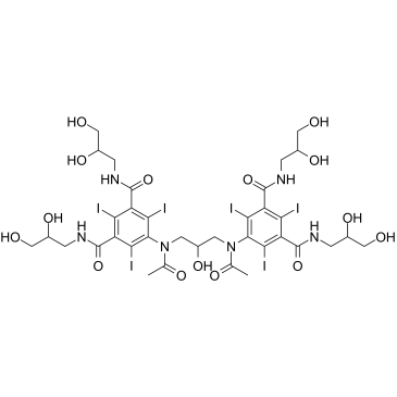
Iodixanol |
رقم الكتالوجGC39741
 One-Click Copy Product Info
|

Iodixanol هو عامل تباين إشعاعي غير أيوني يحتوي على اليود
Products are for research use only. Not for human use. We do not sell to patients.
Cas No.: 92339-11-2
Sample solution is provided at 25 µL, 10mM.
_1-3.$$$39978408@51.jpg');)
_1-9.$$$38538795@65.jpg');)
_1-9.$$$39112701@51.jpg');)
_1-7.$$$37316672@70.jpg');)
_366-372.$$$36198801@70.jpg');)
.$$$38565142@65.jpg');)
_1867-1883.$$$33248023@67.jpg');)
_eads6055.$$$39977546@45.jpg');)
_eadm9805.$$$40080571@45.jpg');)
_eadj3020.$$$39666821@45.jpg');)
_1-20.$$$39757301@28.jpg');)
_1-24.$$$38898113@28.jpg');)
_273-287.$$$36806353@46.jpg');)
_1-16.$$$37156877@44.jpg');)
_1-19.$$$37460805@44.jpg');)
_1291-1307.$$$34518654@46.jpg');)
Iodixanol is a non-ionic, dimeric, iso-osmolar iodinated X-ray contrast agent. Iodixanol can absorb X-rays through the iodine atoms contained in its molecules, thereby creating artificial contrast with surrounding tissues to clearly visualize vascular and visceral structures. Iodixanol reduces adverse reactions such as endothelial damage and nephrotoxicity by maintaining an osmotic pressure equal to that of plasma. Iodixanol can be used in research related to medical imaging modalities such as cardiovascular angiography, cerebrovascular angiography, and CT enhancement[1-4].
In vitro, LLC-PK1 cells (proximal tubular epithelial cells of porcine origin) were treated with Iodixanol (18.75–75mg I/mL) for 1 to 24 hours. Iodixanol significantly induced apoptosis and anti-proliferative effects without causing cellular necrosis[5]. Human LAD2 mast cells were treated with Iodixanol (4–30%vol) for 30 minutes. Iodixanol significantly induced the release of β-hexosaminidase and histamine, promoted intracellular calcium mobilization, and facilitated the synthesis of inflammatory cytokines by activating PLC-γ- and PI3K-related pathways, thereby exacerbating pseudo-allergic reactions[6].
In vivo, C57BL/6J mice were pretreated with dehydration and furosemide, followed by intraperitoneal injection of Iodixanol (15g I/kg; single dose). Iodixanol significantly induced mitochondrial fragmentation, dysfunction, and apoptosis in renal tubular cells, increased the levels of superoxide and malondialdehyde in renal tubules, and led to renal tubular epithelial cell damage and renal dysfunction[7]. Normal female C57/BL6 mice were injected with Iodixanol (10g I/kg; single dose) via the tail vein. Iodixanol significantly elevated serum levels of creatinine, NGAL, and KIM-1, resulting in renal dysfunction and histological damage[8].
References:
[1] Liu ZZ, Viegas VU, Perlewitz A, et al. Iodinated contrast media differentially affect afferent and efferent arteriolar tone and reactivity in mice: a possible explanation for reduced glomerular filtration rate. Radiology. 2012 Dec;265(3):762-71.
[2] Jensen H, Doughty RW, Grant D, et al. The effects of the iodinated X-ray contrast media iodixanol, iohexol, iopromide, and ioversol on the rat kidney epithelial cell line NRK 52-E. Ren Fail. 2011;33(4):426-33.
[3] Zhang J, Jiang Y, Rui Q, et al. Iodixanol versus iopromide in patients with renal insufficiency undergoing coronary angiography with or without PCI. Medicine (Baltimore). 2018 May;97(18):e0617.
[4] Spencer CM, Goa KL. Iodixanol. A review of its pharmacodynamic and pharmacokinetic properties and diagnostic use as an x-ray contrast medium. Drugs. 1996 Dec;52(6):899-927.
[5] Heinrich MC, Scheer M, Heckmann M, et al. Iodixanol induces apoptotic and antiproliferative effects but no necrotic cell death in renal proximal tubular cells in vitro. Rofo. 2009 Apr;181(4):349-54.
[6] Zou W, Yang S, Chen L, et al. Iodixanol activation of mast cells: Implications in the pathogenesis of iodixanol-induced delayed cutaneous adverse reactions. Toxicology. 2022 Jan 15;465:153034.
[7] Ran F, Yang Y, Yang L, et al. Capsaicin Prevents Contrast-Associated Acute Kidney Injury through Activation of Nrf2 in Mice. Oxid Med Cell Longev. 2022 May 16;2022:1763922.
[8] Kim BW, Kim HJ, Kim SH, et al. 15-Hydroxyprostaglandin dehydrogenase inhibitor prevents contrast-induced acute kidney injury. Ren Fail. 2021 Dec;43(1):168-179.
| Cell experiment [1]: | |
Cell lines | LLC-PK1 cells (a proximal tubular epithelial cell line of porcine origin) |
Preparation Method | LLC-PK1 cells were maintained in modified medium 199 (M199) supplemented with 10% fetal bovine serum (FBS), 100U/ml penicillin and 100μg/ml streptomycin at 37°C, 5% CO₂. Confluent LLC-PK1 cell monolayers were treated with Iodixanol at clinically relevant concentrations (18.75, 37.5, and 75mg I/mL) for 1 to 24 hours. |
Reaction Conditions | 18.75–75mg I/ml; 1-24h |
Applications | Iodixanol significantly increased the number of cytoplasmic oligonucleosomes, indicating the induction of apoptosis. Iodixanol also induced a significant, dose- and time-dependent inhibition of BrdU incorporation, indicating an antiproliferative effect. Iodixanol did not induce any significant increase in the number of necrotic cells. |
| Animal experiment [2]: | |
Animal models | Female C57/BL6 mice |
Preparation Method | Mice were injected with 10g I/kg of Iodixanol via the tail vein, and serum and kidney tissues were collected 48 hours after administration. |
Dosage form | 10g I/kg; i.v.; single injection |
Applications | Iodixanol administration successfully induced contrast-induced acute kidney injury (CIAKI), significantly elevating serum levels of renal injury markers (creatinine, NGAL, and KIM-1), exacerbating histologic damage and apoptosis (increased renal injury score and TUNEL-positive cells), reducing the average renal arteriole area in the outer medulla, and decreasing renal blood flow. |
References: | |
| Cas No. | 92339-11-2 | SDF | |
| Canonical SMILES | OC(CN(C1=C(I)C(C(NCC(O)CO)=O)=C(I)C(C(NCC(O)CO)=O)=C1I)C(C)=O)CN(C2=C(I)C(C(NCC(O)CO)=O)=C(I)C(C(NCC(O)CO)=O)=C2I)C(C)=O | ||
| Formula | C35H44I6N6O15 | M.Wt | 1550.18 |
| الذوبان | DMSO: 250 mg/mL (161.27 mM) | Storage | Store at -20°C |
| General tips | Please select the appropriate solvent to prepare the stock solution according to the
solubility of the product in different solvents; once the solution is prepared, please store it in
separate packages to avoid product failure caused by repeated freezing and thawing.Storage method
and period of the stock solution: When stored at -80°C, please use it within 6 months; when stored
at -20°C, please use it within 1 month. To increase solubility, heat the tube to 37°C and then oscillate in an ultrasonic bath for some time. |
||
| Shipping Condition | Evaluation sample solution: shipped with blue ice. All other sizes available: with RT, or with Blue Ice upon request. | ||
| Prepare stock solution | |||

|
1 mg | 5 mg | 10 mg |
| 1 mM | 645.1 μL | 3.2254 mL | 6.4509 mL |
| 5 mM | 129 μL | 645.1 μL | 1.2902 mL |
| 10 mM | 64.5 μL | 322.5 μL | 645.1 μL |
Step 1: Enter information below (Recommended: An additional animal making an allowance for loss during the experiment)
 g
μL
g
μL
Step 2: Enter the in vivo formulation (This is only the calculator, not formulation. Please contact us first if there is no in vivo formulation at the solubility Section.)
Calculation results:
Working concentration: mg/ml;
Method for preparing DMSO master liquid: mg drug pre-dissolved in μL DMSO ( Master liquid concentration mg/mL, Please contact us first if the concentration exceeds the DMSO solubility of the batch of drug. )
Method for preparing in vivo formulation: Take μL DMSO master liquid, next addμL PEG300, mix and clarify, next addμL Tween 80, mix and clarify, next add μL saline, mix and clarify.
Method for preparing in vivo formulation: Take μL DMSO master liquid, next add μL Corn oil, mix and clarify.
Note: 1. Please make sure the liquid is clear before adding the next solvent.
2. Be sure to add the solvent(s) in order. You must ensure that the solution obtained, in the previous addition, is a clear solution before proceeding to add the next solvent. Physical methods such as vortex, ultrasound or hot water bath can be used to aid dissolving.
3. All of the above co-solvents are available for purchase on the GlpBio website.
Quality Control & SDS
- View current batch:
- Purity: >99.50% Appearance: A solid
- COA (Certificate of Analysis)
- SDS (Safety Data Sheet)
- Datasheet
Average Rating: 5 (Based on Reviews and 10 reference(s) in Google Scholar.)