

Nifedipine (Synonyms: BAY 1040) |
رقم الكتالوجGC15024
 One-Click Copy Product Info
|

نيفيديبين (BAY-a-1040) هو مانع قوي لقنوات الكالسيوم والدواء المفضل لقصور القلب
Products are for research use only. Not for human use. We do not sell to patients.
Cas No.: 21829-25-4
Sample solution is provided at 25 µL, 10mM.
_1-3.$$$39978408@51.jpg');)
_1-9.$$$38538795@65.jpg');)
_1-9.$$$39112701@51.jpg');)
_1-7.$$$37316672@70.jpg');)
_366-372.$$$36198801@70.jpg');)
.$$$38565142@65.jpg');)
_1867-1883.$$$33248023@67.jpg');)
_eads6055.$$$39977546@45.jpg');)
_eadm9805.$$$40080571@45.jpg');)
_eadj3020.$$$39666821@45.jpg');)
_1-20.$$$39757301@28.jpg');)
_1-24.$$$38898113@28.jpg');)
_273-287.$$$36806353@46.jpg');)
_1-16.$$$37156877@44.jpg');)
_1-19.$$$37460805@44.jpg');)
_1291-1307.$$$34518654@46.jpg');)
Nifedipine (BAY-a-1040) is a potent calcium channel blocker and drug of choice for cardiac insufficiencies.
Nifedipine (BAY-a-1040) (100 μM) significantly lowers the viability of the WKPT-0293 Cl.2 Cells, and treatment of nifedipine (10 or 100 μM) plus FAC induces a significant reduction in cell viability, but there are no significant differences in viability between the control cells and the cells treated with 100 μM of FAC or 1 and 10 μM of nifedipine.Nifedipine (BAY-a-1040) (1, 10, or 100 μM) significantly increases iron level in WKPT-0293 Cl.2 cells. Nifedipine treatment also increases expression of TfR1, DMT1+IRE and DMT1-IRE in WKPT-0293 Cl.2 cells. In addition, co-treatment with nifedipine (100 μM) and FAC (100 μM) increases TfR1, DMT1+IRE and DMT1-IRE expression in WKPT-0293 Cl.2 cells[2]. Nifedipine plus ritodrine produces a significantly greater inhibition of contractility than each drug alone in the midrange of concentrations. The combination of nifedipine plus nitroglycerin or nifedipine plus atosiban produces a significantly greater inhibition than nitroglycerin or atosiban alone but not greater than nifedipine. The combination of nifedipine plus NS-1619 (Ca2+-activated K+ [BKCa] channel opener) reduces the inhibitory effect of each drug[3]. Nifedipine (BAY-a-1040) (2 μM) significantly inhibits P. capsici mycelial growth and sporulation. Nifedipine (BAY-a-1040)-induced inhibition of mycelial growth is calcium-dependent. Nifedipine (0.5 μM) increases P. capsici sensitivity to H2O2 in a calcium-dependent manner. Nifedipine inhibition of P. capsici virulence and expression of genes involved in pathogenicity[4].
In Nifedipine (BAY-a-1040) (50 mg/kg)- and CsA-treated rats, the BL dimensions (BLi and BLk), MD dimensions (MDk) and vertical dimensions (VHi and VHk) are significantly increased (P < 0.05) at the end of the 4th week[1].
References:
[1]. Ratre MS, et al. Effect of azithromycin on gingival overgrowth induced by cyclosporine A + nifedipine combination therapy: A morphometric analysis in rats. J Indian Soc Periodontol. 2016 Jul-Aug;20(4):396-401.
[2]. Carvajal JA, et al. The Synergic In Vitro Tocolytic Effect of Nifedipine Plus Ritodrine on Human Myometrial Contractility. Reprod Sci. 2017 Apr;24(4):635-640.
[3]. Yu SS, et al. Nifedipine Increases Iron Content in WKPT-0293 Cl.2 Cells via Up-Regulating Iron Influx Proteins. Front Pharmacol. 2017 Feb 13;8:60
[4]. Liu P, et al. The L-type Ca(2+) Channel Blocker Nifedipine Inhibits Mycelial Growth, Sporulation, and Virulence of Phytophthora capsici. Front Microbiol. 2016 Aug 4;7:1236.
|
Cell experiment: |
Cell viability is assessed using an MTT assay. Briefly, a total of 25 μL MTT (1 g/L in PBS) is added to each well before incubation is conducted at 37°C for 4 h. The assay is stopped by the addition of a 100 μL lysis buffer (20% SDS in 50% N’Ndimethylformamide, pH 4.7). Optical density (OD) is measured at the 570 nm wavelength by the use of an ELX-800 microplate assay reader and the results are expressed as a percentage of the absorbance measured in the control cells. |
|
Animal experiment: |
All the 30 rats are randomLy distributed into three equal groups of ten animals each. Group 1 (control) receive olive oil for the 8 weeks. Group 2 and Group 3 receive a combination of CsA (30 mg/kg body weight) and Nf (50 mg/kg body weight) in olive oil for 8 weeks. In Group 3 rats, Azi (10 mg/kg body weight) is added to this regimen, in the 5th week. The total study period is 8 weeks. |
|
References: [1]. Ratre MS, et al. Effect of azithromycin on gingival overgrowth induced by cyclosporine A + nifedipine combination therapy: A morphometric analysis in rats. J Indian Soc Periodontol. 2016 Jul-Aug;20(4):396-401. |
|
| Cas No. | 21829-25-4 | SDF | |
| المرادفات | BAY 1040 | ||
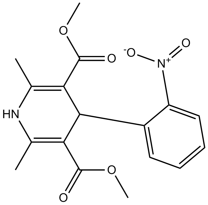
| Chemical Name | dimethyl 2,6-dimethyl-4-(2-nitrophenyl)-1,4-dihydropyridine-3,5-dicarboxylate | ||
| Canonical SMILES | CC1=C(C(C(=C(N1)C)C(=O)OC)C2=CC=CC=C2[N+](=O)[O-])C(=O)OC | ||
| Formula | C17H18N2O6 | M.Wt | 346.33 |
| الذوبان | ≥ 15.75mg/mL in DMSO | Storage | 4°C, protect from light |
| General tips | Please select the appropriate solvent to prepare the stock solution according to the
solubility of the product in different solvents; once the solution is prepared, please store it in
separate packages to avoid product failure caused by repeated freezing and thawing.Storage method
and period of the stock solution: When stored at -80°C, please use it within 6 months; when stored
at -20°C, please use it within 1 month. To increase solubility, heat the tube to 37°C and then oscillate in an ultrasonic bath for some time. |
||
| Shipping Condition | Evaluation sample solution: shipped with blue ice. All other sizes available: with RT, or with Blue Ice upon request. | ||
| Prepare stock solution | |||

|
1 mg | 5 mg | 10 mg |
| 1 mM | 2.8874 mL | 14.4371 mL | 28.8742 mL |
| 5 mM | 577.5 μL | 2.8874 mL | 5.7748 mL |
| 10 mM | 288.7 μL | 1.4437 mL | 2.8874 mL |
Step 1: Enter information below (Recommended: An additional animal making an allowance for loss during the experiment)
 g
μL
g
μL
Step 2: Enter the in vivo formulation (This is only the calculator, not formulation. Please contact us first if there is no in vivo formulation at the solubility Section.)
Calculation results:
Working concentration: mg/ml;
Method for preparing DMSO master liquid: mg drug pre-dissolved in μL DMSO ( Master liquid concentration mg/mL, Please contact us first if the concentration exceeds the DMSO solubility of the batch of drug. )
Method for preparing in vivo formulation: Take μL DMSO master liquid, next addμL PEG300, mix and clarify, next addμL Tween 80, mix and clarify, next add μL saline, mix and clarify.
Method for preparing in vivo formulation: Take μL DMSO master liquid, next add μL Corn oil, mix and clarify.
Note: 1. Please make sure the liquid is clear before adding the next solvent.
2. Be sure to add the solvent(s) in order. You must ensure that the solution obtained, in the previous addition, is a clear solution before proceeding to add the next solvent. Physical methods such as vortex, ultrasound or hot water bath can be used to aid dissolving.
3. All of the above co-solvents are available for purchase on the GlpBio website.
Quality Control & SDS
- View current batch:
- Purity: >98.00% Appearance: A solid
- COA (Certificate of Analysis)
- SDS (Safety Data Sheet)
- Datasheet
Average Rating: 5 (Based on Reviews and 9 reference(s) in Google Scholar.)