

NVP-BGJ398 phosphate |
رقم الكتالوجGC16028
 One-Click Copy Product Info
|

Products are for research use only. Not for human use. We do not sell to patients.
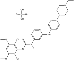
Cas No.: 1310746-10-1
Sample solution is provided at 25 µL, 10mM.
_1-3.$$$39978408@51.jpg');)
_1-9.$$$38538795@65.jpg');)
_1-9.$$$39112701@51.jpg');)
_1-7.$$$37316672@70.jpg');)
_366-372.$$$36198801@70.jpg');)
.$$$38565142@65.jpg');)
_1867-1883.$$$33248023@67.jpg');)
_eads6055.$$$39977546@45.jpg');)
_eadm9805.$$$40080571@45.jpg');)
_eadj3020.$$$39666821@45.jpg');)
_1-20.$$$39757301@28.jpg');)
_1-24.$$$38898113@28.jpg');)
_273-287.$$$36806353@46.jpg');)
_1-16.$$$37156877@44.jpg');)
_1-19.$$$37460805@44.jpg');)
_1291-1307.$$$34518654@46.jpg');)
Kinase experiment: | The enzymatic kinase activity is assessed by measuring the phosphorylation of a synthetic substrate by the purified GST-fusion FGFR3-K650E kinase domain, in the presence of radiolabeled ATP. Enzyme activities are measured by mixing 10 μL of a 3-fold concentrated Infigratinib solution or control with 10 μL of the corresponding substrate mixture (peptidic substrate, ATP and [γ33P]ATP). The reactions are initiated by addition of 10 μL of a 3-fold concentrated solution of the enzyme in assay buffer. The final concentrations of the assay components are as following: 10 ng of GST-FGFR3-K650E, 20 mM Tris-HCl, pH 7.5, 3 mM MnCl2, 3 mM MgCl2, 1 mM DTT, 250 μg/mL PEG 20000, 2 μg/mL poly(EY) 4:1, 1% DMSO and 0.5 μM ATP (γ-[33P]-ATP 0.1 μCi). The assay is carried out according to the filter binding (FB) method in 96-well plates at room temperature for 10 min in a final volume of 30 μL including the components as indicated above. The enzymatic reactions are stopped by the addition of 20 μL of 125 mM EDTA, and the incorporation of 33P into the polypeptidic substrates is quantified as following: 30 μL of the stopped reaction mixture are transferred onto Immobilon-PVDF membranes previously soaked for 5 min with methanol, rinsed with water, soaked for 5 min with 0.5% H3PO4, and mounted on vacuum manifold with disconnected vacuum source. After spotting, vacuum is connected, and each well rinsed with 0.5% H3PO4 (200 μL). Free membranes are removed and washed four times on a shaker with 1% H3PO4 and once with ethanol. Membranes are dried and overlaid with addition of 10 μL/well of a scintillation fluid. The plates are eventually sealed and counted in a microplate scintillation counter. IC50 values are calculated by linear regression analysis of the percentage inhibition of NVP-BGJ398[1]. |
Cell experiment: | Murine BaF3 cell lines are cultured in RPMI-1640 media supplemented with 10% FBS, 4.5 g/L glucose, 1.5 g/L sodium bicarbonate, and Pen/Strep. Cells are passaged twice weekly. Compound-mediated inhibition of BaF3 cell proliferation and viability is assessed using a Luciferase bioluminescent assay. Exponentially growing BaF3 or BaF3 Tel-TK cells are seeded into 384-well plates (4250 cells/well) at 50 μL/well using a μFill liquid dispenser in fresh medium. Infigratinib is serially diluted in DMSO and arrayed in a polypropylene 384-well plate. Then 50 nL of compound are transferred into the plates containing the cells by using the pintool transfer device, and the plates incubated at 37°C (5% CO2) for 48 h. Then 25 μL of Bright-Glo are added, and luminescence is quantified using an Analyst-GT. Custom curve-fitting software is used to produce a logistic fit of percent cell viability as a function of the logarithm of inhibitor concentration. The IC50 value is determined as the concentration of compound needed to reduce cell viability to 50% of a DMSO control[1]. |
Animal experiment: | Mice[1] Female HsdNpa: Athymic Nude-nu mice are used. Infigratinib is formulated as a suspension in PEG300/D5W (2:1, v/v) and administered orally for 12 consecutive days at the doses of 10 and 30 mg/kg/qd. Tumor and body weight data are analyzed by ANOVA with post hoc Dunnett’s test for comparison of treatment versus control group. The post hoc Tukey test is used for intragroup comparison. Statistical analysis is performed using GraphPad prism 4.02. As a measure of efficacy, the T/C (%) value is calculated. Rats[1] Female nude Rowett rats 6-9 weeks of age are used. Infigratinib is formulated as a solution in acetic acid-acetate buffer pH 4.6/PEG300 (1:1, v/v) and applied daily by gavage to the tumor-bearing rats (n=8) for 20 consecutive days at doses of 5, 10, and 15 mg/kg/qd (free base equivalents). The application volume is 5 mL/kg. Tumor volumes are measured with calipers and determined according to the formula: length×width×height×π/6. Antitumor activity is expressed as T/C (%): (mean change of tumor volume of treated animals/mean change of tumor volume of control animals)×100. Regressions (%) are calculated. |
References: [1]. Guagnano V, et al. Discovery of 3-(2,6-Dichloro-3,5-dimethoxy-phenyl)-1-{6-[4-(4-ethyl-piperazin-1-yl)-phenylamino]-pyrimidin-4-yl}-1-me thyl-urea (NVP-BGJ398), A Potent and Selective Inhibitor of the Fibroblast Growth Factor Receptor Family of Receptor T | |
| Cas No. | 1310746-10-1 | SDF | |
| Chemical Name | 3-(2,6-dichloro-3,5-dimethoxyphenyl)-1-(6-((4-(4-ethylpiperazin-1-yl)phenyl)amino)pyrimidin-4-yl)-1-methylurea phosphate | ||
| Canonical SMILES | COC1=C(Cl)C(NC(N(C)C2=CC(NC3=CC=C(N4CCN(CC)CC4)C=C3)=NC=N2)=O)=C(Cl)C(OC)=C1.OP(O)(O)=O | ||
| Formula | C26H34Cl2N7O7P | M.Wt | 658.47 |
| الذوبان | ≥ 95.7 mg/mL in DMSO, ≥ 28.07 mg/mL in Water with ultrasonic and warming | Storage | Store at -20°C |
| General tips | Please select the appropriate solvent to prepare the stock solution according to the
solubility of the product in different solvents; once the solution is prepared, please store it in
separate packages to avoid product failure caused by repeated freezing and thawing.Storage method
and period of the stock solution: When stored at -80°C, please use it within 6 months; when stored
at -20°C, please use it within 1 month. To increase solubility, heat the tube to 37°C and then oscillate in an ultrasonic bath for some time. |
||
| Shipping Condition | Evaluation sample solution: shipped with blue ice. All other sizes available: with RT, or with Blue Ice upon request. | ||
| Prepare stock solution | |||

|
1 mg | 5 mg | 10 mg |
| 1 mM | 1.5187 mL | 7.5934 mL | 15.1867 mL |
| 5 mM | 303.7 μL | 1.5187 mL | 3.0373 mL |
| 10 mM | 151.9 μL | 759.3 μL | 1.5187 mL |
Step 1: Enter information below (Recommended: An additional animal making an allowance for loss during the experiment)
 g
μL
g
μL
Step 2: Enter the in vivo formulation (This is only the calculator, not formulation. Please contact us first if there is no in vivo formulation at the solubility Section.)
Calculation results:
Working concentration: mg/ml;
Method for preparing DMSO master liquid: mg drug pre-dissolved in μL DMSO ( Master liquid concentration mg/mL, Please contact us first if the concentration exceeds the DMSO solubility of the batch of drug. )
Method for preparing in vivo formulation: Take μL DMSO master liquid, next addμL PEG300, mix and clarify, next addμL Tween 80, mix and clarify, next add μL saline, mix and clarify.
Method for preparing in vivo formulation: Take μL DMSO master liquid, next add μL Corn oil, mix and clarify.
Note: 1. Please make sure the liquid is clear before adding the next solvent.
2. Be sure to add the solvent(s) in order. You must ensure that the solution obtained, in the previous addition, is a clear solution before proceeding to add the next solvent. Physical methods such as vortex, ultrasound or hot water bath can be used to aid dissolving.
3. All of the above co-solvents are available for purchase on the GlpBio website.
Quality Control & SDS
- View current batch:
- Purity: >98.00% Appearance: A solid
- COA (Certificate of Analysis)
- SDS (Safety Data Sheet)
- Datasheet
Average Rating: 5 (Based on Reviews and 30 reference(s) in Google Scholar.)