

sodium fluoride |
رقم الكتالوجGC10594
 One-Click Copy Product Info
|

Sodium Fluoride, another form of fluorine, is orally bioavailable and can accumulate in reproductive organs and interfere with hormonal regulation and oxidative stress pathways, contributing to reproductive toxicity.
Products are for research use only. Not for human use. We do not sell to patients.
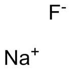
Cas No.: 7681-49-4
Sample solution is provided at 25 µL, 10mM.
_1-3.$$$39978408@51.jpg');)
_1-9.$$$38538795@65.jpg');)
_1-9.$$$39112701@51.jpg');)
_1-7.$$$37316672@70.jpg');)
_366-372.$$$36198801@70.jpg');)
.$$$38565142@65.jpg');)
_1867-1883.$$$33248023@67.jpg');)
_eads6055.$$$39977546@45.jpg');)
_eadm9805.$$$40080571@45.jpg');)
_eadj3020.$$$39666821@45.jpg');)
_1-20.$$$39757301@28.jpg');)
_1-24.$$$38898113@28.jpg');)
_273-287.$$$36806353@46.jpg');)
_1-16.$$$37156877@44.jpg');)
_1-19.$$$37460805@44.jpg');)
_1291-1307.$$$34518654@46.jpg');)
Sodium Fluoride, another form of fluorine, is orally bioavailable and can accumulate in reproductive organs and interfere with hormonal regulation and oxidative stress pathways, contributing to reproductive toxicity[1]. Sodium Fluoride is extensively used for the prevention of dental caries and tooth decay in the form of fluorinated drinking water, salts, or milk, tooth pastes, mouth washes, and fluo-ride tablets[2]. The acute Fluoride toxicosis usually occurs by soluble forms of Sodium Fluoride upon ingestion in large doses[3].
In vitro, bone mesenchymal stem cells (BMSCs) grew well in media containing 0-240μM Sodium Fluoride, whereas exposure to 480μM or 1200μM Sodium Fluoride caused a decline in cell viability—first evident at day 7 for 480μM and from day 3 onward for 1200μM—accompanied by reduced cell density and a marked rise in reactive oxygen species (ROS) on day 7[4]. After 12h of exposure to 0, 0.5, 5 or 10mM Sodium Fluoride, OCCM-30 cells treated with 5 or 10mM Sodium Fluoride exhibited apoptotic morphological changes and DNA fragmentation; 5mM Sodium Fluoride up-regulated Fas-L mRNA, whereas 10mM Sodium Fluoride induced the expression of cleaved caspase-3, -8, -9 and cleaved Poly (ADP-ribose) polymerase (PARP) and provoked a significant rise in intracellular ROS[5].
In vivo, oral administration of Sodium Fluoride to ICR mice at 12, 24, and 48mg/kg body weight for 42 consecutive days elicited dose- and time-dependent histopathological lesions in the liver, and dose-dependent elevate of alanine aminotransferase (ALT), aspartate aminotransferase (AST), alkaline phosphatase (AKP) activities, and total bilirubin (TBIL) contents on days 21 and 42[6]. Intragastric administration of 0, 12, 24 and 48mg/kg Sodium Fluoride to ICR mice, Sodium Fluoride in excess of 12mg/kg activated the mitogen-activated protein kinases (MAPKs) signaling pathway by markedly increasing mRNA and protein levels of apoptosis signal-regulating kinase 1 (ASK1), mitogen-activated protein kinase kinases 1/2 (MEK1/2), extracellular signal-regulated protein kinases 1/2 (Erk1/2), mitogen-activated protein kinase kinases 4/7 (MEK4/7), c-Jun N-terminal kinase (JNK), p38 mitogen-activated protein kinase (p38) and mitogen-activated protein kinase kinases 3/6 (MEK3/6), and the nuclear factor-kappa B (NF-κB) signaling pathway by increasing the production of NF-κB and inhibitor of nuclear factor kappa-B kinase subunit beta (IKK-β) and reducing the production of the inhibitory kappa B (IκB)[7].
References:
[1] Talebi SF, Seify M, Bhandari RK, et al. Fluoride-induced testicular and ovarian toxicity: evidence from animal studies. Biol Res. 2025;58(1):6.
[2] A.A. Oyagbemi, T.O. Omobowale, O.E. Ola-Davies, et al. Luteolin-mediated Kim-1/NF-kB/Nrf2 signaling pathways protects sodium fluoride-induced hypertension and cardiovascular complications. BioFactors. 2018;44(6):518-531.
[3] Giri D K, Ghosh R C, Mondal M, et al. Pathology of acute oral toxicity of sodium fluoride in Wistar rats. Indian Journal of Veterinary Pathology. 2014;38(1):33-38.
[4] Wu S, Xia B, Mai S, et al. Sodium Fluoride under Dose Range of 2.4-24 μM, a Promising Osteoimmunomodulatory Agent for Vascularized Bone Formation. ACS Biomater Sci Eng. 2019;5(2):817-830.
[5] Ni J, Li Y, Zhang W, et al. Sodium fluoride causes oxidative stress and apoptosis in cementoblasts. Chem Biol Interact. 2018;294:34-39.
[6] Lu Y, Luo Q, Cui H, et al. Sodium fluoride causes oxidative stress and apoptosis in the mouse liver. Aging (Albany NY). 2017;9(6):1623-1639.
[7] Chen L, Kuang P, Liu H, et al. Sodium Fluoride (NaF) Induces Inflammatory Responses Via Activating MAPKs/NF-κB Signaling Pathway and Reducing Anti-inflammatory Cytokine Expression in the Mouse Liver. Biol Trace Elem Res. 2019;189(1):157-171.
| Cell experiment [1]: | |
|
Cell lines |
Bone mesenchymal stem cells (BMSCs) |
|
Preparation Method |
A series of concentrations of fluoride was prepared (0, 0.24, 2.4, 24, 120, 240, 480, 1200μM Sodium Fluoride) with complete medium. Cells were plated in 96-well plates at a density of 2,000 per well and incubated overnight. The culture medium was removed and replaced with complete medium with Sodium Fluoride. To evaluate proliferation, at days 1, 3, 5, and 7, the medium was removed, followed by adding 10% cell counting kit-8 (CCK-8) solution. After 2h incubation, the absorbance of each well was detected by a microplate reader at a wavelength of 450nm. The morphologies of BMSCs were also observed using a light microscope. To evaluate reactive oxygen species (ROS) production, at days 3 and 7, the medium was removed, followed by adding 10μM 2,7-dichlorofluorescin diacetate (DCFH-DA) for 3min. The medium was then removed, and the plates were rinsed with phosphate buffer. The cellular fluorescence was measured on a fluorescence microplate reader with excitation and emission wavelengths at 500 and 530nm, respectively. |
|
Reaction Conditions |
0, 0.24, 2.4, 24, 120, 240, 480, 1200μM; 1, 3, 5, and 7 days |
|
Applications |
The BMSCs grew well in the complete medium with 0, 0.24, 2.4, 24, 120, and 240μM Sodium Fluoride. The CCK-8 assay showed significantly decreased cell vitality in the 1200μM group at days 3, 5, and 7, whereas significantly decreased cell vitality was found at day 7 in the 480μM group. The BMSC density decreased in groups of 480 and 1200μM, while the cells grew well in other groups with lower fluoride concentration. The production of ROS was observed in all Sodium Fluoride-treated groups at day 3 and there was a significant increase in the 480 and 1200μM groups at day 7. |
| Animal experiment [2]: | |
|
Animal models |
ICR mice |
|
Preparation Method |
240 ICR mice were randomly divided into 4 equal groups, and housed in separate cages. The control group received distilled water. The low-, medium-, and high-fluoride groups were oral administered with Sodium Fluoride at a dose of 12, 24 and 48mg/kg body weight for consecutively 42 days, and the gavage volume was 1ml/100g body weight respectively. All of the mice had free access to food and water. |
|
Dosage form |
12, 24, and 48mg/kg; orally |
|
Applications |
Sodium Fluoride resulted in histopathological lesions in a dose- and time-dependent manner. Lesions included hepatocellular granular degeneration, vacuolar degeneration, and necrosis. In the granular and vacuolar degenerated hepatocytes, tiny particles and small or large vacuoles appeared in the cytoplasm. Karyorrhexis, karyolysis, and hypochromatosis appeared in the necrotic hepatocytes. Alanine amino transferase (ALT), aspartic acid transferase (AST), alkaline phosphatase (AKP) activities, and TBIL contents were increased in a dose-dependent manner in the Sodium Fluoride groups when compared with those in the control group at 21 and 42 days of the experiment. |
|
References: |
|
| Cas No. | 7681-49-4 | SDF | |
| Chemical Name | sodium fluoride | ||
| Canonical SMILES | [F-].[Na+] | ||
| Formula | FNa | M.Wt | 41.99 |
| الذوبان | ≥ 4.2mg/mL in Water | Storage | Store at RT |
| General tips | Please select the appropriate solvent to prepare the stock solution according to the
solubility of the product in different solvents; once the solution is prepared, please store it in
separate packages to avoid product failure caused by repeated freezing and thawing.Storage method
and period of the stock solution: When stored at -80°C, please use it within 6 months; when stored
at -20°C, please use it within 1 month. To increase solubility, heat the tube to 37°C and then oscillate in an ultrasonic bath for some time. |
||
| Shipping Condition | Evaluation sample solution: shipped with blue ice. All other sizes available: with RT, or with Blue Ice upon request. | ||
| Prepare stock solution | |||

|
1 mg | 5 mg | 10 mg |
| 1 mM | 23.8152 mL | 119.076 mL | 238.1519 mL |
| 5 mM | 4.763 mL | 23.8152 mL | 47.6304 mL |
| 10 mM | 2.3815 mL | 11.9076 mL | 23.8152 mL |
Step 1: Enter information below (Recommended: An additional animal making an allowance for loss during the experiment)
 g
μL
g
μL
Step 2: Enter the in vivo formulation (This is only the calculator, not formulation. Please contact us first if there is no in vivo formulation at the solubility Section.)
Calculation results:
Working concentration: mg/ml;
Method for preparing DMSO master liquid: mg drug pre-dissolved in μL DMSO ( Master liquid concentration mg/mL, Please contact us first if the concentration exceeds the DMSO solubility of the batch of drug. )
Method for preparing in vivo formulation: Take μL DMSO master liquid, next addμL PEG300, mix and clarify, next addμL Tween 80, mix and clarify, next add μL saline, mix and clarify.
Method for preparing in vivo formulation: Take μL DMSO master liquid, next add μL Corn oil, mix and clarify.
Note: 1. Please make sure the liquid is clear before adding the next solvent.
2. Be sure to add the solvent(s) in order. You must ensure that the solution obtained, in the previous addition, is a clear solution before proceeding to add the next solvent. Physical methods such as vortex, ultrasound or hot water bath can be used to aid dissolving.
3. All of the above co-solvents are available for purchase on the GlpBio website.
Quality Control & SDS
- View current batch:
- Purity: >99.00% Appearance: A solid
- COA (Certificate of Analysis)
- SDS (Safety Data Sheet)
- Datasheet
Average Rating: 5 (Based on Reviews and 30 reference(s) in Google Scholar.)