

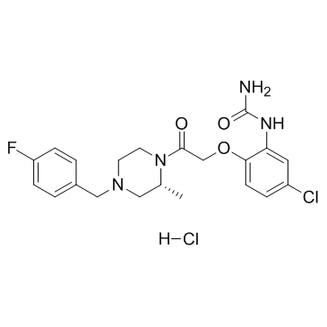
BX471 hydrochloride (Synonyms: ZK-811752 hydrochloride) |
Catalog No.GC35570
 One-Click Copy Product Info
|

El clorhidrato de BX471 (clorhidrato de ZK-811752) es un potente y selectivo antagonista de CCR1 no peptÍdico con Ki de 1 nM para CCR1 humano, y exhibe una selectividad de 250 veces para CCR1 sobre CCR2, CCR5 y CXCR4.
Products are for research use only. Not for human use. We do not sell to patients.
Cas No.: 288262-96-4
Sample solution is provided at 25 µL, 10mM.
_1-3.$$$39978408@51.jpg');)
_1-9.$$$38538795@65.jpg');)
_1-9.$$$39112701@51.jpg');)
_1-7.$$$37316672@70.jpg');)
_366-372.$$$36198801@70.jpg');)
.$$$38565142@65.jpg');)
_1867-1883.$$$33248023@67.jpg');)
_eads6055.$$$39977546@45.jpg');)
_eadm9805.$$$40080571@45.jpg');)
_eadj3020.$$$39666821@45.jpg');)
_1-20.$$$39757301@28.jpg');)
_1-24.$$$38898113@28.jpg');)
_273-287.$$$36806353@46.jpg');)
_1-16.$$$37156877@44.jpg');)
_1-19.$$$37460805@44.jpg');)
_1291-1307.$$$34518654@46.jpg');)
BX471 hydrochloride (ZK-811752 hydrochloride) is a potent, selective non-peptide CCR1 antagonist with Ki of 1 nM for human CCR1, and exhibits 250-fold selectivity for CCR1 over CCR2, CCR5 and CXCR4. MIP-1α-CCR1|1 nM (Ki)|RANTES-CCR1|2.8 nM (Ki)|MCP-3-CCR1|5.5 nM (Ki)
BX471 is a potent functional antagonist based on its ability to inhibit a number of CCR1-mediated effects including Ca2+ mobilization, increase in extracellular acidification rate, CD11b expression, and leukocyte migration. BX471 demonstrats a greater than 10,000-fold selectivity for CCR1 compared with 28 G-protein-coupled receptors[1]. BX471 is also able to displace 125I-MIP-1α/CCL3 binding to mouse CCR1 in a concentration-dependent manner with a Ki of 215±46 nM. Increasing concentrations of BX471 inhibits the Ca2+ transients induced by MIP-1α/CCL3 in both human and mouse CCR1 with IC50 of 5.8±1 nM and 198±7 nM, respectively[2]. BX471 (0.1-10 μM) shows a dose-dependent inhibition of RANTES-mediated and shear-resistant adhesion on IL-1β-activated microvascular endothelium in shear flow in isolated blood monocytes. BX471 also inhibits the RANTES-mediated adhesion of T lymphocytes to activated endothelium[4].
BX471 (4 mg/kg, p.o. or i.v.) is orally active with a bioavailability of 60% in dogs. Furthermore, BX471 effectively reduces disease in a rat experimental allergic encephalomyelitis model of multiple sclerosis[1]. BX471 (20 mg/kg, s.c.) reaches peak plasma levels of 9 μM by around 30 minutes, and this rapidly declines to approximately 0.4 μM after 2 hours. From 4 to 8 hours the drug plasma levels drops to 0.1 μM or lower. Mice treated with 20 mg/kg of BX471 for 10 days shows a reduction of interstitial CD45 positive leukocytes of approximately 55%. BX471 has a borderline significant effect on the number of CCR5-positive CD8 cells in the peripheral blood. BX471 reduces the amount of FSP1-positive cells by 65% in UUO kidneys as compared with vehicle control[2]. Pretreatment witih BX471 reduces macrophage and neutrophil accumulation in kidney after ischemia-reperfusion injury[3].
[1]. Liang M, et al. Identification and characterization of a potent, selective, and orally active antagonist of the CC chemokine receptor-1. J Biol Chem. 2000 Jun 23;275(25):19000-8. [2]. Anders HJ, et al. A chemokine receptor CCR-1 antagonist reduces renal fibrosis after unilateral ureter ligation. J Clin Invest. 2002 Jan;109(2):251-9. [3]. Furuichi K, et al. Chemokine receptor CCR1 regulates inflammatory cell infiltration after renal ischemia-reperfusion injury. J Immunol. 2008 Dec 15;181(12):8670-6. [4]. Horuk R, et al. A non-peptide functional antagonist of the CCR1 chemokine receptor is effective in rat heart transplant rejection. J Biol Chem. 2001 Feb 9;276(6):4199-204.
Cell experiment: | Briefly, dermal microvascular endothelial cells grown to confluence in Petri dishes are stimulated with IL-1β (10 ng/mL) for 12 h followed by pre-incubation with RANTES (10 nM) for 30 min at 37°C just prior to assay. The plates are assembled as the lower wall in a parallel wall flow chamber and mounted on the stage of an Olympus IMT-2 inverted microscope with ×20 and ×40 phase-contrast objectives. Isolated human blood monocytes are isolated and resuspended at 5×10⁵?cells/mL in assay buffer (HBSS) containing 10 mM?HEPES, pH 7.4 and 0.5% human serum albumin. Shortly before the assay, 1 mM Mg2+?and 1 mM?Ca2+?are added. The cell suspensions are kept in a heating block at 37°C during the assay and perfused into the flow chamber at a rate of 1.5 dyn/cm2?for 5 min. For inhibition experiments, monocytes are preincubated with BX471 at different concentrations (0.1-10 μM) or a Me2SO control for 10 min at 37°C. The number of firmLy adherent cells after 5 min is quantitated in multiple fields (at least five per experiment) by analysis of images recorded with a long integration JVC 3CCD video camera and a JVC SR L 900 E video recorder and are expressed as cells/mm2. The type of adhesion analyzed is restricted to primary,?i.e.?direct interactions of monocytes with endothelium. |
Animal experiment: | Fasted male beagle dogs (n=3 per treatment group) are given BX471 either by oral gavage or by intravenous injection via the cephalic vein at a dose of 4 mg/kg. The compound is dissolved in a vehicle of 40% aqueous cyclodextrin. Serial blood samples are collected utilizing an in-dwelling catheter in the jugular vein at the indicated time points up to 6 h post-dosing. EDTA is used as an anticoagulant. The samples are centrifuged (1000× g for 10 min at 4°C), and plasma is stored frozen until analyzed for drug levels by HPLC-MS (electrospray mode operated under a positive ion mode). Plasma samples are thawed and denatured by the addition of four parts of ice-cold methanol containing a fixed amount of an internal standard to one part of plasma. The resulting protein precipitate is removed by centrifugation at 5000× g, and the supernatants are analyzed directly. Concurrently plasma calibration standards of BX471 are prepared over the range of quantification, processed, and analyzed under identical conditions. A FISONS, VG Platform single quadrupole instrument is used in these analyses with an electrospray inlet operated at 3.57 kV. Chromatographic separation is accomplished using a YMC AQ octadecyl silane reversed phase column (4.6×250 mm) following a short isocratic elution method (35% methanol, 65% water containing 0.1% trifluoroacetic acid). The total column flow (1 mL/min) is split post-column to infuse 50 μL/min into the mass spectrometer. The chromatograms are collected over a total run time of 7.5 min/sample following a 50-μL injection on the column. The ions are collected in a single ion positive ionization mode. A calibration curve for quantification is generated by plotting ion current ratios between the internal standard peak and the analyte in the plasma standards over the quantification range. Calculations of percent oral availability is deduced from the area under curve measurements. Pharmacokinetic parameters are calculated using WinNonLin version 3.0. |
References: [1]. Liang M, et al. Identification and characterization of a potent, selective, and orally active antagonist of the CC chemokine receptor-1. J Biol Chem. 2000 Jun 23;275(25):19000-8. | |
| Cas No. | 288262-96-4 | SDF | |
| Sinónimos | ZK-811752 hydrochloride | ||
| Canonical SMILES | O=C(N)NC1=CC(Cl)=CC=C1OCC(N2[C@H](C)CN(CC3=CC=C(F)C=C3)CC2)=O.[H]Cl | ||
| Formula | C21H25Cl2FN4O3 | M.Wt | 471.35 |
| Solubility | DMSO: 150 mg/mL (318.23 mM) | Storage | Store at -20°C |
| General tips | Please select the appropriate solvent to prepare the stock solution according to the
solubility of the product in different solvents; once the solution is prepared, please store it in
separate packages to avoid product failure caused by repeated freezing and thawing.Storage method
and period of the stock solution: When stored at -80°C, please use it within 6 months; when stored
at -20°C, please use it within 1 month. To increase solubility, heat the tube to 37°C and then oscillate in an ultrasonic bath for some time. |
||
| Shipping Condition | Evaluation sample solution: shipped with blue ice. All other sizes available: with RT, or with Blue Ice upon request. | ||
| Prepare stock solution | |||

|
1 mg | 5 mg | 10 mg |
| 1 mM | 2.1216 mL | 10.6078 mL | 21.2157 mL |
| 5 mM | 424.3 μL | 2.1216 mL | 4.2431 mL |
| 10 mM | 212.2 μL | 1.0608 mL | 2.1216 mL |
Step 1: Enter information below (Recommended: An additional animal making an allowance for loss during the experiment)
 g
μL
g
μL
Step 2: Enter the in vivo formulation (This is only the calculator, not formulation. Please contact us first if there is no in vivo formulation at the solubility Section.)
Calculation results:
Working concentration: mg/ml;
Method for preparing DMSO master liquid: mg drug pre-dissolved in μL DMSO ( Master liquid concentration mg/mL, Please contact us first if the concentration exceeds the DMSO solubility of the batch of drug. )
Method for preparing in vivo formulation: Take μL DMSO master liquid, next addμL PEG300, mix and clarify, next addμL Tween 80, mix and clarify, next add μL saline, mix and clarify.
Method for preparing in vivo formulation: Take μL DMSO master liquid, next add μL Corn oil, mix and clarify.
Note: 1. Please make sure the liquid is clear before adding the next solvent.
2. Be sure to add the solvent(s) in order. You must ensure that the solution obtained, in the previous addition, is a clear solution before proceeding to add the next solvent. Physical methods such as vortex, ultrasound or hot water bath can be used to aid dissolving.
3. All of the above co-solvents are available for purchase on the GlpBio website.
Quality Control & SDS
- View current batch:
- Purity: >98.00% Appearance: A solid
- COA (Certificate of Analysis)
- SDS (Safety Data Sheet)
- Datasheet
Average Rating: 5 (Based on Reviews and 26 reference(s) in Google Scholar.)