

Coniferaldehyde |
Catalog No.GC38376
 One-Click Copy Product Info
|

El coniferaldehÍdo (ferulaldehÍdo) es un inductor eficaz de la hemooxigenasa-1 (HO-1).
Products are for research use only. Not for human use. We do not sell to patients.
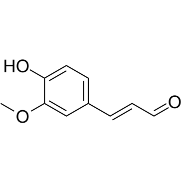
Cas No.: 458-36-6
Sample solution is provided at 25 µL, 10mM.
_1-3.$$$39978408@51.jpg');)
_1-9.$$$38538795@65.jpg');)
_1-9.$$$39112701@51.jpg');)
_1-7.$$$37316672@70.jpg');)
_366-372.$$$36198801@70.jpg');)
.$$$38565142@65.jpg');)
_1867-1883.$$$33248023@67.jpg');)
_eads6055.$$$39977546@45.jpg');)
_eadm9805.$$$40080571@45.jpg');)
_eadj3020.$$$39666821@45.jpg');)
_1-20.$$$39757301@28.jpg');)
_1-24.$$$38898113@28.jpg');)
_273-287.$$$36806353@46.jpg');)
_1-16.$$$37156877@44.jpg');)
_1-19.$$$37460805@44.jpg');)
_1291-1307.$$$34518654@46.jpg');)
Coniferaldehyde is a natural phenolic compound with potent anti-inflammatory activity[1]. Coniferaldehyde reduces hemolytic activity, protease hydrolysis, and bacterial invasion ability by regulating the transcription of main virulence factors, and leads to decreased resistance of S. aureus to adverse stimuli, including heat, acidic pH, high osmotic environment, hydrogen peroxide, and NaClO stress assays[2]. Coniferaldehyde has been widely used in C. elegans models to activate autophagy and enhance oxidative stress resistance[3].
In vitro, Coniferaldehyde treatment (50µM) for 6 hours promoted the neurite growth of Neuro2a cells and increased the expression of phosphorylated ERK[4]. 5µM of Coniferaldehyde pretreatment for 1 hour inhibited the production of NO and cell death induced by lipopolysaccharide (LPS) in RAW 264.7 cells, induced nuclear localization of Nrf-2, and facilitated the expression of HO-1[5]. Incubation with 100µM Coniferaldehyde for 24 hours reduced lipid accumulation in HepG2 cells induced by palmitic acid (PA) and enhanced glucose uptake and intracellular glycogen content[6].
In vivo, Coniferaldehyde treatment via oral administration at a dose of 0.2mmol/kg/day for 3 months effectively abolished Aβ deposits in the brain, reduced the levels of toxic soluble Aβ peptides, and improved the learning and memory abilities in an Alzheimer's disease mouse model[7]. Intraperitoneal injection of Coniferaldehyde (0.05mmol/kg/day) for 6 weeks alleviated cartilage damage and inhibited cartilage degeneration in the experimental osteoarthritis (OA) mouse model[8]. Continuous intraperitoneal injection of Coniferaldehyde at a dose of 20mg/kg/day for 6 consecutive days reversed 3-nitropropionic acid-induced neurological dysfunction and improved motor ability in mice[9].
References:[1] He Y, Li Q, Zhou W, et al. Coniferyl aldehyde alleviates LPS-induced WI-38 cell apoptosis and inflammation injury via JAK2-STAT1 pathway in acute pneumonia[J]. Allergologia et Immunopathologia, 2021, 49(5): 72-77.
[2] Li S, Zhang Y, Wang J, et al. Discovery of coniferaldehyde as an inhibitor of caseinolytic protease to combat Staphylococcus aureus infections[J]. Molecular Medicine, 2025, 31(1): 249.
[3] Ramatchandirane M, Rajendran P, Athira M P, et al. Coniferaldehyde activates autophagy and enhances oxidative stress resistance and lifespan of Caenorhabditis elegans via par-4/aak-2/skn-1 pathway[J]. Biogerontology, 2025, 26(1): 25.
[4] Jeon J, Kwon H, Cho E, et al. The effect of coniferaldehyde on neurite outgrowth in neuroblastoma Neuro2a cells[J]. Neurochemistry international, 2019, 131: 104579.
[5] Kim K M, Heo D R, Kim Y A, et al. Coniferaldehyde inhibits LPS-induced apoptosis through the PKC α/β II/Nrf-2/HO-1 dependent pathway in RAW264. 7 macrophage cells[J]. Environmental Toxicology and Pharmacology, 2016, 48: 85-93.
[6] Gai H, Zhou F, Zhang Y, et al. Coniferaldehyde ameliorates the lipid and glucose metabolism in palmitic acid‐induced HepG2 cells via the LKB1/AMPK signaling pathway[J]. Journal of food science, 2020, 85(11): 4050-4060.
[7] Dong Y, Stewart T, Bai L, et al. Coniferaldehyde attenuates Alzheimer's pathology via activation of Nrf2 and its targets[J]. Theranostics, 2020, 10(1): 179.
[8] Cai D, Wang J, Chen S, et al. Coniferaldehyde prevents articular cartilage destruction in a murine model via Nrf2/HO-1 pathway[J]. Molecular medicine reports, 2021, 23(3): 224.
[9] Ibiayo A G, Varinthra P, Nagarajan M, et al. Coniferaldehyde reverses 3-nitropropionic acid-induced Huntington’s disease pathologies via PKM2 restoration and JAK2/STAT3 inhibition[J]. Molecular Medicine, 2025, 31(1): 271.
| Cell experiment [1]: | |
|
Cell lines |
RAW 264.7 cells |
|
Preparation Method |
RAW 264.7 cells were cultured in DMEM medium, supplemented with 5% fetal bovine serum, 2mM glutamine, and 1% penicillin-streptomycin, at 37°C in an incubator with 5% CO2. Cells were seeded at a density of approximately 6×10³ cells per well in 6-well plates overnight and were pretreated with different concentrations of Coniferaldehyde (0, 0.1, 0.5, 1, 2, and 5µM) for 1h, and then stimulated by LPS (200ng/ml) for 24h, the NO production was measured. |
|
Reaction Conditions |
0, 0.1, 0.5, 1, 2, and 5µM; 1h |
|
Applications |
Coniferaldehyde treatment markedly decreased LPS-induced NO production in RAW 264.7 cells in a dose-dependent manner. |
| Animal experiment [2]: | |
|
Animal models |
Male C57BL/6 mice |
|
Preparation Method |
Male C57BL/6 mice (8 weeks old; 26.40±3.92g) were housed in a normal atmosphere under a 12h light-dark cycle at 24°C with 50% humidity and provided with standard laboratory food and water ad libitum. Mice were divided into four groups [sham surgery+saline, sham surgery+Coniferaldehyde, destabilization of the medial meniscus (DMM) surgery+saline, and DMM surgery+Coniferaldehyde). OA was induced by DMM surgery on the right knee joints. The delivery of Coniferaldehyde (0.05mmol/kg/day) was performed via i.p. at 2 weeks following surgery. The mice were sacrificed at 8 weeks post-OA surgery for histological assessment. |
|
Dosage form |
0.05mmol/kg/day; 6 weeks; i.p. |
|
Applications |
Coniferaldehyde treatment alleviated cartilage destruction and suppressed cartilage degradation in OA mice. |
| References: [1] Kim K M, Heo D R, Kim Y A, et al. Coniferaldehyde inhibits LPS-induced apoptosis through the PKC α/β II/Nrf-2/HO-1 dependent pathway in RAW264. 7 macrophage cells[J]. Environmental Toxicology and Pharmacology, 2016, 48: 85-93. [2] Cai D, Wang J, Chen S, et al. Coniferaldehyde prevents articular cartilage destruction in a murine model via Nrf2/HO-1 pathway[J]. Molecular medicine reports, 2021, 23(3): 224. | |
| Cas No. | 458-36-6 | SDF | |
| Canonical SMILES | O=C/C=C/C1=CC=C(O)C(OC)=C1 | ||
| Formula | C10H10O3 | M.Wt | 178.18 |
| Solubility | DMSO: 120 mg/mL (673.48 mM) | Storage | 4°C, protect from light |
| Shipping Condition | Evaluation sample solution: shipped with blue ice. All other sizes available: with RT, or with Blue Ice upon request. | ||
| Prepare stock solution | |||

|
1 mg | 5 mg | 10 mg |
| 1 mM | 5.6123 mL | 28.0615 mL | 56.123 mL |
| 5 mM | 1.1225 mL | 5.6123 mL | 11.2246 mL |
| 10 mM | 561.2 μL | 2.8062 mL | 5.6123 mL |
Step 1: Enter information below (Recommended: An additional animal making an allowance for loss during the experiment)
 g
μL
g
μL
Step 2: Enter the in vivo formulation (This is only the calculator, not formulation. Please contact us first if there is no in vivo formulation at the solubility Section.)
Calculation results:
Working concentration: mg/ml;
Method for preparing DMSO master liquid: mg drug pre-dissolved in μL DMSO ( Master liquid concentration mg/mL, Please contact us first if the concentration exceeds the DMSO solubility of the batch of drug. )
Method for preparing in vivo formulation: Take μL DMSO master liquid, next addμL PEG300, mix and clarify, next addμL Tween 80, mix and clarify, next add μL saline, mix and clarify.
Method for preparing in vivo formulation: Take μL DMSO master liquid, next add μL Corn oil, mix and clarify.
Note: 1. Please make sure the liquid is clear before adding the next solvent.
2. Be sure to add the solvent(s) in order. You must ensure that the solution obtained, in the previous addition, is a clear solution before proceeding to add the next solvent. Physical methods such as vortex, ultrasound or hot water bath can be used to aid dissolving.
3. All of the above co-solvents are available for purchase on the GlpBio website.
Quality Control & SDS
- View current batch:
- Purity: >99.50% Appearance: A solid
- COA (Certificate of Analysis)
- SDS (Safety Data Sheet)
- Datasheet
Average Rating: 5 (Based on Reviews and 24 reference(s) in Google Scholar.)