

Hypaphorine (Synonyms: L-Hypaphorine, (+)-Tryptophan, Tryptophan Betaine) |
رقم الكتالوجGC38797
 One-Click Copy Product Info
|

Hypaphorine عبارة عن قلويد إندولي معزول من Pisolithus tinctorius ، وله تأثيرات عصبية وخفض الجلوكوز في القوارض
Products are for research use only. Not for human use. We do not sell to patients.
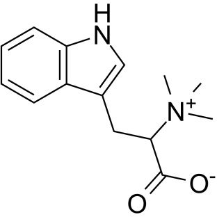
Cas No.: 487-58-1
Sample solution is provided at 25 µL, 10mM.
_1-3.$$$39978408@51.jpg');)
_1-9.$$$38538795@65.jpg');)
_1-9.$$$39112701@51.jpg');)
_1-7.$$$37316672@70.jpg');)
_366-372.$$$36198801@70.jpg');)
.$$$38565142@65.jpg');)
_1867-1883.$$$33248023@67.jpg');)
_eads6055.$$$39977546@45.jpg');)
_eadm9805.$$$40080571@45.jpg');)
_eadj3020.$$$39666821@45.jpg');)
_1-20.$$$39757301@28.jpg');)
_1-24.$$$38898113@28.jpg');)
_273-287.$$$36806353@46.jpg');)
_1-16.$$$37156877@44.jpg');)
_1-19.$$$37460805@44.jpg');)
_1291-1307.$$$34518654@46.jpg');)
Hypaphorine, an indole alkaloid extracted from leguminous plants, exerts anti-inflammatory, antioxidant, and neuroprotective effects by modulating multiple signaling pathways, including PI3K/Akt/mTOR and p38/JNK. Hypaphorine is frequently investigated in studies related to neurodegenerative diseases and metabolic disorders[1-2]. Additionally, Hypaphorine has been shown to effectively prevent osteoporosis[3], and exhibits antiviral activity against Dengue virus[4].
In vitro, pretreatment of human microvascular endothelial cells (HMEC-1) with Hypaphorine (0–100μM) for 6 hours, followed by lipopolysaccharide (LPS, 500ng/mL) stimulation for 48 hours, significantly suppressed LPS-induced expression of pro-inflammatory cytokines (TNF-α, IL-1β, MCP-1, VCAM-1). Hypaphorine also reversed LPS-induced upregulation of TLR4 protein and mRNA levels and downregulation of PPAR-γ. Furthermore, it blocked LPS-activated PI3K/Akt/mTOR signaling[5]. In RAW 264.7 macrophages, Hypaphorine (12.5–50μM) pretreatment for 1 hour, followed by LPS (1μg/mL) stimulation for 24 hours, markedly inhibited LPS-induced pro-inflammatory cytokine (TNF-α, IL-1β, IL-6, IL-10, MCP-1) expression and NO/PGE₂ release, while downregulating COX-2 and iNOS protein expression. Hypaphorine also suppressed LPS-induced phosphorylation of ERK, IκBα, IKKβ, and NF-κB, and inhibited NF-κB nuclear translocation[6].
In vivo, Hypaphorine (10mg/kg; intraperitoneal injection) administered 2 hours after LPS (20mg/kg) significantly attenuated LPS-induced acute lung injury (ALI) in male Sprague-Dawley (SD) rats. Hypaphorine reduced lung histopathological damage, decreased total cell counts and neutrophil proportions in bronchoalveolar lavage fluid (BALF), and lowered lung wet/dry weight ratio, microvascular permeability, and lactate dehydrogenase (LDH)/myeloperoxidase (MPO) activity. Mechanistically, Hypaphorine upregulated DUSP1 expression and inhibited p38/JNK activation, thereby suppressing pro-inflammatory cytokines (TNF-α, IL-6, IL-1β, IL-18) [7]. Hypaphorine (10mg/kg; intraperitoneal injection) was found to selectively inhibit acetylcholinesterase (AChE) activity across all brain regions (cortex, cerebellum, striatum, hippocampus) in normal rat after 24 hours, with the D-enantiomer demonstrating potent inhibition in key brain areas[8].
References:
[1] Qin P, Li Y, Su Y, et al. Bifidobacterium adolescentis-derived hypaphorine alleviates acetaminophen hepatotoxicity by promoting hepatic Cry1 expression. J Transl Med. 2024 May 31;22(1):525.
[2] Chen H, Guo T, Wang D, et al. Vaccaria hypaphorine impairs RANKL-induced osteoclastogenesis by inhibition of ERK, p38, JNK and NF-κB pathway and prevents inflammatory bone loss in mice. Biomed Pharmacother. 2018 Jan;97:1155-1163.
[3] Zhang T, Yan Y, Xue Y, et al. Pharmacokinetic Study of Hypaphorine, a Potential Agent for Treating Osteoclast-based Bone Loss, on Rats Using LC-MS/MS. Comb Chem High Throughput Screen. 2022;25(11):1889-1896.
[4] Bai S, Liang H, Jiang W, et al. Identifying hypaphorine as a novel antiviral compound against dengue virus. Antiviral Res. 2025 Jun 24;240:106220.
[5] Sun H, Zhu X, Cai W, et al. Hypaphorine Attenuates Lipopolysaccharide-Induced Endothelial Inflammation via Regulation of TLR4 and PPAR-γ Dependent on PI3K/Akt/mTOR Signal Pathway. Int J Mol Sci. 2017 Apr 17;18(4):844.
[6] Sun H, Cai W, Wang X, et al. Vaccaria hypaphorine alleviates lipopolysaccharide-induced inflammation via inactivation of NFκB and ERK pathways in Raw 264.7 cells. BMC Complement Altern Med. 2017 Feb 20;17(1):120.
[7] Ding YH, Miao RX, Zhang Q. Hypaphorine exerts anti-inflammatory effects in sepsis induced acute lung injury via modulating DUSP1/p38/JNK pathway. Kaohsiung J Med Sci. 2021 Oct;37(10):883-893.
[8] Yonekawa MKA, Penteado BB, Dal'Ongaro Rodrigues A, et al. l-Hypaphorine and d-hypaphorine: Specific antiacetylcholinesterase activity in rat brain tissue. Bioorg Med Chem Lett. 2021 Sep 1;47:128206.
| Cell experiment [1]: | |
Cell lines | Human microvascular endothelial cells (HMEC-1) |
Preparation Method | HMEC-1 cells were seeded into 6-well plates at a density of 5×10⁴ cells per well and cultured until 80% confluence. Cells were pretreated with Hypaphorine (0, 25, 50, or 100μM) for 6 hours, followed by lipopolysaccharide (LPS, 500ng/mL) stimulation for an additional 48 hours to establish the endothelial inflammation model. |
Reaction Conditions | Hypaphorine (0–100μM); 6-hour pretreatment+48-hour LPS stimulation. |
Applications | Hypaphorine downregulated pro-inflammatory cytokines (TNF-α, IL-1β, MCP-1, VCAM-1) at both mRNA and protein levels, inhibited PI3K/Akt/mTOR pathway activation, and blocked NF-κB nuclear translocation and ERK phosphorylation. |
| Animal experiment [2]: | |
Animal models | Sprague-Dawley rats |
Preparation Method | Lipopolysaccharide (LPS, 20mg/kg) was administered intratracheally to induce sepsis-associated acute lung injury (ALI). Hypaphorine (10mg/kg) was injected intraperitoneally 2 hours post-LPS. Control groups received saline or LPS alone. Rats were sacrificed after 24 hours for tissue and fluid collection. |
Dosage form | 10mg/kg; i.p. |
Applications | Hypaphorine exerted anti-inflammatory and anti-apoptotic effects by upregulating DUSP1 to inactivate the p38/JNK pathway, thereby mitigating LPS-induced ALI in rats. |
References: | |
| Cas No. | 487-58-1 | SDF | |
| المرادفات | L-Hypaphorine, (+)-Tryptophan, Tryptophan Betaine | ||
| Canonical SMILES | C[N+](C)(C(C([O-])=O)CC1=CNC2=C1C=CC=C2)C | ||
| Formula | C14H18N2O2 | M.Wt | 246.3 |
| الذوبان | H2O : 100 mg/mL (406.01 mM; Need ultrasonic); DMSO : < 1 mg/mL (insoluble or slightly soluble) | Storage | Store at -20°C,protect from light |
| General tips | Please select the appropriate solvent to prepare the stock solution according to the
solubility of the product in different solvents; once the solution is prepared, please store it in
separate packages to avoid product failure caused by repeated freezing and thawing.Storage method
and period of the stock solution: When stored at -80°C, please use it within 6 months; when stored
at -20°C, please use it within 1 month. To increase solubility, heat the tube to 37°C and then oscillate in an ultrasonic bath for some time. |
||
| Shipping Condition | Evaluation sample solution: shipped with blue ice. All other sizes available: with RT, or with Blue Ice upon request. | ||
| Prepare stock solution | |||

|
1 mg | 5 mg | 10 mg |
| 1 mM | 4.0601 mL | 20.3004 mL | 40.6009 mL |
| 5 mM | 812 μL | 4.0601 mL | 8.1202 mL |
| 10 mM | 406 μL | 2.03 mL | 4.0601 mL |
Step 1: Enter information below (Recommended: An additional animal making an allowance for loss during the experiment)
 g
μL
g
μL
Step 2: Enter the in vivo formulation (This is only the calculator, not formulation. Please contact us first if there is no in vivo formulation at the solubility Section.)
Calculation results:
Working concentration: mg/ml;
Method for preparing DMSO master liquid: mg drug pre-dissolved in μL DMSO ( Master liquid concentration mg/mL, Please contact us first if the concentration exceeds the DMSO solubility of the batch of drug. )
Method for preparing in vivo formulation: Take μL DMSO master liquid, next addμL PEG300, mix and clarify, next addμL Tween 80, mix and clarify, next add μL saline, mix and clarify.
Method for preparing in vivo formulation: Take μL DMSO master liquid, next add μL Corn oil, mix and clarify.
Note: 1. Please make sure the liquid is clear before adding the next solvent.
2. Be sure to add the solvent(s) in order. You must ensure that the solution obtained, in the previous addition, is a clear solution before proceeding to add the next solvent. Physical methods such as vortex, ultrasound or hot water bath can be used to aid dissolving.
3. All of the above co-solvents are available for purchase on the GlpBio website.
Quality Control & SDS
- View current batch:
- Purity: >99.50% Appearance: A solid
- COA (Certificate of Analysis)
- SDS (Safety Data Sheet)
- Datasheet
Average Rating: 5 (Based on Reviews and 23 reference(s) in Google Scholar.)