Bafilomycin A1 (Synonyms: NSC 381866) |
رقم الكتالوجGC17597 |
البافيلومايسين A1، وهو مضاد حيوي ماكروليد (معزول) مأخوذ من سلالة الستريبتوميسيس، هو مثبط لأدينوزين ثلاثي الفوسفات في الفجوة الحبيبية (V-ATPase).
Products are for research use only. Not for human use. We do not sell to patients.
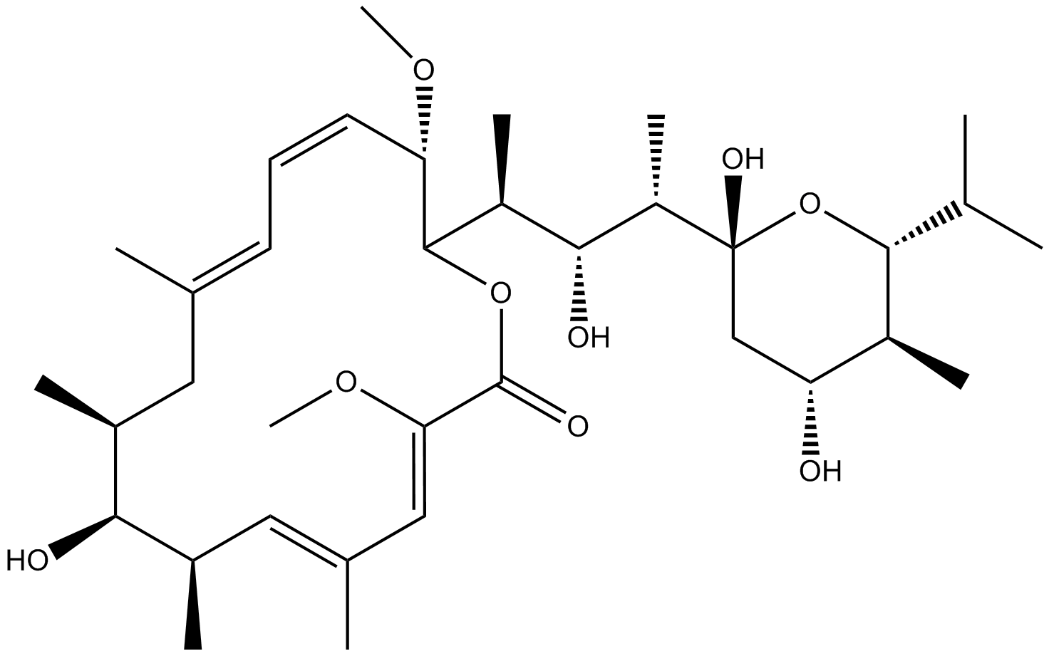
Cas No.: 88899-55-2
Sample solution is provided at 25 µL, 10mM.
Bafilomycin A1, a macrolide antibiotic isolated from the Streptomyces species, is an inhibitor of vacuolar H ATPase (V-ATPase). Bafilomycin A1 has been used to study the autophagy as an inhibitor of autophagosomes-lysosomes fusion and as an inhibitor of lysosomal degradation.[1] It binds to the V0 sector subunit c of the V-ATPase complex and inhibits H translocation which cause an accumulation of H in the cytoplasm of treated cells. Bafilomycin A1 inhibits cell growth and leads to apoptosis and differentiation. These anticancer effects of Bafilomycin A1 are considered to be attributable to the intracellular acidosis caused by V-ATPase inhibition.[2]
In vitro study of Bafilomycin A1 indicated that Bafilomycin A1 preferentially inhibits in vitro growth of pediatric B-cell acute lymphoblastic leukemia cells. A various of leukemia cell lines including 697, Nalm-6, RS4;11, NB4, HL-60, K562 and BV173 representing B-ALL (697, Nalm-6, RS4;11), acute myeloid leukemia (NB4, HL-60), and chronic myeloid leukemia (K562, BV173) were used to investigate the effect of Bafilomycin A1. Cells were cultured in the presence of increasing concentrations of Bafilomycin A1 (0 nM, 0.5 nM, 1 nM). Cell proliferation was measured using an MTT assay. Results showed that Bafilomycin A1 has frequently been used as an inhibitor to block the autophagosomes-lysosomes fusion, or to inhibit lysosomal activity at high doses (0.1–1 mμ). While low concentration of Bafilomycin A1 (1 nM) cpuld effectively and specifically inactivate autophagy and activate apoptosis at multiple targets in pediatric B-ALL cells.[3]
In vivo study demonstrated that Bafilomycin A1 can extend survival in the Bafilomycin A1-treated B-ALL mice with advanced disease compared with control mice. The red blood cell counts, hemoglobin concentration, and platelet counts in the B-ALL model group were extremely reduced, however they were normalized in the Bafilomycin A1-treated group. The mice in the B-ALL model group had severe hepatosplenomegaly compared with the control group. However, the size and weight of livers and spleens in the Bafilomycin A1-treated group were normalized compared with those in the disease model group. Results showed that Bafilomycin A1 dramatically inhibits pediatric B-ALL cell engraftment by targeting leukemia cells in vivo.[3]
References:
[1]. Yamamoto A, et al. Bafilomycin A1 prevents maturation of autophagic vacuoles by inhibiting fusion between autophagosomes and lysosomes in rat hepatoma cell line, H-4-II-E cells. Cell Struct Funct. 1998;23(1):33–42.
[2] Bowman EJ, et al. The bafilomycin/concanamycin binding site in subunit c of the V-ATPases from Neurospora crassa and Saccharomyces cerevisiae. J Biol Chem. 2004;279(32):33131–33138.
[3]. Yuan N, et al. Bafilomycin A1 targets both autophagy and apoptosis pathways in pediatric B-cell acute lymphoblastic leukemia. Haematologica. 2015 Mar;100(3):345-56.
Cell experiment [1, 2]: | |
Cell lines |
Eosinophils |
Preparation Method |
Circulating eosinophils were purified from donors with eosinophils >35 per μL of blood. Eosinophil preparations with purity >98% and viability ~98% were used. |
Reaction Conditions |
Eosinophils were cultured at 1 × 10⁶/mL in complete medium (RPMI 1640 plus 10% fetal bovine serum) with IL3 (2 ng/mL) or IL5 (2 ng/mL) both cytokines from BD Biosciences for 20 h, and were then washed and cultured with complete medium (no IL3 or IL5) on coated heat-aggregated immunoglobulins-G (HA-IgG or IgG) or without IgG as previously described. |
Applications |
Bafilomycin-A1 could be used to show the impact of autophagolysosome formation on eosinophil cytolysis. Treated IL3- and IL5-primed eosinophils with bafilomycin-A1 before interaction with IgG, and then measured cytolysis using rhodamine-110 (R110). Treatment with bafilomycin-A1 increased IL3-primed and IL5-primed eosinophil lysis by 50% and 100%, respectively, suggesting that autophagolysosome formation protects from cytolysis for both IL3- and IL5-primed eosinophils. |
Animal experiment [3]: | |
Animal models |
Lck-mTOR + CLP mice, WT + CLP mice, Lck-TSC1 mice |
Preparation Method |
Crossed TSC1loxp/loxp and mTORloxp/loxp mice with Lck-Cre mice (Cre expression under the control of lymphocyte-specific protein tyrosine kinase, Lck) to obtain the F1 generation Lck-Cre:mTORloxp/− mice and Lck-Cre:TSC1loxp/− mice, respectively. We interbred F1 generation mice in each group and obtained F2 generation Lck-Cre: mTORloxp/loxp and Lck-Cre;TSC1loxp/loxp mice. Littermates lacking Lck-Cre served as controls. Then mice were anesthetized, and the abdomen was opened surgically and the cecum was ligated halfway between the distal pole and the base. The cecal stump was punctured once with a 22-gauge needle. Put back the cecum and closed the abdomen. Administered 1 mL saline subcutaneously to each animal for fluid resuscitation. Animals in the sham group underwent the same surgical procedure without ligation or puncture. Bafilomycin A1 was administered intraperitoneally 1 h after the CLP operation. |
Dosage form |
1 mg/kg |
Applications |
Bafilomycin A1 could specifically block fusion of autophagosomes and lysosomes and attenuate the protective effects of mTOR deletion against CD4 + T cell apoptosis. In Lck-mTOR + CLP mice treated with bafilomycin A1, the apoptosis rate of CD4 + T cells was higher compared with mice that did not receive bafilomycin A1 and lower compared with mice in WT + CLP + bafilomycin A1 group. |
References: |
Cas No. | 88899-55-2 | SDF | |
المرادفات | NSC 381866 | ||
Chemical Name | (3Z,5E,7R,8S,9S,11E,13E,15S,16R)-16-[(2S,3R,4S)-4-[(2R,4R,5S,6R)-2,4-dihydroxy-5-methyl-6-propan-2-yloxan-2-yl]-3-hydroxypentan-2-yl]-8-hydroxy-3,15-dimethoxy-5,7,9,11-tetramethyl-1-oxacyclohexadeca-3,5,11,13-tetraen-2-one | ||
Canonical SMILES | CC1CC(=CC=CC(C(OC(=O)C(=CC(=CC(C1O)C)C)OC)C(C)C(C(C)C2(CC(C(C(O2)C(C)C)C)O)O)O)OC)C | ||
Formula | C35H58O9 | M.Wt | 622.84 |
الذوبان | 5 mg/ml in DMSO or in menthanol | Storage | Desiccate at -20°C, protect from light |
General tips | Please select the appropriate solvent to prepare the stock solution according to the
solubility of the product in different solvents; once the solution is prepared, please store it in
separate packages to avoid product failure caused by repeated freezing and thawing.Storage method
and period of the stock solution: When stored at -80°C, please use it within 6 months; when stored
at -20°C, please use it within 1 month. To increase solubility, heat the tube to 37°C and then oscillate in an ultrasonic bath for some time. |
||
Shipping Condition | Evaluation sample solution: shipped with blue ice. All other sizes available: with RT, or with Blue Ice upon request. |
Prepare stock solution | |||
![]() |
1 mg | 5 mg | 10 mg |
1 mM | 1.6055 mL | 8.0277 mL | 16.0555 mL |
5 mM | 0.3211 mL | 1.6055 mL | 3.2111 mL |
10 mM | 0.1606 mL | 0.8028 mL | 1.6055 mL |
Step 1: Enter information below (Recommended: An additional animal making an allowance for loss during the experiment)
Step 2: Enter the in vivo formulation (This is only the calculator, not formulation. Please contact us first if there is no in vivo formulation at the solubility Section.)
Calculation results:
Working concentration: mg/ml;
Method for preparing DMSO master liquid: mg drug pre-dissolved in μL DMSO ( Master liquid concentration mg/mL, Please contact us first if the concentration exceeds the DMSO solubility of the batch of drug. )
Method for preparing in vivo formulation: Take μL DMSO master liquid, next addμL PEG300, mix and clarify, next addμL Tween 80, mix and clarify, next add μL ddH2O, mix and clarify.
Method for preparing in vivo formulation: Take μL DMSO master liquid, next add μL Corn oil, mix and clarify.
Note: 1. Please make sure the liquid is clear before adding the next solvent.
2. Be sure to add the solvent(s) in order. You must ensure that the solution obtained, in the previous addition, is a clear solution before proceeding to add the next solvent. Physical methods such as vortex, ultrasound or hot water bath can be used to aid dissolving.
3. All of the above co-solvents are available for purchase on the GlpBio website.
Quality Control & SDS
- View current batch:
- Purity: >95.00%
- COA (Certificate Of Analysis)
- SDS (Safety Data Sheet)
- Datasheet
-
Related Biological Data
Autophagy is activated in GLY-treated TM3 cells. (A) Cells were incubated with 1 mM GLY for 24 h and then the protein levels of LC3-Ⅱ and ATG5 were determined by immunoblotting (n = 3). (B) Cells were pretreated with BA for 4 h and then treated with GLY for 24 h to detect the protein levels of LC3-Ⅱ (n = 3).
Cells were pretreated with BA for 4 h and then treated with GLY for 24 h to detect the protein levels of LC3-Ⅱ (n = 3).
Science of The Total Environment (2024): 169927. PMID: 38199345 IF: 9.8003 -
Related Biological Data
Mitophagy is activated in low-dose GLY-treated TM3 cells.Cells were pretreated with 25 nM BA for 4 h, and then incubated with 10 μM GLY for another 24 h to analyze the protein levels of HSP60, COX IV and LC3.
Cells were pretreated with 25 nM BA for 4 h, and then incubated with 10 μM GLY for another 24 h to analyze the protein levels of HSP60, COX IV and LC3.
Environmental Pollution 314 (2022): 120314. PMID: 36183875 IF: 8.9 -
Related Biological Data
The enhanced autophagic activity promoted NLRP3 degradation. (E) The levels of NLRP3 and cleaved Casp1 (p10) in BMDMs after treatment with LPS/ATP/RAPA in presence or absence of BafA1 determined by western blot. n=4.
To further evaluate the effect of autophagy on corneal transplantation, other autophagy inducers 5-Aminoimidazole-4-carboxamide ribonucleotide (AICAR, 6.5 µg/µL, 6µL, MCE) and inhibitor Bafilomycin-A1 (BafA1, 0.25 µg/µL, 6µL, GLPBIO) was subconjunctivally injected into the recipient mice.
American Journal of Transplantation. PMID: 35092164 IF: 8.086 -
Related Biological Data
Knockout of TNPO3 does not interfere with endocytosis and acidification of IAV. (C). Baf-A1 was used as a specific inhibitor of vacuolar-type H(+)- ATPase to inhibit acidification and protein degradation in lysosomes of cultured cells. WT cells and TNPO3 KO cells were incubated either with or without Baf-A1.
The cells were treated with Bafilomycin A1 (100 nM) or DMSO at 37 ◦C in an incubator for 2 h and then washed with PBS three times.
Int J Mol Sci 23.8 (2022): 4128. PMID: 35456945 IF: 5.9238 -
Related Biological Data
RGS20 promotes NSCLC cell proliferation and activates autophagy in vitro. CCK-8 assay was performed to assess the proliferation of H1299 and Anip973 cells in the RGS20-OE group treated with Baf-A1 (50 nM, 2 h).
Representative western blot results show the protein levels of autophagy related proteins after blockage of autophagy flux by Baf-A1 (50 nM, 2 h) in three groups (CTRL, RGS20-KD, and RGS20-OE) of H1299 and Anip973 cells.
Cancer Cell International 24.1 (2024): 1-13. PMID: 38431606 IF: 5.8 -
Related Biological Data
Resveratrol controlled autophagosome-lysosome fusion. (A–C) The protein levels of P62 and Lc3B II/I were detected usingWestern blotting and normalized to GAPDH (n = 3).
Luteinized granulosa cells were treated with 100 nM Baf A1 with or without 200 µM H2O2 and 20 µM resveratrol, and the dose of Baf A1 was based on a previous study
Int J Mol Sci, 2023, 24(13): 10914. PMID: 37446088 IF: 5.5999
Average Rating: 5
(Based on Reviews and 39 reference(s) in Google Scholar.)GLPBIO products are for RESEARCH USE ONLY. Please make sure your review or question is research based.
Required fields are marked with *