

DPDPE |
رقم الكتالوجGC10333
 One-Click Copy Product Info
|

DPDPE ، ببتيد أفيوني ، هو ناهض انتقائي لمستقبلات أفيونية المفعول (DOR) مع تأثيرات مضادة للاختلاج
Products are for research use only. Not for human use. We do not sell to patients.
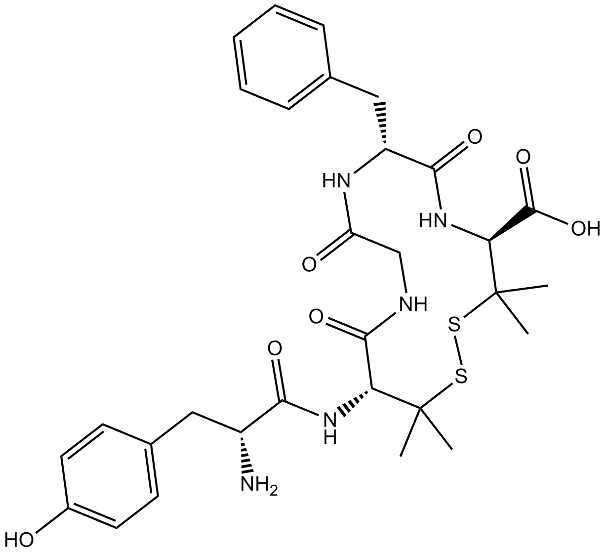
Cas No.: 88373-73-3
Sample solution is provided at 25 µL, 10mM.
_1-3.$$$39978408@51.jpg');)
_1-9.$$$38538795@65.jpg');)
_1-9.$$$39112701@51.jpg');)
_1-7.$$$37316672@70.jpg');)
_366-372.$$$36198801@70.jpg');)
.$$$38565142@65.jpg');)
_1867-1883.$$$33248023@67.jpg');)
_eads6055.$$$39977546@45.jpg');)
_eadm9805.$$$40080571@45.jpg');)
_eadj3020.$$$39666821@45.jpg');)
_1-20.$$$39757301@28.jpg');)
_1-24.$$$38898113@28.jpg');)
_273-287.$$$36806353@46.jpg');)
_1-16.$$$37156877@44.jpg');)
_1-19.$$$37460805@44.jpg');)
_1291-1307.$$$34518654@46.jpg');)
DPDPE is a selective and anticonvulsant δ-opioid receptor (DOR) agonist[1]. DOR is a G protein-coupled receptor (GPCR) primarily distributed in the central nervous system, which mediates various physiological effects such as analgesia and antidepressant actions of both endogenous enkephalins and exogenous ligands[2]. DPDPE is commonly used in studies involving opioid receptor signaling, pain pathways, and addiction mechanisms[3].
In vitro, treatment of HEK293 cells overexpressing or with knocked-down GRK2 with DPDPE (2μM) for 30min showed that GRK2 overexpression enhanced internalization and subsequent recycling of the wild-type receptor, whereas GRK2 knockdown inhibited these processes[4]. Pretreatment of HEK293 cells with DPDPE (1μM) for 60min induced only mild DOR desensitization, which was significantly enhanced in the presence of monensin[5]. Treatment of CHO cells stably expressing human DOR (CHO/DOR) with DPDPE (100nM) for 5-30min rapidly and significantly induced phosphorylation of glycogen synthase kinase-3β (GSK-3β) at Ser9, accompanied by a marked inhibition (~35%) of kinase activity[6].
In vivo, subcutaneous injection of DPDPE (0.5, 1, 3, 5mg/kg) in streptozotocin (STZ)-induced diabetic neuropathic mice produced dose-dependent inhibition of mechanical allodynia, thermal hyperalgesia, and thermal allodynia 30min after administration[7]. In Sprague Dawley rats, intrathecal pretreatment with DPDPE (100nM; 10μL) 10min before hindpaw compression significantly inhibited noxious stimulation-induced neurokinin 1 receptor (NK1R) internalization in lamina I of the spinal dorsal horn[8].
References:
[1] TORTELLA F C, ECHEVARRIA E, ROBLES L, et al. Anticonvulsant effects of mu (DAGO) and delta (DPDPE) enkephalins in rats[J]. Peptides, 1988, 9(5): 1177-1181.
[2] NIETO M M, GUEN S L E, KIEFFER B L, et al. Physiological control of emotion-related behaviors by endogenous enkephalins involves essentially the delta opioid receptors[J]. Neuroscience, 2005, 135(2): 305-313.
[3] ABDALLAH K, GENDRON L. The delta opioid receptor in pain control[J]. Delta Opioid Receptor Pharmacology and Therapeutic Applications. 2017: 147-177.
[4] ZHANG X, WANG F, CHEN X, et al. Post-endocytic fates of δ-opioid receptor are regulated by GRK2-mediated receptor phosphorylation and distinct β-arrestin isoforms[J]. Journal of Neurochemistry, 2008, 106(2): 781-792.
[5] AUDET N, CHARFI I, MNIE-FILALI O, et al. Differential association of receptor-Gβγ complexes with β-arrestin2 determines recycling bias and potential for tolerance of delta opioid receptor agonists[J]. Journal of Neuroscience, 2012, 32(14): 4827-4840.
[6] OLIANAS M C, DEDONI S, ONALI P. Signaling pathways mediating phosphorylation and inactivation of glycogen synthase kinase-3β by the recombinant human δ-opioid receptor stably expressed in Chinese hamster ovary cells[J]. Neuropharmacology, 2011, 60(7-8): 1326-1336.
[7] CASTANY S, CARCOLÉ M, LEÁNEZ S, et al. The antinociceptive effects of a δ-opioid receptor agonist in mice with painful diabetic neuropathy: Involvement of heme oxygenase 1[J]. Neuroscience Letters, 2016, 614: 49-54.
[8] KONDO I, MARVIZON J C G, SONG B, et al. Inhibition by spinal μ- and δ-opioid agonists of afferent-evoked substance P release[J]. Journal of Neuroscience, 2005, 25(14): 3651-3660.
| Cell experiment [1]: | |
Cell lines | CHO cells stably expressing the DOR (CHO/DOR) |
Preparation Method | Serum-starved CHO/DOR cells were incubated for 5-30min with 100nM DPDPE. Zero time samples were treated with vehicle. Cell extracts were analyzed for the levels of phosphoGSK-3b (pGSK), total GSK-3b (GSK) and actin. |
Reaction Conditions | 100nM; 5-30min |
Applications | Treatment of CHO/DOR cells with DPDPE (100nM) caused a rapid increase in phospho-Ser9-GSK-3β levels, which was significant after 5min, peaked at 10min and remained above basal levels up to 30min. |
| Animal experiment [2]: | |
Animal models | Sprague Dawley rats |
Preparation Method | Rats received intrathecal injections of DPDPE (100nM; 10μL) 10min before hindpaw compression. Rats were anesthetized with sodium pentobarbital, one of the hindpaws was positioned perpendicularly across the jaws of a 6 inch mosquito forceps with nonserrated jaws, and the jaws were closed to the first click of the hemostat ratchet. Compression was applied for 60s. Five minutes after compression, the rats were fixed by aortal perfusion, and the spinal cords were harvested for NK1R immunocytochemistry. |
Dosage form | 100nM; 10μL; i.t. |
Applications | DPDPE treatment significantly inhibited NK1R internalization in the dorsal horn I layer of the spinal cord induced by hind claw compression injury. |
References: | |
| Cas No. | 88373-73-3 | SDF | |
| Chemical Name | (4S,7R,13R)-13-((R)-2-amino-3-(4-hydroxyphenyl)propanamido)-7-benzyl-3,3,14,14-tetramethyl-6,9,12-trioxo-1,2-dithia-5,8,11-triazacyclotetradecane-4-carboxylic acid | ||
| Canonical SMILES | O=C1NCC(N[C@H](CC2=CC=CC=C2)C(N[C@@H](C(O)=O)C(C)(C)SSC(C)(C)[C@@H]1NC([C@@H](CC(C=C3)=CC=C3O)N)=O)=O)=O | ||
| Formula | C30H39N5O7S2 | M.Wt | 645.79 |
| الذوبان | 10mg/mL in Water | Storage | Desiccate at -20°C |
| General tips | Please select the appropriate solvent to prepare the stock solution according to the
solubility of the product in different solvents; once the solution is prepared, please store it in
separate packages to avoid product failure caused by repeated freezing and thawing.Storage method
and period of the stock solution: When stored at -80°C, please use it within 6 months; when stored
at -20°C, please use it within 1 month. To increase solubility, heat the tube to 37°C and then oscillate in an ultrasonic bath for some time. |
||
| Shipping Condition | Evaluation sample solution: shipped with blue ice. All other sizes available: with RT, or with Blue Ice upon request. | ||
| Prepare stock solution | |||

|
1 mg | 5 mg | 10 mg |
| 1 mM | 1.5485 mL | 7.7425 mL | 15.4849 mL |
| 5 mM | 309.7 μL | 1.5485 mL | 3.097 mL |
| 10 mM | 154.8 μL | 774.2 μL | 1.5485 mL |
Step 1: Enter information below (Recommended: An additional animal making an allowance for loss during the experiment)
 g
μL
g
μL
Step 2: Enter the in vivo formulation (This is only the calculator, not formulation. Please contact us first if there is no in vivo formulation at the solubility Section.)
Calculation results:
Working concentration: mg/ml;
Method for preparing DMSO master liquid: mg drug pre-dissolved in μL DMSO ( Master liquid concentration mg/mL, Please contact us first if the concentration exceeds the DMSO solubility of the batch of drug. )
Method for preparing in vivo formulation: Take μL DMSO master liquid, next addμL PEG300, mix and clarify, next addμL Tween 80, mix and clarify, next add μL saline, mix and clarify.
Method for preparing in vivo formulation: Take μL DMSO master liquid, next add μL Corn oil, mix and clarify.
Note: 1. Please make sure the liquid is clear before adding the next solvent.
2. Be sure to add the solvent(s) in order. You must ensure that the solution obtained, in the previous addition, is a clear solution before proceeding to add the next solvent. Physical methods such as vortex, ultrasound or hot water bath can be used to aid dissolving.
3. All of the above co-solvents are available for purchase on the GlpBio website.
Quality Control & SDS
- View current batch:
- Purity: >98.00% Appearance: A solid
- COA (Certificate of Analysis)
- SDS (Safety Data Sheet)
- Datasheet
Average Rating: 5 (Based on Reviews and 1 reference(s) in Google Scholar.)