

DPDPE |
カタログ番号GC10333
 One-Click Copy Product Info
|

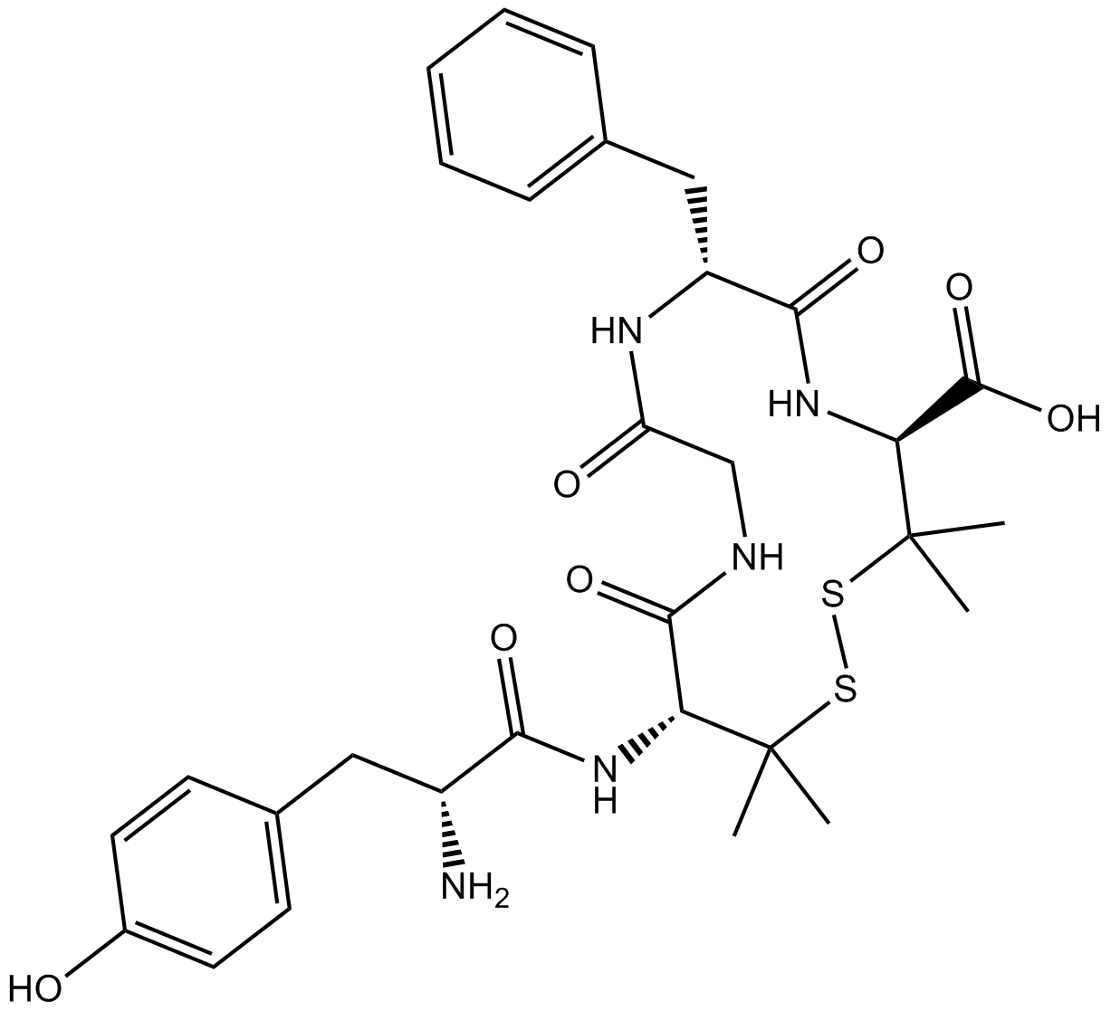
DPDPEは選択性で抗痙攣のδ-オピオイド受容体(DOR)作動剤である。
Products are for research use only. Not for human use. We do not sell to patients.
Cas No.: 88373-73-3
Sample solution is provided at 25 µL, 10mM.
_1-3.$$$39978408@51.jpg');)
_1-9.$$$38538795@65.jpg');)
_1-9.$$$39112701@51.jpg');)
_1-7.$$$37316672@70.jpg');)
_366-372.$$$36198801@70.jpg');)
.$$$38565142@65.jpg');)
_1867-1883.$$$33248023@67.jpg');)
_eads6055.$$$39977546@45.jpg');)
_eadm9805.$$$40080571@45.jpg');)
_eadj3020.$$$39666821@45.jpg');)
_1-20.$$$39757301@28.jpg');)
_1-24.$$$38898113@28.jpg');)
_273-287.$$$36806353@46.jpg');)
_1-16.$$$37156877@44.jpg');)
_1-19.$$$37460805@44.jpg');)
_1291-1307.$$$34518654@46.jpg');)
DPDPEは選択性で抗痙攣のδ-オピオイド受容体(DOR)作動剤である[1]。DORは、主に中枢神経系に分布しているGタンパク質共役受容体(GPCR)であり、内因性エンケファリンと外因性リガンドの両方の鎮静や抗抑鬱作用などの様々な生理作用を媒介する[2]。DPDPEはよく、オピオイド受容体のシグナル伝達、疼痛経路、依存症メカニズムに関連する研究に使われる[3]。
体外で、過剰発見、またはノックダウンGRK2のHEK293細胞をDPDPE(2μM)で30分間処理した結果、GRK2の過剰発見が野生型受容体の内在化と後続するリサイクルを増強し、GRK2のノックダウンがこれらのプロセスを阻害したことを示した[4]。DPDPE(1μM)でHEK293細胞を60分間事前処理した結果、軽度のDOR脱感受性のみを誘発し、それがmonensinの存在下では著しく増強された[5]。ヒトDOR(CHO/DOR)を安定的に発見するCHO細胞をDPDPE(100nM)で5-30分間処理した結果、Ser9でグリコーゲンシンターゼキナーゼ-3β(GSK-3β)のリン酸化を速やかに著しく誘発し、同時にキナーゼの活性が著しく阻害(~35%)された[6]。
体内で、streptozotocin(STZ)誘発の糖尿病神経障害マウスにDPDPE(0.5、1、3、5mg/kg)を皮下注射し、投与の30分後に、機械的アロディニア、温熱性痛覚過敏や温熱性アロディニアを用量依存的に阻害した[7]。Sprague Dawleyラットで、後肢の圧迫の前に、DPDPE(100nM;10μL)で髄腔内事前処理した結果、脊髄後角のI層で、有害刺激誘発性ニューロキニン1受容体(NK1R)の内在化を著しく阻害した[8]。
References:
[1] TORTELLA F C, ECHEVARRIA E, ROBLES L, et al. Anticonvulsant effects of mu (DAGO) and delta (DPDPE) enkephalins in rats[J]. Peptides, 1988, 9(5): 1177-1181.
[2] NIETO M M, GUEN S L E, KIEFFER B L, et al. Physiological control of emotion-related behaviors by endogenous enkephalins involves essentially the delta opioid receptors[J]. Neuroscience, 2005, 135(2): 305-313.
[3] ABDALLAH K, GENDRON L. The delta opioid receptor in pain control[J]. Delta Opioid Receptor Pharmacology and Therapeutic Applications. 2017: 147-177.
[4] ZHANG X, WANG F, CHEN X, et al. Post-endocytic fates of δ-opioid receptor are regulated by GRK2-mediated receptor phosphorylation and distinct β-arrestin isoforms[J]. Journal of Neurochemistry, 2008, 106(2): 781-792.
[5] AUDET N, CHARFI I, MNIE-FILALI O, et al. Differential association of receptor-Gβγ complexes with β-arrestin2 determines recycling bias and potential for tolerance of delta opioid receptor agonists[J]. Journal of Neuroscience, 2012, 32(14): 4827-4840.
[6] OLIANAS M C, DEDONI S, ONALI P. Signaling pathways mediating phosphorylation and inactivation of glycogen synthase kinase-3β by the recombinant human δ-opioid receptor stably expressed in Chinese hamster ovary cells[J]. Neuropharmacology, 2011, 60(7-8): 1326-1336.
[7] CASTANY S, CARCOLÉ M, LEÁNEZ S, et al. The antinociceptive effects of a δ-opioid receptor agonist in mice with painful diabetic neuropathy: Involvement of heme oxygenase 1[J]. Neuroscience Letters, 2016, 614: 49-54.
[8] KONDO I, MARVIZON J C G, SONG B, et al. Inhibition by spinal μ- and δ-opioid agonists of afferent-evoked substance P release[J]. Journal of Neuroscience, 2005, 25(14): 3651-3660.
| 細胞実験[1]: | |
細胞株 | DOR(CHO/DOR)を安定的に発見するCHO細胞 |
準備方法 | 血清飢餓のCHO/DOR細胞を100nMのDPDPEで5-30分間インキュベートした。ゼロ時間サンプルを溶媒で処理した。リン酸化GSK-3b(pGSK)、総GSK-3b(GSK)とアクチンのレベルについて、細胞抽出物を分析した。 |
反応条件 | 100nM;5-30分間 |
アプリケーション | CHO/DOR細胞をDPDPE(100nM)で処理した結果、リン酸化-Ser9-GSK-3βのレベルを速やかに増やし、その効果は5分後に著しくなり、10分後にピークに達し、最大30分間は基礎レベルを上回る状態を保った。 |
| 動物実験 [2]: | |
動物モデル | Sprague Dawleyラット |
準備方法 | 後肢の圧迫の10分前に、ラットにDPDPE(100nM;10μL)を髄腔内注射した。ラットをペントバルビタールナトリウムで麻酔し、片方の後肢を、鋸歯のない顎の6インチの蚊鉗子の顎に垂直に配置し、止血鉗子のラチェットが最初のクリックまで顎を閉じた。60秒間圧迫を行った。その5分後に、ラットを大動脈潅流で固定し、NK1R免疫細胞化学の為に、脊髄を採取した。 |
投与形態 | 100nM;10μL;i.t. |
アプリケーション | DPDPEの処理は、後肢の爪の圧迫損傷による脊髄の背角I層で、NK1Rの内在化を著しく阻害した。 |
References: | |
| Cas No. | 88373-73-3 | SDF | |
| Chemical Name | (4S,7R,13R)-13-((R)-2-amino-3-(4-hydroxyphenyl)propanamido)-7-benzyl-3,3,14,14-tetramethyl-6,9,12-trioxo-1,2-dithia-5,8,11-triazacyclotetradecane-4-carboxylic acid | ||
| Canonical SMILES | O=C1NCC(N[C@H](CC2=CC=CC=C2)C(N[C@@H](C(O)=O)C(C)(C)SSC(C)(C)[C@@H]1NC([C@@H](CC(C=C3)=CC=C3O)N)=O)=O)=O | ||
| Formula | C30H39N5O7S2 | M.Wt | 645.79 |
| 溶解度 | 10mg/mL in Water | Storage | Desiccate at -20°C |
| General tips | Please select the appropriate solvent to prepare the stock solution according to the
solubility of the product in different solvents; once the solution is prepared, please store it in
separate packages to avoid product failure caused by repeated freezing and thawing.Storage method
and period of the stock solution: When stored at -80°C, please use it within 6 months; when stored
at -20°C, please use it within 1 month. To increase solubility, heat the tube to 37°C and then oscillate in an ultrasonic bath for some time. |
||
| Shipping Condition | Evaluation sample solution: shipped with blue ice. All other sizes available: with RT, or with Blue Ice upon request. | ||
| Prepare stock solution | |||

|
1 mg | 5 mg | 10 mg |
| 1 mM | 1.5485 mL | 7.7425 mL | 15.4849 mL |
| 5 mM | 309.7 μL | 1.5485 mL | 3.097 mL |
| 10 mM | 154.8 μL | 774.2 μL | 1.5485 mL |
Step 1: Enter information below (Recommended: An additional animal making an allowance for loss during the experiment)
 g
μL
g
μL
Step 2: Enter the in vivo formulation (This is only the calculator, not formulation. Please contact us first if there is no in vivo formulation at the solubility Section.)
Calculation results:
Working concentration: mg/ml;
Method for preparing DMSO master liquid: mg drug pre-dissolved in μL DMSO ( Master liquid concentration mg/mL, Please contact us first if the concentration exceeds the DMSO solubility of the batch of drug. )
Method for preparing in vivo formulation: Take μL DMSO master liquid, next addμL PEG300, mix and clarify, next addμL Tween 80, mix and clarify, next add μL saline, mix and clarify.
Method for preparing in vivo formulation: Take μL DMSO master liquid, next add μL Corn oil, mix and clarify.
Note: 1. Please make sure the liquid is clear before adding the next solvent.
2. Be sure to add the solvent(s) in order. You must ensure that the solution obtained, in the previous addition, is a clear solution before proceeding to add the next solvent. Physical methods such as vortex, ultrasound or hot water bath can be used to aid dissolving.
3. All of the above co-solvents are available for purchase on the GlpBio website.
Quality Control & SDS
- View current batch:
- Purity: >98.00% Appearance: A solid
- COA (Certificate of Analysis)
- SDS (Safety Data Sheet)
- Datasheet
Average Rating: 5 (Based on Reviews and 1 reference(s) in Google Scholar.)