

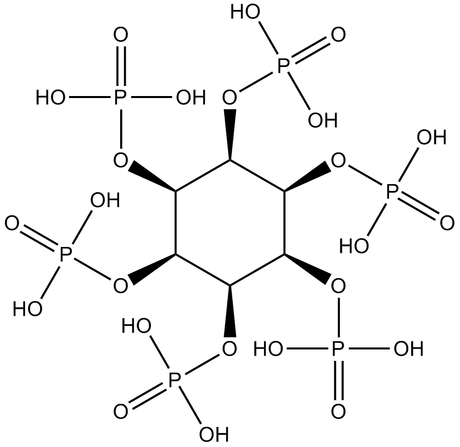
Phytic acid |
رقم الكتالوجGN10554
 One-Click Copy Product Info
|

Phytic acid is an antinutrient found naturally in legume seeds.
Products are for research use only. Not for human use. We do not sell to patients.
Cas No.: 83-86-3
Sample solution is provided at 25 µL, 10mM.
_1-3.$$$39978408@51.jpg');)
_1-9.$$$38538795@65.jpg');)
_1-9.$$$39112701@51.jpg');)
_1-7.$$$37316672@70.jpg');)
_366-372.$$$36198801@70.jpg');)
.$$$38565142@65.jpg');)
_1867-1883.$$$33248023@67.jpg');)
_eads6055.$$$39977546@45.jpg');)
_eadm9805.$$$40080571@45.jpg');)
_eadj3020.$$$39666821@45.jpg');)
_1-20.$$$39757301@28.jpg');)
_1-24.$$$38898113@28.jpg');)
_273-287.$$$36806353@46.jpg');)
_1-16.$$$37156877@44.jpg');)
_1-19.$$$37460805@44.jpg');)
_1291-1307.$$$34518654@46.jpg');)
Phytic acid is an antinutrient found naturally in legume seeds [1]. Phytic acid and its degradation products can scavenge free radicals and reduce oxidative stress [2]. Phytic acid chelation of metal ions (such as iron) can reduce metal-catalyzed oxidative reactions [3]. Phytic acid exhibits anticancer and antioxidant properties [4].
In MC65 cells, Cell survival rate was significantly improved after Phytic acid (100μM; 4d) treatment [5]. In HepG2 cells, Phytic acid (0.1-4mM; 24-48h) treatment inhibited cell growth [6].
In dextran sulfate sodium (DSS)-induced UC mice model, Phytic acid (0.25g/kg; ig; 14d) treatment improved weight loss, shortened colon length, improved clinical scores, and reduced the release of proinflammatory factors (Il-1β, Il-6, and TNF-α) [7]. In a high-fat diet mice model, Phytic acid (0.25-2mg/mL; po; 13 weeks) significantly inhibited obesity and alleviated hepatic steatosis [8].
References:
[1]. Oatway L, Vasanthan T, Helm J H. Phytic acid[J]. Food Reviews International, 2001, 17(4): 419-431.
[2]. Graf E, Empson K L, Eaton J W. Phytic acid. A natural antioxidant[J]. Journal of Biological Chemistry, 1987, 262(24): 11647-11650.
[3]. Lee B J, Hendricks D G. Metal‐catalyzed oxidation of ascorbate, deoxyribose and linoleic acid as affected by phytic acid in a model system[J]. Journal of Food Science, 1997, 62(5): 935-984.
[4]. Bloot A P M, Kalschne D L, Amaral J A S, et al. A review of phytic acid sources, obtention, and applications[J]. Food Reviews International, 2023, 39(1): 73-92.
[5]. Anekonda T S, Wadsworth T L, Sabin R, et al. Phytic acid as a potential treatment for Alzheimer's pathology: evidence from animal and in vitro models[J]. Journal of Alzheimer’s disease, 2011, 23(1): 21-35.
[6]. Al-Fatlawi A A, Al-Fatlawi A A, Irshad M, et al. Rice bran phytic acid induced apoptosis through regulation of Bcl-2/Bax and p53 genes in HepG2 human hepatocellular carcinoma cells[J]. Asian Pacific Journal of Cancer Prevention, 2014, 15(8): 3731-3736.
[7]. Hou X, Sang Y, Dong L. The improved effect and its mechanism of phytic acid on DSS-induced UC mice[J]. Life sciences, 2022, 311: 121139.
[8]. Ran X, Hu G, He F, et al. Phytic acid improves hepatic steatosis, inflammation, and oxidative stress in high-fat diet (HFD)-fed mice by modulating the gut–liver axis[J]. Journal of Agricultural and Food Chemistry, 2022, 70(36): 11401-11411.
| Cell experiment [1]: | |
Cell lines | MC65 cells |
Preparation Method | Cell viability was determined using an MTS assay and a calcium ion assay. At 90% confluence, cells were harvested from TC flasks using 0.05% trypsin/EDTA, washed twice with PBS, and seeded at 15 × 103 cells/well in 96-well plates in OPTIMem medium containing (Tet+) or (Tet-) tetracycline (1μg/mL) and excipients, or phytic acid (100μM). Cells grown under Tet- conditions typically died after approximately 3–4 days of culture, accompanied by expression of endogenous A oligomers, whereas cells grown under Tet+ conditions remained viable during this period. Wells containing growth medium without cells served as background controls, and wells containing vehicle served as positive controls. Each treatment was replicated in 3–6 wells, and the treatment duration for each experiment was 72 hours. Absorbance was measured at a wavelength of 490nm using a Spectra Max PLUS microplate reader. The cell viability under different treatments was determined relative to the cell viability under Tet + treatment. The experiments were repeated at least three times. |
Reaction Conditions | 100μM; 4d |
Applications | Cell survival rate was significantly improved after Phytic acid treatment. |
| Animal experiment [2]: | |
Animal models | Dextran sulfate sodium (DSS)-induced UC mice model |
Preparation Method | The C57BL/6 mice were randomly divided into four groups of five mice each. The specific groups were as follows: no treatment group (NT group), Phytic acid feeding group (Phytic acid group), DSS modeling group (DSS group), and Phytic acid treatment group (Phytic acid + DSS group). The mice in the Phytic acid and Phytic acid + DSS groups were gavaged with Phytic acid (0.25g/kg/day) for 21days. After receiving Phytic acid for 14days, mice in the DSS and Phytic acid + DSS groups were given 2.5% DSS aqueous solution for 7days to develop the mouse model of UC. The body weights of all mice were recorded from day 1 of the DSS feeding. Following the completion of the DSS induction period, all mice were euthanized, and colon samples were collected for subsequent studies. |
Dosage form | 0.25g/kg; ig; 14d |
Applications | Phytic acid treatment improved weight loss, shortened colon length, improved clinical scores, and reduced the release of proinflammatory factors (Il-1β, Il-6, and TNF-α). |
References: | |
| Cas No. | 83-86-3 | SDF | |
| Chemical Name | (2,3,4,5,6-pentaphosphonooxycyclohexyl) dihydrogen phosphate | ||
| Canonical SMILES | C1(C(C(C(C(C1OP(=O)(O)O)OP(=O)(O)O)OP(=O)(O)O)OP(=O)(O)O)OP(=O)(O)O)OP(=O)(O)O | ||
| Formula | C6H18O24P6 | M.Wt | 660.04 |
| الذوبان | Water : ≥ 250 mg/mL | Storage | Store at -20°C |
| General tips | Please select the appropriate solvent to prepare the stock solution according to the
solubility of the product in different solvents; once the solution is prepared, please store it in
separate packages to avoid product failure caused by repeated freezing and thawing.Storage method
and period of the stock solution: When stored at -80°C, please use it within 6 months; when stored
at -20°C, please use it within 1 month. To increase solubility, heat the tube to 37°C and then oscillate in an ultrasonic bath for some time. |
||
| Shipping Condition | Evaluation sample solution: shipped with blue ice. All other sizes available: with RT, or with Blue Ice upon request. | ||
| Prepare stock solution | |||

|
1 mg | 5 mg | 10 mg |
| 1 mM | 1.5151 mL | 7.5753 mL | 15.1506 mL |
| 5 mM | 303 μL | 1.5151 mL | 3.0301 mL |
| 10 mM | 151.5 μL | 757.5 μL | 1.5151 mL |
Step 1: Enter information below (Recommended: An additional animal making an allowance for loss during the experiment)
 g
μL
g
μL
Step 2: Enter the in vivo formulation (This is only the calculator, not formulation. Please contact us first if there is no in vivo formulation at the solubility Section.)
Calculation results:
Working concentration: mg/ml;
Method for preparing DMSO master liquid: mg drug pre-dissolved in μL DMSO ( Master liquid concentration mg/mL, Please contact us first if the concentration exceeds the DMSO solubility of the batch of drug. )
Method for preparing in vivo formulation: Take μL DMSO master liquid, next addμL PEG300, mix and clarify, next addμL Tween 80, mix and clarify, next add μL saline, mix and clarify.
Method for preparing in vivo formulation: Take μL DMSO master liquid, next add μL Corn oil, mix and clarify.
Note: 1. Please make sure the liquid is clear before adding the next solvent.
2. Be sure to add the solvent(s) in order. You must ensure that the solution obtained, in the previous addition, is a clear solution before proceeding to add the next solvent. Physical methods such as vortex, ultrasound or hot water bath can be used to aid dissolving.
3. All of the above co-solvents are available for purchase on the GlpBio website.
Quality Control & SDS
- View current batch:
- Purity: >98.00% Appearance: A solid
- COA (Certificate of Analysis)
- SDS (Safety Data Sheet)
- Datasheet
Average Rating: 5 (Based on Reviews and 30 reference(s) in Google Scholar.)