

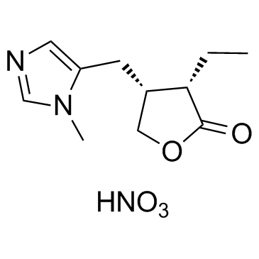
Pilocarpine nitrate |
Catalog No.GC31001
 One-Click Copy Product Info
|

Le nitrate de pilocarpine est un puissant agoniste des récepteurs muscariniques de l'acétylcholine de type M3 (récepteur muscarinique M3).
Products are for research use only. Not for human use. We do not sell to patients.
Cas No.: 148-72-1
Sample solution is provided at 25 µL, 10mM.
_1-3.$$$39978408@51.jpg');)
_1-9.$$$38538795@65.jpg');)
_1-9.$$$39112701@51.jpg');)
_1-7.$$$37316672@70.jpg');)
_366-372.$$$36198801@70.jpg');)
.$$$38565142@65.jpg');)
_1867-1883.$$$33248023@67.jpg');)
_eads6055.$$$39977546@45.jpg');)
_eadm9805.$$$40080571@45.jpg');)
_eadj3020.$$$39666821@45.jpg');)
_1-20.$$$39757301@28.jpg');)
_1-24.$$$38898113@28.jpg');)
_273-287.$$$36806353@46.jpg');)
_1-16.$$$37156877@44.jpg');)
_1-19.$$$37460805@44.jpg');)
_1291-1307.$$$34518654@46.jpg');)
Pilocarpine nitrate is a selective M3-type muscarinic acetylcholine receptor (M3 muscarinic receptor) agonist.
To evaluate the cytotoxicity of Pilocarpine, the morphology and viability of human corneal stromal (HCS) cells are examined by light microscopy and MTT assay, respectively. Morphological observations show that HCS cells exposed to Pilocarpine at a concentration from 0.625 to 20 g/L exhibit dose- and time-dependent proliferation retardation and morphological abnormality such as cellular shrinkage, cytoplasmic vacuolation, detachment from culture matrix, and eventually death, while no obvious difference is observed between those exposed to Pilocarpine below the concentration of 0.625 g/L and controls. Results of MTT assay reveal that the cell viability of HCS cells decrease with time and concentration after exposing to Pilocarpine above the concentration of 0.625 g/L (P<0.01 or 0.05), while that of HCS cells treated with Pilocarpine below the concentration of 0.625 g/L show no significant difference to controls[2]. The partial muscarinic agonist, Pilocarpine, evokes concentration-dependent relaxation with an EC50 of 2.4 mM in isolated segments of rat tail artery that were constricted with Penylephrine (10 to 200 nM)[3].
The Pilocarpine-induced saliva secretion of the control rats (CN) and exercised (EX) rats is examined. A significantly greater amount of saliva is induced by Pilocarpine in the EX rats than in the CN rats (P<0.01). Conversely, the Na+ concentration in the saliva of the EX rats is significantly lower than that of the CN rats (P<0.05)[1].
[1]. Matsuzaki K, et al. Daily voluntary exercise enhances pilocarpine-induced saliva secretion and aquaporin 1 expression in rat submandibular glands. FEBS Open Bio. 2017 Dec 7;8(1):85-93. [2]. Yuan XL, et al. Cytotoxicity of pilocarpine to human corneal stromal cells and its underlying cytotoxic mechanisms. Int J Ophthalmol. 2016 Apr 18;9(4):505-11. [3]. Tonta MA, et al. Pilocarpine-induced relaxation of rat tail artery by a non-cholinergic mechanism and in the absence of an intact endothelium. Br J Pharmacol. 1994 Jun;112(2):525-32.
|
Cell experiment: |
Cell viability is determined by MTT assay. Briefly, HCS cells are inoculated into a 96-well culture plate (Nunc) at a density of 1×10⁴ cells/100 µL/well, and are cultured and treated. At a 4h interval, the Pilocarpine (0.625 to 20 g/L)-containing medium is replaced entirely with 100 µL serum-free DMEM/F12 medium containing 1.0 g/L MTT, and the cells are incubated at 37°C in the dark for 4h. After the MTT-containing medium is discarded with caution, 150 µL DMSO is added to dissolve the produced formazan crystals at 37°C in the dark for 15 min, and the absorbance at 490 nm is measured with a Multiskan GO microplate reader[2]. |
|
Animal experiment: |
Rats[1]Male, 10-week-old Wistar rats are assigned to one of two groups, exercise (EX, n=6) and control (CN, n=6). The EX rats are kept for 40 days in cages with a running wheel (SN-451), allowing them to undertake voluntary exercise, while the CN rats are kept in cages with the running wheel locked. On the 40th day, Pilocarpine-induced saliva is measured as follows. Briefly, the rats are anesthetized, preweighed cotton was placed in their mouths sublingually, and Pilocarpine (0.5 mg/kg) is intraperitoneally injected to induce saliva secretion. Each cotton ball is then changed every 10 min for 1 h. The collected cotton balls are weighed again, and the mass of saliva secreted is calculated by subtracting the initial from the final weight. |
|
References: [1]. Matsuzaki K, et al. Daily voluntary exercise enhances pilocarpine-induced saliva secretion and aquaporin 1 expression in rat submandibular glands. FEBS Open Bio. 2017 Dec 7;8(1):85-93. |
|
| Cas No. | 148-72-1 | SDF | |
| Canonical SMILES | [O-][N+](O)=O.O=C1OC[C@H](CC2=CN=CN2C)[C@@H]1CC | ||
| Formula | C11H17N3O5 | M.Wt | 271.27 |
| Solubility | Soluble in DMSO | Storage | Store at -20°C |
| General tips | Please select the appropriate solvent to prepare the stock solution according to the
solubility of the product in different solvents; once the solution is prepared, please store it in
separate packages to avoid product failure caused by repeated freezing and thawing.Storage method
and period of the stock solution: When stored at -80°C, please use it within 6 months; when stored
at -20°C, please use it within 1 month. To increase solubility, heat the tube to 37°C and then oscillate in an ultrasonic bath for some time. |
||
| Shipping Condition | Evaluation sample solution: shipped with blue ice. All other sizes available: with RT, or with Blue Ice upon request. | ||
| Prepare stock solution | |||

|
1 mg | 5 mg | 10 mg |
| 1 mM | 3.6864 mL | 18.4318 mL | 36.8636 mL |
| 5 mM | 737.3 μL | 3.6864 mL | 7.3727 mL |
| 10 mM | 368.6 μL | 1.8432 mL | 3.6864 mL |
Step 1: Enter information below (Recommended: An additional animal making an allowance for loss during the experiment)
 g
μL
g
μL
Step 2: Enter the in vivo formulation (This is only the calculator, not formulation. Please contact us first if there is no in vivo formulation at the solubility Section.)
Calculation results:
Working concentration: mg/ml;
Method for preparing DMSO master liquid: mg drug pre-dissolved in μL DMSO ( Master liquid concentration mg/mL, Please contact us first if the concentration exceeds the DMSO solubility of the batch of drug. )
Method for preparing in vivo formulation: Take μL DMSO master liquid, next addμL PEG300, mix and clarify, next addμL Tween 80, mix and clarify, next add μL saline, mix and clarify.
Method for preparing in vivo formulation: Take μL DMSO master liquid, next add μL Corn oil, mix and clarify.
Note: 1. Please make sure the liquid is clear before adding the next solvent.
2. Be sure to add the solvent(s) in order. You must ensure that the solution obtained, in the previous addition, is a clear solution before proceeding to add the next solvent. Physical methods such as vortex, ultrasound or hot water bath can be used to aid dissolving.
3. All of the above co-solvents are available for purchase on the GlpBio website.
Quality Control & SDS
- View current batch:
- Purity: >98.00% Appearance: A solid
- COA (Certificate of Analysis)
- SDS (Safety Data Sheet)
- Datasheet
Average Rating: 5 (Based on Reviews and 1 reference(s) in Google Scholar.)