

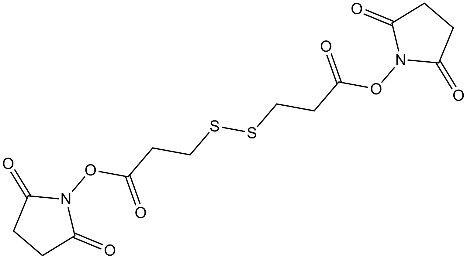
DSP (Synonyms: 3,3′-Dithiodipropionic acid di) |
رقم الكتالوجGC18378
 One-Click Copy Product Info
|

DSP هو رابط ADC قابل للانقسام ، يستخدم في تخليق اتحادات الأجسام المضادة والأدوية (ADCs).
Products are for research use only. Not for human use. We do not sell to patients.
Cas No.: 57757-57-0
Sample solution is provided at 25 µL, 10mM.
_1-3.$$$39978408@51.jpg');)
_1-9.$$$38538795@65.jpg');)
_1-9.$$$39112701@51.jpg');)
_1-7.$$$37316672@70.jpg');)
_366-372.$$$36198801@70.jpg');)
.$$$38565142@65.jpg');)
_1867-1883.$$$33248023@67.jpg');)
_eads6055.$$$39977546@45.jpg');)
_eadm9805.$$$40080571@45.jpg');)
_eadj3020.$$$39666821@45.jpg');)
_1-20.$$$39757301@28.jpg');)
_1-24.$$$38898113@28.jpg');)
_273-287.$$$36806353@46.jpg');)
_1-16.$$$37156877@44.jpg');)
_1-19.$$$37460805@44.jpg');)
_1291-1307.$$$34518654@46.jpg');)
DSP, a homobifunctional cross-linker that reacts with amino groups such as lysine, and is widely used for surface functionalization, is the preferred linker for immobilizing enzymes and antibodies on gold surfaces[1-3]. DSP has a disulfide bridge within its structure, which cleaves upon binding to gold[2]. By immobilizing DSP on a gold thin film (GTF), dopamine (DA) can be selectively captured via the specific reaction between its primary amine and the NHS ester of DSP, enabling sensitive detection of DA in human cerebrospinal fluid (CSF) samples[4-5]. DSP can be employed in irreversible biotinylation procedures (IBP) to eliminate interference from intermolecular disulfide bonds during S-nitrosation detection[6]. DSP can also be used to isolate weak protein complexes and to prepare Au-TiO2 thin films for immunoassay experiments[3, 7].
References:
[1] Darder M, Takada K, Pariente F, et al. Dithiobissuccinimidyl propionate as an anchor for assembling peroxidases at electrodes surfaces and its application in a H2O2 biosensor. Anal Chem. 1999;71(24):5530-5537
[2] Ataman Sadık D, Boyacı İH, et al. Mixed monolayer decorated SPR sensing surface for thrombin detection. J Pharm Biomed Anal. 2019;176:112822.
[3] Akaki K, Mino T, Takeuchi O. DSP-crosslinking and Immunoprecipitation to Isolate Weak Protein Complex. Bio Protoc. 2022;12(15):e4478.
[4] Zhang K, Liu Y, Wang Y, et al. Quantitative SERS Detection of Dopamine in Cerebrospinal Fluid by Dual-Recognition-Induced Hot Spot Generation. ACS Appl Mater Interfaces. 2018;10(18):15388-15394.
[5] Gu H, Guo Y, Xiao X, et al. Double molecular recognition strategy based on boronic acid-diol and NHS ester-amine for selective electrochemical detection of cerebral dopamine. Anal Bioanal Chem. 2020;412(15):3727-3736.
[6] Huang B, Chen C. Detection of protein S-nitrosation using irreversible biotinylation procedures (IBP). Free Radic Biol Med. 2010;49(3):447-456.
[7] Meira DI, Barbosac AI, Proença M, et al. Immobilizing antibody biorecognition layers on Au–TiO2 thin films: direct (physisorption) vs. DSP-crosslinking (chemisorption) surface functionalization. Journal of Physics D: Applied Physics. 2024;415401.
This plan only provides a guide, please modify it to meet your specific needs.
(1) DSP-crosslinking and Immunoprecipitation to Isolate Weak Protein Complex[1]:
A. Preparing cells expressing proteins of interests (POIs)
1. Plate 8.0×10⁵ HeLa cells in 10cm dish per sample (10mL of DMEM containing 10% FBS per dish).
2. Incubate the cells at 37°C, 5% CO2 for 24h.
3. Transfect plasmids for FLAG-HA-tagged POI expression.
4. Incubate the cells at 37°C, 5% CO2 overnight.
B. Preparing Wash buffer and IP buffer
1. On the day of DSP-crosslinking and immunoprecipitation, prepare Wash buffer and IP buffer before the crosslinking. Keep them on ice.
C. DSP-crosslinking
1. Prepare 100mM DSP just prior to DSP-crosslinking.
2. Dilute 100mM DSP to 0.1mM DSP in pre-warmed (37°C) PBS.
3. Rinse the dishes twice with 5mL of pre-warmed (37°C) PBS/dish.
4. Discard the PBS from the dishes.
5. Add 5mL of 0.1mM DSP (prepared in step C2) in each dish.
6. Incubate the dishes at 37°C for 30min in a CO2 incubator.
D. Preparing beads for the first IP
1. During the DSP-crosslinking (step C6), prepare 40μL of Dynabeads Protein G/sample in one 1.5mL tube.
2. Wash the Dynabeads Protein G with the same amount of Wash buffer three times: Set the tube containing Dynabeads Protein G on a magnetic stand and let the beads accumulate onto the magnet. Discard the supernatant with a pipette. Add ice-cold Wash buffer. Remove the tube from the magnetic stand. Resuspend the beads by pipetting up and down. Repeat step D2 twice more.
3. Set the tube containing the washed beads on a magnetic stand and discard the supernatant.
4. Resuspend the beads with ice-cold IP buffer.
5. Add 1μL of anti-FLAG antibody per sample.
6. Incubate the beads on a rotating incubator at 4°C for 1h.
E. Stopping DSP-crosslinking
1. After the 30min incubation at step C6, discard the 0.1mM DSP and rinse the dishes once with 5mL of pre-warmed (37°C) PBS/dish.
2. Discard the PBS and add 5mL of STOP solution (room temperature)/dish.
3. Incubate the dishes at room temperature for 15min.
F. First IP
1. Discard the STOP solution from the dishes.
2. Rinse the dishes twice with 5mL of ice-cold PBS/dish.
3. Discard the PBS from the dishes.
4. Add 500μL of ice-cold IP buffer/dish.
5. Scrape off the cells with a scraper and transfer them with all the IP buffer on the dish into a 1.5mL tube.
6. Resuspend the cells by pipetting up and down.
7. Incubate the tubes on ice for 10min to complete cell lysis.
8. Centrifuge the tubes containing the lysates at 20,000×g at 4°C for 5min.
9. Transfer 500μL of the supernatant to a new 1.5mL tube for IP.
10. Add 40μL of anti-FLAG-antibody-bound beads (prepared at step D6) into the tube containing the supernatant.
11. Incubate the tubes on a rotating incubator at 4°C for 2h.
G. Preparing beads for the second IP
1. Approximately 30min before finishing the 2h incubation at step F11, prepare and wash new Dynabeads Protein G, as in steps D1 to D4.
2. Add 1μL of anti-HA antibody per sample.
3. Incubate the beads on a rotating incubator at 4°C for 1h.
H. Protein elution with FLAG peptides
1. Prepare FLAG-elution buffer by diluting FLAG peptide stock solution with TBS and keep it on ice.
2. After the 2h incubation at step F11, wash the beads with ice-cold 700μL of Wash buffer three times.
3. Discard the Wash buffer from the beads.
4. Resuspend the beads with 100μL of FLAG-elution buffer (prepared at step H1) gently by pipetting up and down.
5. Incubate the beads with FLAG-elution buffer on a rotating incubator at 4°C for 10min.
6. Set the tubes on a magnetic stand and transfer the supernatant (containing POI and POI-bound proteins) to new 1.5mL tubes.
7. Repeat steps H4 to H5 once more.
8. Set the tubes on a magnetic stand and transfer the supernatant to the tubes containing the first eluted proteins at step H6.
9. Add 300μL of ice-cold IP buffer to the collected supernatant.
I. Second IP
1. Mix the 500μL of supernatant at step H9 with 40μL of anti-HA-antibody-bound beads (prepared at step G3).
2. Incubate the beads on a rotating incubator at 4°C for 2h.
J. Final elution
1. After the 2h incubation at step I2, wash the beads with 700μL of ice-cold Wash buffer three times.
(2) DSP treatment of lysates of mouse brain[2]:
1. The lysates of mouse brain were treated with 5mM DSP for 30min to form intermolecular disulfide bonds, Pr-S-S-Pr in vitro and was stopped by addition of Tris buffer.
2. The lysates were then analyzed by the original biotin switch method and irreversible biotinylation procedures (IBP).
(3) Use DSP to prepare Au-TiO2 thin films for immunoassay experiments[3]:
1. Plasma-activated Au-TiO2 thin films were functionalized with DSP solutions, using various concentrations (0.4, 1, 4, and 10mg/ml) dissolved in DMSO solvent (dimethyl sulfoxide, anhydrous, ≥99.9%).
2. The immobilization time was 30min, followed by cleaning steps, including DMSO, PBS (Phosphate-Buffered Saline; pH 7.4; 10mM), and ethanol 96% solutions to remove any unbounded cross-linker molecules.
References:
[1] Akaki K, Mino T, Takeuchi O. DSP-crosslinking and Immunoprecipitation to Isolate Weak Protein Complex. Bio Protoc. 2022;12(15):e4478.
[2] Huang B, Chen C. Detection of protein S-nitrosation using irreversible biotinylation procedures (IBP). Free Radic Biol Med. 2010;49(3):447-456.
[3] Meira DI, Barbosac AI, Proença M, et al. Immobilizing antibody biorecognition layers on Au–TiO2 thin films: direct (physisorption) vs. DSP-crosslinking (chemisorption) surface functionalization. Journal of Physics D: Applied Physics. 2024;415401.
| Cas No. | 57757-57-0 | SDF | |
| المرادفات | 3,3′-Dithiodipropionic acid di | ||
| Formula | C14H16N2O8S2 | M.Wt | 404.41 |
| الذوبان | DMF: 30 mg/ml,DMSO: 30 mg/ml,DMSO:PBS (pH 7.2) (1:4): 0.20 mg/ml | Storage | Store at -20°C, protect from light, stored under nitrogen |
| General tips | Please select the appropriate solvent to prepare the stock solution according to the
solubility of the product in different solvents; once the solution is prepared, please store it in
separate packages to avoid product failure caused by repeated freezing and thawing.Storage method
and period of the stock solution: When stored at -80°C, please use it within 6 months; when stored
at -20°C, please use it within 1 month. To increase solubility, heat the tube to 37°C and then oscillate in an ultrasonic bath for some time. |
||
| Shipping Condition | Evaluation sample solution: shipped with blue ice. All other sizes available: with RT, or with Blue Ice upon request. | ||
| Prepare stock solution | |||

|
1 mg | 5 mg | 10 mg |
| 1 mM | 2.4727 mL | 12.3637 mL | 24.7274 mL |
| 5 mM | 494.5 μL | 2.4727 mL | 4.9455 mL |
| 10 mM | 247.3 μL | 1.2364 mL | 2.4727 mL |
Step 1: Enter information below (Recommended: An additional animal making an allowance for loss during the experiment)
 g
μL
g
μL
Step 2: Enter the in vivo formulation (This is only the calculator, not formulation. Please contact us first if there is no in vivo formulation at the solubility Section.)
Calculation results:
Working concentration: mg/ml;
Method for preparing DMSO master liquid: mg drug pre-dissolved in μL DMSO ( Master liquid concentration mg/mL, Please contact us first if the concentration exceeds the DMSO solubility of the batch of drug. )
Method for preparing in vivo formulation: Take μL DMSO master liquid, next addμL PEG300, mix and clarify, next addμL Tween 80, mix and clarify, next add μL saline, mix and clarify.
Method for preparing in vivo formulation: Take μL DMSO master liquid, next add μL Corn oil, mix and clarify.
Note: 1. Please make sure the liquid is clear before adding the next solvent.
2. Be sure to add the solvent(s) in order. You must ensure that the solution obtained, in the previous addition, is a clear solution before proceeding to add the next solvent. Physical methods such as vortex, ultrasound or hot water bath can be used to aid dissolving.
3. All of the above co-solvents are available for purchase on the GlpBio website.
Quality Control & SDS
- View current batch:
- Purity: >97.00% Appearance: A solid
- COA (Certificate of Analysis)
- SDS (Safety Data Sheet)
- Datasheet
Average Rating: 5 (Based on Reviews and 25 reference(s) in Google Scholar.)