

DSP (Synonyms: 3,3′-Dithiodipropionic acid di) |
カタログ番号GC18378
 One-Click Copy Product Info
|

DSPは、リシンなどのアミノ基と反応するホモ二官能性架橋剤であり、表面の機能化に広く使われ、金の表面上に酵素や抗体を固定化するのに好ましいリンカーである。
Products are for research use only. Not for human use. We do not sell to patients.
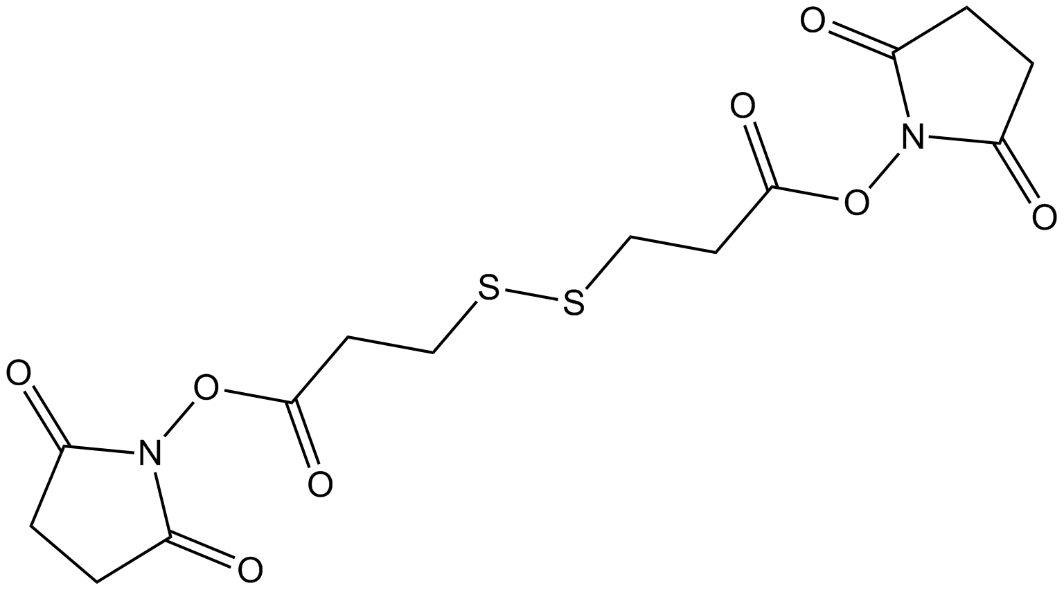
Cas No.: 57757-57-0
Sample solution is provided at 25 µL, 10mM.
_1-3.$$$39978408@51.jpg');)
_1-9.$$$38538795@65.jpg');)
_1-9.$$$39112701@51.jpg');)
_1-7.$$$37316672@70.jpg');)
_366-372.$$$36198801@70.jpg');)
.$$$38565142@65.jpg');)
_1867-1883.$$$33248023@67.jpg');)
_eads6055.$$$39977546@45.jpg');)
_eadm9805.$$$40080571@45.jpg');)
_eadj3020.$$$39666821@45.jpg');)
_1-20.$$$39757301@28.jpg');)
_1-24.$$$38898113@28.jpg');)
_273-287.$$$36806353@46.jpg');)
_1-16.$$$37156877@44.jpg');)
_1-19.$$$37460805@44.jpg');)
_1291-1307.$$$34518654@46.jpg');)
DSPは、リシンなどのアミノ基と反応するホモ二官能性架橋剤であり、表面の機能化に広く使われ、金の表面上に酵素や抗体を固定化するのに好ましいリンカーである[1-3]。DSPは構造内でジスルフィド架橋を持ち、金と結合する時は切断する[2]。金薄膜(GTF)でDSPを固定することで、ドーパミン(DA)は、その初代アミンとDSPのNHSエステルとの特異性の反応を通じて捕えることができ、ヒト脳脊髄液(CSF)のサンプルで、DAの選択的な検出を可能にした[4-5]。DSPは、不可逆的なビオチン化手順(IBP)に使われ、S-ニトロソ化検出の間に、分子間ジスルフィド結合による干渉を排除する[6]。DSPは、弱いタンパク質複合体の分離に使われ、免疫分析実験のために、Au-TiO2薄膜を調製できる[3, 7]。
References:
[1] Darder M, Takada K, Pariente F, et al. Dithiobissuccinimidyl propionate as an anchor for assembling peroxidases at electrodes surfaces and its application in a H2O2 biosensor. Anal Chem. 1999;71(24):5530-5537
[2] Ataman Sadık D, Boyacı İH, et al. Mixed monolayer decorated SPR sensing surface for thrombin detection. J Pharm Biomed Anal. 2019;176:112822.
[3] Akaki K, Mino T, Takeuchi O. DSP-crosslinking and Immunoprecipitation to Isolate Weak Protein Complex. Bio Protoc. 2022;12(15):e4478.
[4] Zhang K, Liu Y, Wang Y, et al. Quantitative SERS Detection of Dopamine in Cerebrospinal Fluid by Dual-Recognition-Induced Hot Spot Generation. ACS Appl Mater Interfaces. 2018;10(18):15388-15394.
[5] Gu H, Guo Y, Xiao X, et al. Double molecular recognition strategy based on boronic acid-diol and NHS ester-amine for selective electrochemical detection of cerebral dopamine. Anal Bioanal Chem. 2020;412(15):3727-3736.
[6] Huang B, Chen C. Detection of protein S-nitrosation using irreversible biotinylation procedures (IBP). Free Radic Biol Med. 2010;49(3):447-456.
[7] Meira DI, Barbosac AI, Proença M, et al. Immobilizing antibody biorecognition layers on Au–TiO2 thin films: direct (physisorption) vs. DSP-crosslinking (chemisorption) surface functionalization. Journal of Physics D: Applied Physics. 2024;415401.
このプランはガイドのみを提供し、ご自分のニーズに合わせてご調整願います。
(1) 弱いタンパク質複合体の分離のためのDSP架橋と免疫沈澱[1]:
A.希望のタンパク質(POIs)を発見する細胞の調製
1.サンプルごとに、8.0×10⁵ HeLa細胞を10cmの皿に播種した(皿ごとに10% FBSを含有する10mLのDMEM)。
2.細胞を37°C, 5% CO2で24時間培養した。
3.FLAG-HA-タグのPOI発見のプラスミドを遺伝子導入する。
4.37°C, 5% CO2で細胞を一晩中培養する。
B.洗浄緩衝液とIP緩衝液の調製
1.DSP架橋と免疫沈澱の当日に、架橋の前に、洗浄緩衝液とIP緩衝液を調製し、氷の上に静置する。
C.DSP架橋
1.DSP架橋前に、100mM DSPを調製する。
2.事前加温の(37°C) PBSで、100mM DSPを0.1mM DSPに希釈する。
3.5mLの事前加温(37°C) PBS/皿で、皿を二回すすぐ。
4.PBSを皿から廃棄する。
5.各皿に、5mLの0.1mM DSP (ステップC2に調製)を添加する。
6.CO2インキュベーターで、37℃、皿を30分間培養する。
D.最初のIP用のビーズの調製
1.DSP架橋(ステップC6)の間、40μLのDynabeads Protein G/サンプルを1.5mLのチューブに調製する。
2.同量の洗浄緩衝液でDynabeads Protein Gを三回洗浄する:Dynabeads Protein Gを含有するチューブをマグネットスタンドに設置し、ビーズをマグネットに蓄積させる。ピペットで上清を廃棄する。ピペッティングでビーズを上下に動かして再懸濁する。ステップD2をさらに2回繰り返し行う。
3.洗浄されたビーズを含有するチューブをマグネットスタンドに設置し、上清を廃棄する。
4.氷冷IP緩衝液でビーズを再懸濁する。
5.サンプルごとに、1μLの抗FLAG抗体を添加する。
6.4℃、回転するインキュベーターでビーズを培養する。
E.DSP架橋の停止
1.ステップC6で30分間の培養後、0.1mM DSPを廃棄し、5mLの事前加温の(37°C) PBS/皿で、皿を一回すすぐ。
2.PBSを廃棄し、5mLのSTOP溶液(室温)/皿を添加する。
3.室温で皿を15分間培養する。
F.最初のIP
1.皿からSTOP溶液を廃棄する。
2.5mLの氷冷PBS/皿で皿を二回すすぐ。
3.皿からPBSを廃棄する。
4.500μLの氷冷IP緩衝液/皿を添加する。
5.細胞をスクレーパーで削ぎとり、皿上のIP緩衝液をすべて1.5mLチューブに移す。
6.ピペットを上下に動かすことで細胞を再懸濁する。
7.氷でチューブを10分間培養することで、細胞の溶解を完成する。
8.4℃、20,000×gで溶解物を含有するチューブを5分間遠心分離する。
9.IPのために、500μLの上清を新しい1.5mLのチューブに移す。
10.上清を含有するチューブに、40μLの抗FLAG抗体結合ビーズ(ステップD6で調製)を添加する。
11.4℃で、回転インキュベーターの上でチューブを2時間培養する。
G.二回目のIPのためのビーズの調製
1.ステップF11で、2時間の培養の終了する約30分前に、ステップD1からD4の通りに、新しいDynabeads Protein Gを調製し洗浄する。
2.サンプルごとに、1μLの抗HA抗体を添加する。
3.4℃で、ビーズを回転インキュベーターで1時間培養する。
H.FLAGペプチドでのタンパク質の溶離
1.TBSでFLAGペプチド貯蔵液を希釈することで、FLAG-溶離緩衝液を調製し、氷の上に静置する。
2.ステップF11で2時間の培養に、氷冷700μLの洗浄緩衝液でビーズを三回洗浄する。
3.ビーズから洗浄緩衝液を廃棄する。
4.ゆっくりピペットを上下にすることで、100μLのFLAG-溶離緩衝液(ステップH1で調製)でビーズを再懸濁する。
5.4℃、回転のインキュベーターでFLAG溶離緩衝液と共にビーズを10分間培養する。
6.チューブをマグネットスタンドに設置し、上清(POIとPOI結合のタンパク質を含有)を新しい1.5mLのチューブに移す。
7.H4からH5までのステップをもう一度繰り返す。
8.チューブをマグネットスタンドに設置し、ステップH6での最初の溶出タンパク質を含有するチューブに上清を移す。
9.300μLの氷冷IP緩衝液を、採取された上清に添加する。
I.二回目のIP
1.ステップH9での500μLの上清と40μLの抗HA抗体結合ビーズ(ステップG3で調製)を混合させる。
2.4℃、回転インキュベーターでビーズを2時間培養する。
J.最後の溶出
1.ステップI2での2時間の培養の後、700μLの氷冷洗浄緩衝液でビーズを三回洗浄する。
(2)マウス脳の溶解液のDSP処理[2]:
1.マウス脳の溶解液を5mM DSPで30分間処理し、体外で分子間ジスルフィド結合Pr-S-S-Prを形成し、Tris緩衝液の添加で停めた。
2.その後、独自のビオチンスイッチ法と不可逆的なビオチン化手順 (IBP) で、溶解物を分析した。
(3)免疫分析実験のため、DSPでAu-TiO2薄膜を調製[3]:
1.プラズマ活性化のAu-TiO2薄膜を、DMSO溶媒(ジメチルスルホキシド、無水、≥99.9%)に溶解された様々な濃度(0.4, 1, 4と10mg/ml)のDSP溶液で機能化した。
2.固定の時間は30分間で、その後は、DMSO、PBS(リン酸緩衝の生理食塩水;pH 7.4;10mM)と96%のエタノール溶液などの清掃手順で、未結合の架橋分子を除去する。
References:
[1] Akaki K, Mino T, Takeuchi O. DSP-crosslinking and Immunoprecipitation to Isolate Weak Protein Complex. Bio Protoc. 2022;12(15):e4478.
[2] Huang B, Chen C. Detection of protein S-nitrosation using irreversible biotinylation procedures (IBP). Free Radic Biol Med. 2010;49(3):447-456.
[3] Meira DI, Barbosac AI, Proença M, et al. Immobilizing antibody biorecognition layers on Au–TiO2 thin films: direct (physisorption) vs. DSP-crosslinking (chemisorption) surface functionalization. Journal of Physics D: Applied Physics. 2024;415401.
| Cas No. | 57757-57-0 | SDF | |
| 同義語 | 3,3′-Dithiodipropionic acid di | ||
| Formula | C14H16N2O8S2 | M.Wt | 404.41 |
| 溶解度 | DMF: 30 mg/ml,DMSO: 30 mg/ml,DMSO:PBS (pH 7.2) (1:4): 0.20 mg/ml | Storage | Store at -20°C, protect from light, stored under nitrogen |
| General tips | Please select the appropriate solvent to prepare the stock solution according to the
solubility of the product in different solvents; once the solution is prepared, please store it in
separate packages to avoid product failure caused by repeated freezing and thawing.Storage method
and period of the stock solution: When stored at -80°C, please use it within 6 months; when stored
at -20°C, please use it within 1 month. To increase solubility, heat the tube to 37°C and then oscillate in an ultrasonic bath for some time. |
||
| Shipping Condition | Evaluation sample solution: shipped with blue ice. All other sizes available: with RT, or with Blue Ice upon request. | ||
| Prepare stock solution | |||

|
1 mg | 5 mg | 10 mg |
| 1 mM | 2.4727 mL | 12.3637 mL | 24.7274 mL |
| 5 mM | 494.5 μL | 2.4727 mL | 4.9455 mL |
| 10 mM | 247.3 μL | 1.2364 mL | 2.4727 mL |
Step 1: Enter information below (Recommended: An additional animal making an allowance for loss during the experiment)
 g
μL
g
μL
Step 2: Enter the in vivo formulation (This is only the calculator, not formulation. Please contact us first if there is no in vivo formulation at the solubility Section.)
Calculation results:
Working concentration: mg/ml;
Method for preparing DMSO master liquid: mg drug pre-dissolved in μL DMSO ( Master liquid concentration mg/mL, Please contact us first if the concentration exceeds the DMSO solubility of the batch of drug. )
Method for preparing in vivo formulation: Take μL DMSO master liquid, next addμL PEG300, mix and clarify, next addμL Tween 80, mix and clarify, next add μL saline, mix and clarify.
Method for preparing in vivo formulation: Take μL DMSO master liquid, next add μL Corn oil, mix and clarify.
Note: 1. Please make sure the liquid is clear before adding the next solvent.
2. Be sure to add the solvent(s) in order. You must ensure that the solution obtained, in the previous addition, is a clear solution before proceeding to add the next solvent. Physical methods such as vortex, ultrasound or hot water bath can be used to aid dissolving.
3. All of the above co-solvents are available for purchase on the GlpBio website.
Quality Control & SDS
- View current batch:
- Purity: >97.00% Appearance: A solid
- COA (Certificate of Analysis)
- SDS (Safety Data Sheet)
- Datasheet
Average Rating: 5 (Based on Reviews and 25 reference(s) in Google Scholar.)