

DSP (Synonyms: 3,3′-Dithiodipropionic acid di) |
Catalog No.GC18378
 One-Click Copy Product Info
|

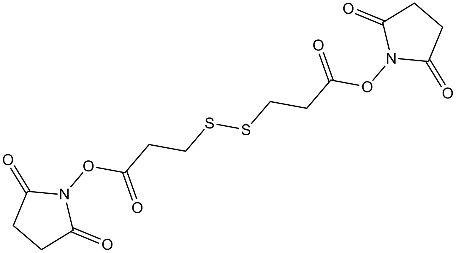
DSP는 라이신과 같은 아미노 기와 반응하는 동형 이기능성 교차결합제로 표면 기능화에 널리 사용되고 금 표면에 효소와 항체를 고정하기 위해 선호되는 연결 분자이다.
Products are for research use only. Not for human use. We do not sell to patients.
Cas No.: 57757-57-0
Sample solution is provided at 25 µL, 10mM.
_1-3.$$$39978408@51.jpg');)
_1-9.$$$38538795@65.jpg');)
_1-9.$$$39112701@51.jpg');)
_1-7.$$$37316672@70.jpg');)
_366-372.$$$36198801@70.jpg');)
.$$$38565142@65.jpg');)
_1867-1883.$$$33248023@67.jpg');)
_eads6055.$$$39977546@45.jpg');)
_eadm9805.$$$40080571@45.jpg');)
_eadj3020.$$$39666821@45.jpg');)
_1-20.$$$39757301@28.jpg');)
_1-24.$$$38898113@28.jpg');)
_273-287.$$$36806353@46.jpg');)
_1-16.$$$37156877@44.jpg');)
_1-19.$$$37460805@44.jpg');)
_1291-1307.$$$34518654@46.jpg');)
DSP는 라이신과 같은 아미노 기와 반응하는 동형 이기능성 교차결합제로 표면 기능화에 널리 사용되고 금 표면에 효소와 항체를 고정하기 위해 선호되는 연결 분자이다. DSP는 구조 내에 이황화물 다리가 있고 금과 결합할 때 분리된다[2]. 금박막 (GTF)에 DSP를 고정하면 일차아민과 DSP의 NHS 에스테르 간의 특이 반응을 통해 DA를 선택적으로 포획할 수 있고 인간 뇌척수액(CSF) 시료에서 DA를 민감하게 검출할 수 있게 한다[4-5]. DSP는 불가역적 바이오틴화 절차 (IBP)에서 사용되고 S-니트로소화 검출 중 분자 간 이황화물 결합의 간섭을 제거할 수 있다.게다가 DSP는 약한 단백질 복합체를 분리하고 Au-TiO₂ 박막을 준비해서 면역 분석 실험에 사용할 수도 있다[3, 7].
References:
[1] Darder M, Takada K, Pariente F, et al. Dithiobissuccinimidyl propionate as an anchor for assembling peroxidases at electrodes surfaces and its application in a H2O2 biosensor. Anal Chem. 1999;71(24):5530-5537
[2] Ataman Sadık D, Boyacı İH, et al. Mixed monolayer decorated SPR sensing surface for thrombin detection. J Pharm Biomed Anal. 2019;176:112822.
[3] Akaki K, Mino T, Takeuchi O. DSP-crosslinking and Immunoprecipitation to Isolate Weak Protein Complex. Bio Protoc. 2022;12(15):e4478.
[4] Zhang K, Liu Y, Wang Y, et al. Quantitative SERS Detection of Dopamine in Cerebrospinal Fluid by Dual-Recognition-Induced Hot Spot Generation. ACS Appl Mater Interfaces. 2018;10(18):15388-15394.
[5] Gu H, Guo Y, Xiao X, et al. Double molecular recognition strategy based on boronic acid-diol and NHS ester-amine for selective electrochemical detection of cerebral dopamine. Anal Bioanal Chem. 2020;412(15):3727-3736.
[6] Huang B, Chen C. Detection of protein S-nitrosation using irreversible biotinylation procedures (IBP). Free Radic Biol Med. 2010;49(3):447-456.
[7] Meira DI, Barbosac AI, Proença M, et al. Immobilizing antibody biorecognition layers on Au–TiO2 thin films: direct (physisorption) vs. DSP-crosslinking (chemisorption) surface functionalization. Journal of Physics D: Applied Physics. 2024;415401.
이 계획은 단지 가이드를 제공하고 필요에 따라 수정하시기 바랍니다.
(1) DSP-교차결합과 면역침전을 통해 약한 단백질 복합체를 분리[1] :
A. 관심 단백질(POIs)을 발현하는 세포 준비
1.각 시료당 10cm 접시에 8.0×10⁵개의 HeLa 세포를 배치(접시당 10% FBS를 포함한 10mL DMEM).
2.세포를 37°C, 5% CO₂에서 24시간 동안 배양합니다.
3.FLAG-HA 태그가 부착된 POI 발현을 위한 플라스미드를 변환합니다.
4.세포를 37°C, 5% CO₂에서 하루 동안 배양합니다.
B. 세척 버퍼와 IP 버퍼 준비
1.DSP-교차결합과 면역침전 당일, 교차결합 전에 세척 버퍼와 IP 버퍼를 준비합니다. 얼음 위에 보관하세요.
C. DSP-교차결합
1.DSP-교차결합 전에 100mM DSP를 준비합니다.
2.100mM DSP를 37°C로 예열한 (37°C)PBS로 0.1mM DSP로 희석합니다.
3. 미리 데운 (37°C) PBS/접시 5mL로 접시를 두 번 헹구합니다.
4.접시에서 PBS를 버립니다.
5.각 접시에 0.1mM DSP (단계 C2에서 준비) 5mL를 추가합니다.
6.접시를 CO₂ 배양기 내 37°C에서 30분간 배양합니다.
D. 첫 번째 IP를 위한 구슬 준비
1.DSP-교차결합 중 (단계 C6)에 1.5mL 튜브 하나에 시료당 40μL의 Dynabeads Protein G를 준비합니다.
2.세척 버퍼와 같은 양으로 Dynabeads Protein G를 세 번 세척합니다: Dynabeads Protein G가 들어 있는 튜브를 자기 스탠드에 놓고 비드가 자석에 축적되도록 합니다. 펑크트로 상청액을 버립니다. 얼음물 세척 버퍼를 넣습니다. 튜브를 자기 스탠드에서 제거합니다. 펑크트로 위아래로 흔들어 구슬을 소생시킵니다. 단계 D2를 두 번 더 반복합니다.
3.세척된 구슬가 들어 있는 튜브를 자기 스탠드에 놓고 상청액을 버립니다.
4.얼음물 IP 버퍼로 구슬을 소생시킵니다.
5.시료당 anti-FLAG 항체 1μL를 추가합니다.
6.회전 배양기에서 4°C에서 1시간 동안 구슬를 배양합니다.
E. DSP-교차결합 중지
1.단계 C6에서 30분간 배양한 후 0.1mM DSP를 버리고 접시당 37°C로 예열한 PBS 5mL로 접시를 한 번 헹구십시오.
2.PBS를 버리고 각 접시에 실온 STOP 용액 5mL를 추가합니다.
3.접시를 실온에서 15분간 배양합니다.
F. 첫 번째 IP
1.접시에서 STOP 용액을 버립니다.
2. 5mL의 얼음장 PBS/접시로 접시를 두 번 헹구합니다.
3.접시에서 PBS를 버립니다.
4.각 접시에 얼음물 IP 버퍼 500μL를 추가합니다.
5.스크래퍼로 세포를 긁어내고 접시에 있는 모든 IP 버퍼와 함께 세포를 1.5mL 튜브로 옮깁니다.
6.위아래로 펑크트로를 사용해서 세포를 소생시킵니다.
7.세포를 완전히 파괴하기 위해 튜브를 얼음 위에서 10분 동안 배양합니다.
8.4°C에서 20,000×g로 5분 동안 세포 파괴물이 담긴 튜브를 원심분리합니다.
9.상청액 500μL를 새로운 1.5mL 튜브로 옮겨 IP를 위해 사용합니다.
10.상청액이 있는 튜브에 D6 단계에서 준비한 anti-FLAG 항체가 결합된 구슬 40μL를 추가합니다.
11.튜브를 4°C에서 2시간 동안 회전 배양기에서 배양합니다.
G. 두 번째 IP를 위한 구슬 준비
1.F11 단계의 2시간 배양이 끝나기 30분 전경에 D1에서 D4까지의 단계를 따라 새로운 Dynabeads Protein G를 준비하고 세척합니다.
2.시료당 anti-HA 항체 1μL를 추가합니다.
3.4°C에서 1시간 동안 회전 배양기에서 구슬을 배양합니다.
H. FLAG 펩타이드로 단백질 용출
1.FLAG 펩타이드 스톡 용액을 TBS로 희석해서 FLAG 용출 버퍼를 준비하고 얼음 위에 보관합니다.
2.F11 단계의 2시간 배양 후 얼음물 세척 버퍼 700μL로 구슬을 세척합니다.
3.구슬에서 세척 버퍼를 버립니다.
4.H1 단계에서 준비한 FLAG 용출 버퍼 100μL를 사용해서 위아래로 펑크트로를 사용하고 부드럽게 구슬을 소생시킵니다.
5.4°C에서 회전 배양기에서 FLAG 용출 버퍼와 함께 구슬를 10분간 배양합니다.
6.튜브를 자기 스탠드에 놓고 상청액(관심 단백질과 결합된 단백질을 포함)을 새로운 1.5mL 튜브로 옮깁니다.
7.H4에서 H5까지의 단계를 한 번 더 반복합니다.
8.튜브를 자기 스탠드에 놓고 상청액을 H6 단계에서 처음 용출된 단백질이 있는 튜브로 옮깁니다.
9.수집된 상청액에 얼음물 IP 버퍼 300μL를 추가합니다.
I. 두 번째 IP
1.H9 단계의 상청액 500μL를 G3 단계에서 준비한 anti-HA 항체가 결합된 구슬 40μL과 혼합합니다.
2.4°C에서 2시간 동안 회전 배양기에서 구슬을 배양합니다.
J. 최종 용출
1.I2 단계의 2시간 배양 후 얼음물 세척 버퍼 700μL로 구슬을 세 번 세척합니다.
(2) 쥐 뇌 엑스트랙트의 DSP 처리[2]:
1.쥐 뇌 엑스트랙트는 분자 간 이황화물 결합(Pr-S-S-Pr)을 체외 실험에서 형성하기 위해 5mM DSP로 30분간 처리되었고 Tris 버퍼를 추가해서 중지되었습니다.
2.엑스트랙트는 원래 바이오틴 전환 방법과 불가역적 바이오틴화 절차(IBP)를 통해 분석되었습니다.
(3) 면역 분석 실험을 위한 Au-TiO₂ 박막을 DSP로 준비[3] :
1.플라즈마 활성화된 Au-TiO₂ 박막은 DMSO 용매(디메틸술폰, 무수, ≥99.9%)에 다양한 농도(0.4, 1, 4, 10mg/ml)로 녹인 DSP 용액으로 기능화되었습니다.
2.고정 시간은 30분이고 DMSO, PBS(Phosphate-Buffered Saline; pH 7.4; 10mM), 96% 에탄올 용액을 사용한 세척 과정을 거쳐 결합되지 않은 교차결합 분자를 제거합니다.
References:
[1] Akaki K, Mino T, Takeuchi O. DSP-crosslinking and Immunoprecipitation to Isolate Weak Protein Complex. Bio Protoc. 2022;12(15):e4478.
[2] Huang B, Chen C. Detection of protein S-nitrosation using irreversible biotinylation procedures (IBP). Free Radic Biol Med. 2010;49(3):447-456.
[3] Meira DI, Barbosac AI, Proença M, et al. Immobilizing antibody biorecognition layers on Au–TiO2 thin films: direct (physisorption) vs. DSP-crosslinking (chemisorption) surface functionalization. Journal of Physics D: Applied Physics. 2024;415401.
| Cas No. | 57757-57-0 | SDF | |
| Synonyms | 3,3′-Dithiodipropionic acid di | ||
| Formula | C14H16N2O8S2 | M.Wt | 404.41 |
| Solubility | DMF: 30 mg/ml,DMSO: 30 mg/ml,DMSO:PBS (pH 7.2) (1:4): 0.20 mg/ml | Storage | Store at -20°C, protect from light, stored under nitrogen |
| General tips | Please select the appropriate solvent to prepare the stock solution according to the
solubility of the product in different solvents; once the solution is prepared, please store it in
separate packages to avoid product failure caused by repeated freezing and thawing.Storage method
and period of the stock solution: When stored at -80°C, please use it within 6 months; when stored
at -20°C, please use it within 1 month. To increase solubility, heat the tube to 37°C and then oscillate in an ultrasonic bath for some time. |
||
| Shipping Condition | Evaluation sample solution: shipped with blue ice. All other sizes available: with RT, or with Blue Ice upon request. | ||
| Prepare stock solution | |||

|
1 mg | 5 mg | 10 mg |
| 1 mM | 2.4727 mL | 12.3637 mL | 24.7274 mL |
| 5 mM | 494.5 μL | 2.4727 mL | 4.9455 mL |
| 10 mM | 247.3 μL | 1.2364 mL | 2.4727 mL |
Step 1: Enter information below (Recommended: An additional animal making an allowance for loss during the experiment)
 g
μL
g
μL
Step 2: Enter the in vivo formulation (This is only the calculator, not formulation. Please contact us first if there is no in vivo formulation at the solubility Section.)
Calculation results:
Working concentration: mg/ml;
Method for preparing DMSO master liquid: mg drug pre-dissolved in μL DMSO ( Master liquid concentration mg/mL, Please contact us first if the concentration exceeds the DMSO solubility of the batch of drug. )
Method for preparing in vivo formulation: Take μL DMSO master liquid, next addμL PEG300, mix and clarify, next addμL Tween 80, mix and clarify, next add μL saline, mix and clarify.
Method for preparing in vivo formulation: Take μL DMSO master liquid, next add μL Corn oil, mix and clarify.
Note: 1. Please make sure the liquid is clear before adding the next solvent.
2. Be sure to add the solvent(s) in order. You must ensure that the solution obtained, in the previous addition, is a clear solution before proceeding to add the next solvent. Physical methods such as vortex, ultrasound or hot water bath can be used to aid dissolving.
3. All of the above co-solvents are available for purchase on the GlpBio website.
Quality Control & SDS
- View current batch:
- Purity: >97.00% Appearance: A solid
- COA (Certificate of Analysis)
- SDS (Safety Data Sheet)
- Datasheet
Average Rating: 5 (Based on Reviews and 25 reference(s) in Google Scholar.)