

PY-60 |
Catalog No.GC62424
 One-Click Copy Product Info
|

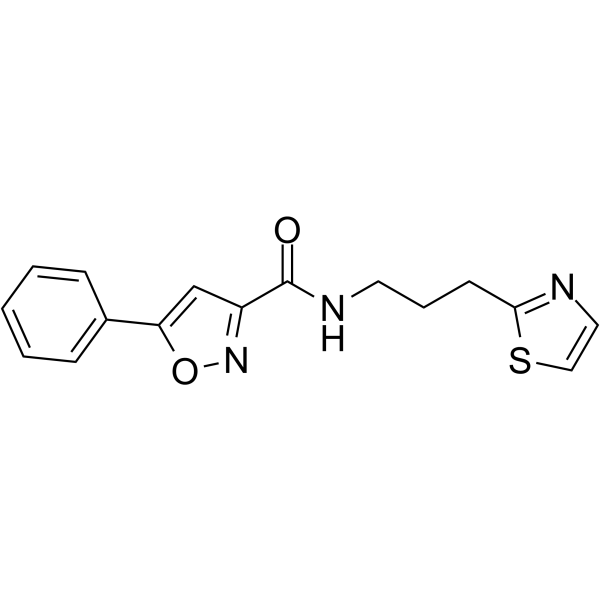
PY-60 est un dérivé substitué par un thiazole avec 5-phénylisoxazole comme structure de base, qui peut activer de manière potentielle et spécifique l'activité transcriptionnelle de YAP (EC50 = 1.6µM).
Products are for research use only. Not for human use. We do not sell to patients.
Cas No.: 2765218-56-0
Sample solution is provided at 25 µL, 10mM.
PY-60 est un dérivé substitué par un thiazole avec 5-phénylisoxazole comme structure de base, qui peut activer de manière potentielle et spécifique l'activité transcriptionnelle de YAP (EC50 = 1.6µM) [1]. PY-60 promeut la libération d' ANXA2 à partir de la membrane cellulaire, affaiblit sa restriction de liaison à YAP, permettant ainsi d'améliorer la déphosphorylation de YAP et de favoriser sa transclocation dans le noyau, activant ainsi la fonction de co-activateur transcriptionnel de YAP [1-2]. PY-60 est communément utilisé pour la régénération et la réparation tissulaires [3].
Dans les cellules de rétinoblastome humain (RB) Y79 et RB3823, le traitement avec PY-60 (0, 1, 5, 10μM; 24h) a entraîné une YAP transcriptionnellement active, ainsi donc, a inhibé la prolifération des cellules RB Y79 et RB3823[4]. Dans les cellules MDA-MB-231, PY-60 (0, 2, 4, 8μM; 24h) a inversé l'effet inhibiteur de la surexpression de RFX2 sur les cellules LUAD [6]. Dans les cellules HuCCT1, PY-60 (10μM; 24h) a inversé les effets inhibiteurs de Knockdown de PIN1 sur la migration, l'adhésion et l'invasion cellulaires [7].
Dans les rats C57BL/6 WT, le traitement avec PY-60 (10mg/mL; application topique; 10j) améliore le potentiel de formation de colonies des kératinocytes épidermiques primaires dans les rats[1]. Dans le modèle d'hémorragie sous-arachnoÏdienne chez le rat, PY-60 (0.2mg/kg; administration intraventriculaire; injection seule) a partiellement éliminé l'effet neuroprotecteur de NE-52-QQ57 [8].
References:
[1]. Shalhout SZ, Yang PY, Grzelak EM, et al. YAP-dependent proliferation by a small molecule targeting annexin A2. Nature Chemical Biology. 2021 Jul; 17(7): 767-775.
[2]. Martin JF. Regeneration and rejuvenation of skin by a topical YAP activator. Proceedings of the National Academy of Sciences. 2023 Aug 8; 120(32): e2309991120.
[3]. Grzelak EM, Elshan ND, Shao S, et al. Pharmacological YAP activation promotes regenerative repair of cutaneous wounds. Proceedings of the National Academy of Sciences. 2023 Jul 11; 120(28): e2305085120.
[4]. Zhong L, Meng X, Huang J, et al. Expression of yap suppresses cell proliferation and elevates the sensitivity of chemotherapy in retinoblastoma cells through lipid-peroxidation induced ferroptosis. Chinese Clinical Oncology. 2023 Oct 31; 12(5): 52.
[5]. Chen J, Su X, Tan Q, et al. Effect of cell density on the malignant biological behavior of breast cancer by altering the subcellular localization of ANXA2 and its clinical implications. Clinical and Translational Oncology. 2022 Nov; 24(11): 2136-2145.
[6]. Kong Z, Zhou P, Xu J, et al. RFX2 downregulates RASSF1 expression and YAP phosphorylation through Hippo signaling to promote immune escape in lung adenocarcinoma. Cell Division. 2025 Mar 11; 20(1): 7.
[7]. Wang Y, Liu Y, Chen H, et al. PIN1 promotes the metastasis of cholangiocarcinoma cells by RACK1-mediated phosphorylation of ANXA2. Cellular Oncology. 2024 Aug; 47(4): 1233-1252.
[8]. He Q, Zhou Y, Wu L, et al. Inhibition of acid-sensing receptor GPR4 attenuates neuronal ferroptosis via RhoA/YAP signaling in a rat model of subarachnoid hemorrhage. Free Radical Biology and Medicine. 2024 Nov 20; 225: 333-345.
| Expériences cellulaires [1]: | |
Lignées cellulaires | Cellules de rétinoblastome humain Y79 et RB3823 |
Méthode de préparation | La viabilité cellulaire des cellules de rétinoblastome humain Y79 et RB3823 a été détectée à l'aide de Kit MTS. Les cellules ont été ensemencées dans une plaque à 96 puits à la densité de [1-2]×103 cellules par puits. Après 24 heures de culture, PY-60 à différentes concentrations (0, 1, 5, 10μM) ont été ajouté pour traiter les cellules, et 20µL réactif MTS a été ajouté à chaque puits pendant les 4 dernières heures. |
Conditions de réaction | 0, 1, 5, 10μM; 24h |
Domaines d'application | La prolifération cellulaire des cellules Y79 et RB3823 a été supprimée par PY-60 à 10µM, confirmé par le test MTS. |
| Expériences animales [2]: | |
Modèles animaux | Rats C57BL/6 |
Méthode de préparation | Les rats C57BL/6 WT âgés de 8 semaines ont été traités une fois par jour (de manière topique, sur la peau) pendant dix jours consécutifs soit avec seulement le véhicule (acétone) soit avec PY-60 (10mg/mL). L'épiderme a été évalué pour son épaisseur à l'aide de la coloration H&E. Les colorations immunofluorescentes anti-K14 et anti-K167 ont été utilisées pour évaluer l'expansion cellulaire. Dans tous les modèles, les animaux ont été aléatoirement divisés en groupes de poids corporel moyen égal avant l'administration topique. |
Forme de dosage | 10mg/mL; application topique; 10j |
Domaines d'application | le traitement avec PY-60 améliore le potentiel de formation de colonies des kératinocytes épidermiques primaires dans les rats. |
References: | |
| Cas No. | 2765218-56-0 | SDF | |
| Formula | C16H15N3O2S | M.Wt | 313.37 |
| Solubility | DMSO : 100 mg/mL (319.11 mM; Need ultrasonic) | Storage | Store at -20°C |
| General tips | Please select the appropriate solvent to prepare the stock solution according to the
solubility of the product in different solvents; once the solution is prepared, please store it in
separate packages to avoid product failure caused by repeated freezing and thawing.Storage method
and period of the stock solution: When stored at -80°C, please use it within 6 months; when stored
at -20°C, please use it within 1 month. To increase solubility, heat the tube to 37°C and then oscillate in an ultrasonic bath for some time. |
||
| Shipping Condition | Evaluation sample solution: shipped with blue ice. All other sizes available: with RT, or with Blue Ice upon request. | ||
| Prepare stock solution | |||

|
1 mg | 5 mg | 10 mg |
| 1 mM | 3.1911 mL | 15.9556 mL | 31.9112 mL |
| 5 mM | 638.2 μL | 3.1911 mL | 6.3822 mL |
| 10 mM | 319.1 μL | 1.5956 mL | 3.1911 mL |
Step 1: Enter information below (Recommended: An additional animal making an allowance for loss during the experiment)
 g
μL
g
μL
Step 2: Enter the in vivo formulation (This is only the calculator, not formulation. Please contact us first if there is no in vivo formulation at the solubility Section.)
Calculation results:
Working concentration: mg/ml;
Method for preparing DMSO master liquid: mg drug pre-dissolved in μL DMSO ( Master liquid concentration mg/mL, Please contact us first if the concentration exceeds the DMSO solubility of the batch of drug. )
Method for preparing in vivo formulation: Take μL DMSO master liquid, next addμL PEG300, mix and clarify, next addμL Tween 80, mix and clarify, next add μL saline, mix and clarify.
Method for preparing in vivo formulation: Take μL DMSO master liquid, next add μL Corn oil, mix and clarify.
Note: 1. Please make sure the liquid is clear before adding the next solvent.
2. Be sure to add the solvent(s) in order. You must ensure that the solution obtained, in the previous addition, is a clear solution before proceeding to add the next solvent. Physical methods such as vortex, ultrasound or hot water bath can be used to aid dissolving.
3. All of the above co-solvents are available for purchase on the GlpBio website.
Quality Control & SDS
- View current batch:
- Purity: >98.00% Appearance: A solid
- COA (Certificate of Analysis)
- SDS (Safety Data Sheet)
- Datasheet
Average Rating: 5 (Based on Reviews and 27 reference(s) in Google Scholar.)