

Calcitriol (Synonyms: 1,25-dihydroxy Cholecalciferol, 1α,25-dihydroxy Cholecalciferol, 1,25-dihydroxy Vitamin D3, 1α,25-dihydroxy Vitamin D3) |
Catalog No.GC13852
 One-Click Copy Product Info
|

Calcitriol, como el metabolito más biológicamente activo derivado de la hormona secosteroide vitamina D, la inhibición de calcitriol causó una disminución en los efectos de los fármacos anticancerígenos.
Products are for research use only. Not for human use. We do not sell to patients.
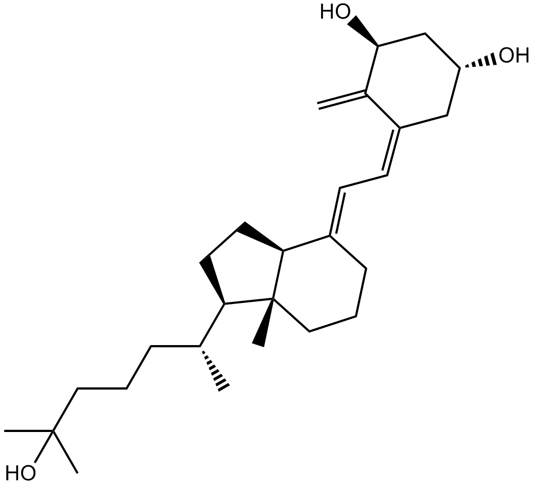
Cas No.: 32222-06-3
Sample solution is provided at 25 µL, 10mM.
Calcitriol, como el metabolito más biológicamente activo derivado de la hormona secosteroide vitamina D, la inhibición de calcitriol causó una disminución en los efectos de los fármacos anticancerígenos.[1]
En una prueba de eficacia in vitro se mostró que la administración oral semanal de calcitriol permitió alcanzar concentraciones séricas máximas de calcitriol muy superiores a 1 nM, una concentración que inhibe más del 50% de la proliferación del cáncer de próstata. [2] Con 10 nM de calcitriol se redujo notablemente la expresión de la proteína RAGE y aumentaron las concentraciones de sRAGE en cardiomiocitos HL-1 en comparación con las células de control.[3] El calcitriol exhibió efectos antiproliferativos contra T47D, MCF-7 y MDA-MB-231 con valores de IC50 en el rango de 0,05-0,25 μM.[4] Además, el calcitriol inhibe la proliferación de células de melanoma con un IC₅₀ de 0,24 μM.[5] Las células BT-474 fueron inhibidas en su crecimiento de manera dosis-dependiente por calcitriol con un IC50 de 2,9 nM. Con 1 nM de calcitriol se mejoraron sinérgicamente los efectos antiproliferativos de AZD4547, permitiendo una reducción de la dosis de AZD4547 a la mitad. [6]
En una prueba in vivo se demostró que el calcitriol (0,03 μg/kg) 5 veces/semana por vía intraperitoneal durante 10 semanas en ratones UNX ApoE-/- causó una calcificación vascular significativa y una mayor expresión de proteínas relacionadas (BMP2, RANKL y Runx2).[8] Con 0,5 ug/día de calcitriol administrado oralmente se mejoró la resistencia a la insulina y el HOMA-β después de 6 meses en pacientes con ND, sin embargo, solo se mejoró el HOMA-β en los pacientes en diálisis, sin un efecto obvio sobre la resistencia a la insulina.[7]
References:
[1]Ma J, et al. The mechanism of calcitriol in cancer prevention and treatment. Curr Med Chem. 2013;20(33):4121-30.
[2]Beer TM. Development of weekly high-dose calcitriol based therapy for prostate cancer. Urol Oncol. 2003 Sep-Oct;21(5):399-405.
[3]Lee TW, et al. ADAM10 modulates calcitriol-regulated RAGE in cardiomyocytes. Eur J Clin Invest. 2017 Sep;47(9):675-683.
[4]Aljunidee KA, et al. Combination therapy of calcitriol inhibits the proliferation of breast cancer cells: new concept of nonclassical function of calcitriol. Horm Mol Biol Clin Investig. 2021 Nov 15.
[5]Sutedja EK, et al. Calcitriol Inhibits Proliferation and Potentially Induces Apoptosis in B16-F10 Cells. Med Sci Monit Basic Res. 2022 May 5;28:e935139.
[6]Morales-Guadarrama G, et al. AZD4547 and calcitriol synergistically inhibited BT-474 cell proliferation while modified stemness and tumorsphere formation. J Steroid Biochem Mol Biol. 2022 May 31;223:106132.
[7]Lu Y, et al. Effects of active vitamin D on insulin resistance and islet β-cell function in non-diabetic chronic kidney disease patients: a randomized controlled study. Int Urol Nephrol. 2022 Jul;54(7):1725-1732.
[8]Becker LE, et al. Effect of paricalcitol and calcitriol on aortic wall remodeling in uninephrectomized ApoE knockout mice. Am J Physiol Renal Physiol. 2011 Mar;300(3):F772-82.
| Experimento celular [1]: | |
Líneas celulares | HL-1 cells |
Método de preparación | Calcitriol (1 y 10 nM) se utilizó para tratar las células HL-1 durante 48 horas para determinar el efecto inicial de la dosis-respuesta. Para estudiar la relevancia funcional de la modulación de FGFR1, se administró FGF-2 recombinante de ratón (25 ng/mL) (durante 0,5 o 48 horas) en células HL-1 control y tratadas con calcitriol. |
Condiciones de reacción | 1 y 10 nM; 48 horas |
Aplicaciones | En comparación con las células control, como se muestra en la Figura 1a, el calcitriol (1 y 10 nM) redujo de manera dosis-dependiente la expresión de la proteína FGFR1 en células HL-1 en un 31% y 62%, respectivamente. De manera similar, las células HL-1 tratadas con calcitriol (10 nM) tenían una expresión de mRNA de FGFR1 más baja que las células HL-1 control. |
| Experimento animal [2]: | |
Modelos animales | Ratones macho C57BL/6J de 4 a 5 semanas de edad y un peso de ~21-27 g |
Método de preparación | Se mantuvieron cuatro grupos de ratones (n = 11 cada uno) con una dieta baja en grasas (LFD) o una dieta alta en grasas (HFD) con y sin 150 UI/kg/día de calcitriol administrado oralmente durante 16 semanas. |
Forma de dosificación | 150 UI/kg/día; vía oral |
Aplicaciones | Se observó una disminución gradual significativa en el peso de los ratones alimentados con HFD y tratados con calcitriol en comparación con un aumento constante en los controles. Además, el tratamiento con calcitriol redujo las concentraciones de varios marcadores inflamatorios, incluyendo TNF-α, CRP e IL-6. |
Referencias: [1]. Lee TW, et al. Calcitriol downregulates fibroblast growth factor receptor 1 through histone deacetylase activation in HL-1 atrial myocytes. J Biomed Sci. 2018 May 18;25(1):42. [2]. Alkharfy KM, et al. Calcitriol attenuates weight-related systemic inflammation and ultrastructural changes in the liver in a rodent model. Basic Clin Pharmacol Toxicol. 2013 Jan;112(1):42-9. | |
| Cas No. | 32222-06-3 | SDF | |
| Sinónimos | 1,25-dihydroxy Cholecalciferol, 1α,25-dihydroxy Cholecalciferol, 1,25-dihydroxy Vitamin D3, 1α,25-dihydroxy Vitamin D3 | ||
| Chemical Name | (1R,3S,5Z)-5-[(2E)-2-[(1R,3aS,7aR)-1-[(2R)-6-hydroxy-6-methylheptan-2-yl]-7a-methyl-2,3,3a,5,6,7-hexahydro-1H-inden-4-ylidene]ethylidene]-4-methylidenecyclohexane-1,3-diol | ||
| Canonical SMILES | CC(CCCC(C)(C)O)C1CCC2C1(CCCC2=CC=C3CC(CC(C3=C)O)O)C | ||
| Formula | C27H44O3 | M.Wt | 416.64 |
| Solubility | ≥ 20.832mg/mL in DMSO | Storage | -20°C, protect from light, stored under nitrogen,unstable in solution, ready to use. |
| Shipping Condition | Evaluation sample solution: shipped with blue ice. All other sizes available: with RT, or with Blue Ice upon request. | ||
| Prepare stock solution | |||

|
1 mg | 5 mg | 10 mg |
| 1 mM | 2.4002 mL | 12.0008 mL | 24.0015 mL |
| 5 mM | 480 μL | 2.4002 mL | 4.8003 mL |
| 10 mM | 240 μL | 1.2001 mL | 2.4002 mL |
Step 1: Enter information below (Recommended: An additional animal making an allowance for loss during the experiment)
 g
μL
g
μL
Step 2: Enter the in vivo formulation (This is only the calculator, not formulation. Please contact us first if there is no in vivo formulation at the solubility Section.)
Calculation results:
Working concentration: mg/ml;
Method for preparing DMSO master liquid: mg drug pre-dissolved in μL DMSO ( Master liquid concentration mg/mL, Please contact us first if the concentration exceeds the DMSO solubility of the batch of drug. )
Method for preparing in vivo formulation: Take μL DMSO master liquid, next addμL PEG300, mix and clarify, next addμL Tween 80, mix and clarify, next add μL saline, mix and clarify.
Method for preparing in vivo formulation: Take μL DMSO master liquid, next add μL Corn oil, mix and clarify.
Note: 1. Please make sure the liquid is clear before adding the next solvent.
2. Be sure to add the solvent(s) in order. You must ensure that the solution obtained, in the previous addition, is a clear solution before proceeding to add the next solvent. Physical methods such as vortex, ultrasound or hot water bath can be used to aid dissolving.
3. All of the above co-solvents are available for purchase on the GlpBio website.
Quality Control & SDS
- View current batch:
- Purity: >99.00% Appearance: A solid
- COA (Certificate of Analysis)
- SDS (Safety Data Sheet)
- Datasheet
-
Related Biological Data

VD reduces ferroptosis induced by high glucose in HK-2 cells and down-regulation of Klotho attenuates the protective effect of VD on ferroptosis in HK-2 cells. (F-K) Protein levels of SLC7A11, GPX4, TFR1, Klotho and p53 were detected by western blot.
HG + VD group (30 mmol/L D-glucose + 10 nmol/L 1,25(OH)2D3);1,25(OH)2D3 (GLPBIO, Beijing, China) was pretreated for 2 h before intervention.
Apoptosis (2024): 1-13. PMID: 38558206 IF: 7.2002 -
Related Biological Data

Effect of vitamin D deficiency on migration of human trophoblast cells. D&E: Cell migration was measured using wound-healing assay. After 12 and 24 h migration, the scratches were photographed and wounded areas were calculated.
And calcitriol (GLPBIO, China) was dissolved in DMSO at the final concentration of 1 mM, and the final concentration of calcitriol were 0.1 μM.
Placenta (2022). PMID: 36103800 IF: 3.2867
Average Rating: 5 (Based on Reviews and 24 reference(s) in Google Scholar.)