

CycLuc1 |
Catalog No.GC34894
 One-Click Copy Product Info
|

CycLuc1 es un sustrato de luciferasa sintético, permeable al cerebro, con una longitud de onda máxima de emisión λmax=604nm
Products are for research use only. Not for human use. We do not sell to patients.
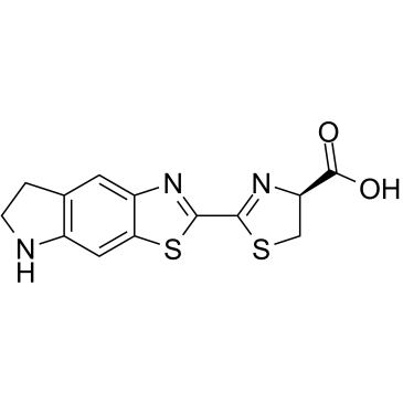
Cas No.: 1247879-16-8
Sample solution is provided at 25 µL, 10mM.
_1-3.$$$39978408@51.jpg');)
_1-9.$$$38538795@65.jpg');)
_1-9.$$$39112701@51.jpg');)
_1-7.$$$37316672@70.jpg');)
_366-372.$$$36198801@70.jpg');)
.$$$38565142@65.jpg');)
_1867-1883.$$$33248023@67.jpg');)
_eads6055.$$$39977546@45.jpg');)
_eadm9805.$$$40080571@45.jpg');)
_eadj3020.$$$39666821@45.jpg');)
_1-20.$$$39757301@28.jpg');)
_1-24.$$$38898113@28.jpg');)
_273-287.$$$36806353@46.jpg');)
_1-16.$$$37156877@44.jpg');)
_1-19.$$$37460805@44.jpg');)
_1291-1307.$$$34518654@46.jpg');)
CycLuc1 es un sustrato de luciferasa sintético, permeable al cerebro, con una longitud de onda máxima de emisión λmax=604nm[1]. CycLuc1 se puede utilizar para el análisis de imágenes de fluorescencia in vivo y estudios de caracterización in vitro de luciferasa mutante[2].
CycLuc1 se ha utilizado para monitorear el crecimiento tumoral del xenoinjerto por glioblastoma intracraneal (GBM) en ratones desnudos atímicos a dosis de 5-25mg/kg[3]. La inyección intraperitoneal de CycLuc1 a 15mg/kg mejoró significativamente las imágenes de bioluminiscencia in vivo de la expresión génica en el órgano subforniceal (SFO) o el núcleo paraventricular hiprtalámico (PVN) en ratones machos C57BL/6 (8-10 semanas de edad) [4]. Después de la inyección intravenosa (iv) de CycLuc1 (5mmol/L) en Pathogen-free BALB/c, FVB/N, C57BL/6 y ratones transgénicos que expresan luciferasa (FVB-Tg(CAG-luc,-GFP)L2G85Chco/FathJ), la señal CycLuc1 alcanzó su punto máximo de 4-5 minutos y luego produjo una señal estable que duró más de 60 minutos[5].
References:
[1] Kuchimaru T, Iwano S, Kiyama M, et al. A luciferin analogue generating near-infrared bioluminescence achieves highly sensitive deep-tissue imaging[J]. Nature communications, 2016, 7(1): 11856.
[2] Harwood K R, Mofford D M, Reddy G R, et al. Identification of mutant firefly luciferases that efficiently utilize aminoluciferins[J]. Cell Chemical Biology, 2011, 18(12): 1649-1657.
[3] Gupta S K, Kim M, Marin B M, et al. Synthetic luciferin, CycLuc1, improves bioluminescence imaging for intracranial glioblastoma xenografts[J]. Cancer Research, 2018, 78(13_Supplement): 4112-4112.
[4] Simonyan H, Hurr C, Young C N. A synthetic luciferin improves in vivo bioluminescence imaging of gene expression in cardiovascular brain regions[J]. Physiological Genomics, 2016, 48(10): 762-770.
[5] Evans M S, Chaurette J P, Adams Jr S T, et al. A synthetic luciferin improves bioluminescence imaging in live mice[J]. Nature methods, 2014, 11(4): 393-395.
Experimentos de imagen de bioluminiscencia basados en CycLuc1 en ratones vivos:
1.Prepare la solución CycLuc1:
La solución de tabajo 50μmol/L-5mmol/L CycLuc1 se prepara en tampón de PBS (pH7.4). Una vez completada la configuración, la esterilización de filtración se lleva a cabo utilizando una membrana de filtro de 0.22μm.
2. Alimentación de ratones:
A los ratones transgénicos sin patógenos BALB/c, FVB/N, C57BL/6 y que expresan luciferasa (FVB-Tg(CAG-luc,-GFP)L2G85Chco/FathJ) recibieron acceso a alimentos y agua ad libitum.
3. Inoculación de células tumorales:
La inoculación subcutánea de ratones involucró 1×10⁶ células CMT-64 o DB7 que expresan luciferasa suspendidas en un medio RPMI libre de suero. Los tumores alcanzaron su fase de crecimiento durante un lapso de 1 a 2 semanas antes de que comenzaran los procedimientos de imagen. Para establecer tumores 4T1, se mezclaron alícuotas de las células con la mezcla de células con Matrigel en una proporción de volumen 1:1 (5×10⁵ células con 50μl de medio DMEM sin suero combinado con 50μl de Matrigel), se implantaron en la segunda o cuarta almohadillas de grasa mamaria en los lados izquierdo o derecho. Los quelos Después de administrar 100μl de solución de D-luciferin o CycLuc1 a concentraciones que oscilaban entre 50μmol/L y 5mmol/L, los ratones recibieron imágenes 10 minutos después de la inyección.
4. Imágenes de bioluminiscenciag:
Administre anestesia con isoflurano (concentración al 2% en 1L/min de oxígeno) a los animales antes de capturar imágenes de bioluminiscencia utilizando el sistema IVIS Lumina. El protocolo de imágenes implicaba capturar imágenes cada 2 minutos durante un período de 30 minutos, cada uno con 10 segundos de tiempo de exposición. Adquiera imágenes de los ratones tanto 1 hora como 2 horas después de la inyección con cada imagen expuesta durante 10 segundos. Las imágenes se analizaron utilizando el Living Image software. Esbozar las regiones de interés (ROIs) alrededor de cada grupo celular para medir el recuento total de fotones dentro de cada área definida. Todas las imágenes mantuvieron el mismo tamaño de ratorno de la inversión.
Reference:
[1] Evans M S, Chaurette J P, Adams Jr S T, et al. A synthetic luciferin improves bioluminescence imaging in live mice[J]. Nature methods, 2014, 11(4): 393-395.
| Cas No. | 1247879-16-8 | SDF | |
| Canonical SMILES | O=C([C@@H]1N=C(C2=NC3=CC4=C(NCC4)C=C3S2)SC1)O | ||
| Formula | C13H11N3O2S2 | M.Wt | 305.38 |
| Solubility | DMSO : ≥ 83.33 mg/mL (272.87 mM) | Storage | Store at -20°C,protect from light |
| General tips | Please select the appropriate solvent to prepare the stock solution according to the
solubility of the product in different solvents; once the solution is prepared, please store it in
separate packages to avoid product failure caused by repeated freezing and thawing.Storage method
and period of the stock solution: When stored at -80°C, please use it within 6 months; when stored
at -20°C, please use it within 1 month. To increase solubility, heat the tube to 37°C and then oscillate in an ultrasonic bath for some time. |
||
| Shipping Condition | Evaluation sample solution: shipped with blue ice. All other sizes available: with RT, or with Blue Ice upon request. | ||
| Prepare stock solution | |||

|
1 mg | 5 mg | 10 mg |
| 1 mM | 3.2746 mL | 16.373 mL | 32.7461 mL |
| 5 mM | 654.9 μL | 3.2746 mL | 6.5492 mL |
| 10 mM | 327.5 μL | 1.6373 mL | 3.2746 mL |
Step 1: Enter information below (Recommended: An additional animal making an allowance for loss during the experiment)
 g
μL
g
μL
Step 2: Enter the in vivo formulation (This is only the calculator, not formulation. Please contact us first if there is no in vivo formulation at the solubility Section.)
Calculation results:
Working concentration: mg/ml;
Method for preparing DMSO master liquid: mg drug pre-dissolved in μL DMSO ( Master liquid concentration mg/mL, Please contact us first if the concentration exceeds the DMSO solubility of the batch of drug. )
Method for preparing in vivo formulation: Take μL DMSO master liquid, next addμL PEG300, mix and clarify, next addμL Tween 80, mix and clarify, next add μL saline, mix and clarify.
Method for preparing in vivo formulation: Take μL DMSO master liquid, next add μL Corn oil, mix and clarify.
Note: 1. Please make sure the liquid is clear before adding the next solvent.
2. Be sure to add the solvent(s) in order. You must ensure that the solution obtained, in the previous addition, is a clear solution before proceeding to add the next solvent. Physical methods such as vortex, ultrasound or hot water bath can be used to aid dissolving.
3. All of the above co-solvents are available for purchase on the GlpBio website.
Quality Control & SDS
- View current batch:
- Purity: >99.00% Appearance: A solid
- COA (Certificate of Analysis)
- SDS (Safety Data Sheet)
- Datasheet
Average Rating: 5 (Based on Reviews and 32 reference(s) in Google Scholar.)