

THK5351 |
Catalog No.GC30814
 One-Click Copy Product Info
|

THK5351, une fois marqué radioactivement, est un traceur d'imagerie de la protéine tau en tomographie par émission de positions (PET).
Products are for research use only. Not for human use. We do not sell to patients.
Cas No.: 1707147-26-9
Sample solution is provided at 25 µL, 10mM.
_1-3.$$$39978408@51.jpg');)
_1-9.$$$38538795@65.jpg');)
_1-9.$$$39112701@51.jpg');)
_1-7.$$$37316672@70.jpg');)
_366-372.$$$36198801@70.jpg');)
.$$$38565142@65.jpg');)
_1867-1883.$$$33248023@67.jpg');)
_eads6055.$$$39977546@45.jpg');)
_eadm9805.$$$40080571@45.jpg');)
_eadj3020.$$$39666821@45.jpg');)
_1-20.$$$39757301@28.jpg');)
_1-24.$$$38898113@28.jpg');)
_273-287.$$$36806353@46.jpg');)
_1-16.$$$37156877@44.jpg');)
_1-19.$$$37460805@44.jpg');)
_1291-1307.$$$34518654@46.jpg');)
THK5351, une fois marqué radioactivement, est un traceur d'imagerie de la protéine tau en tomographie par émission de positions (PET). THK5351 visualise les lésions pathologiques de la protéine tau dans le cerveau, par voie de connecter la structure en feuillet β de la protéine tau présente dans les enchevêtrements neurofibrillaires[1-4].
References:
[1] Brendel M, Wagner L, Levin J, et al. Perfusion-Phase [18F]THK5351 Tau-PET Imaging as a Surrogate Marker for Neurodegeneration. J Alzheimers Dis Rep. 2017 Sep 28;1(1):109-113.
[2] Kang JM, Lee SY, Seo S, et al. Tau positron emission tomography using [18F]THK5351 and cerebral glucose hypometabolism in Alzheimer's disease. Neurobiol Aging. 2017 Nov;59:210-219.
[3] Kikuchi A, Okamura N, Hasegawa T, et al. In vivo visualization of tau deposits in corticobasal syndrome by 18F-THK5351 PET. Neurology. 2016 Nov 29;87(22):2309-2316.
[4] Harada R, Ishiki A, Kai H, Sato N, et al. Correlations of 18F-THK5351 PET with Postmortem Burden of Tau and Astrogliosis in Alzheimer Disease. J Nucl Med. 2018 Apr;59(4):671-674.
Méthodes de chimie radiologique et d'imagerie TEP clinique pour 18F-THK5351[1-2]
Ce protocole est adapté de données de recherche et est fourni à titre indicatif uniquement. Des ajustements peuvent être nécessaires en fonction des exigences expérimentales spécifiques.
1.Synthèse radiochimique de 8F-THK5351 à partir de THK535:
(1)L'eau cible contenant du fluor-18 (18F-), produite par un cyclotron, passe dans une cartouche QMA Sep-Pak Light Accell Plus. On capte 18F- sur la cartouche, puis on l'élue dans un récipient de réaction à l'aide d'une solution de Kryptofix. On mélange l'éluat à de l'acétonitrile, et on élimine l'eau par distillation azéotropique pour obtenir un complexe sec : 18F-/Kryptofix 222/K₂CO₃.
(2)On ajoute THK5352 (3mg, 5.3μmol) dissous dans 0,7ml de DMSO au complexe sec dans le récipient de réaction. On agite le mélange à 110°C pendant 10 minutes. Ensuite, on ajoute 0,2mL de HCI aqueux à 2mol/L, et on continue de agiter à 110°C pendant encore 3 minutes. Par la suite, on ajoute 1mL d' AcOK aqueux à 0,8mol/L et 7mL d'eau distillée pour arrêter la réaction.
(3)On élue le produit d'une cartouche C18 avec 60% d'éthanol. On mélange l'éluat éthanolique à l'eau, puis on le purifie par CLHP semi-préparative. Conditions de CLHP : colonne Inertsil ODS-4, phase mobile : 20mmol/L NaH₂PO₄ / acétonitrile (75/25, v/v), débit : 5,0mL/min. On collecte la fraction de CLHP correspondant à 18F-THK5351.
2.Procédure d'imagerie TEP : On injecte par voie intraveineuse une dose de 185MBq de 18F-THK5351. On réalise une analyse TEP dynamique pendant 90 minutes avec un scanner TEP. On reconstruit les images TEP et on calcule les valeurs de captation standardisées (SUV). On superpose les images IRM pondérées en T1 de l'individu avec les images TEP des premières minutes (0-10 minutes après l'injection). On normalise spatialement les images IRM et TEP superposées dans un espace de Talairach. On place manuellement ou semi-automatiquement des régions d'intérêt (ROIs) sur les images normalisées. On calcule le rapport de SUV de chaque ROI par rapport à SUV du cortex cérébelleux (SUVR), qui sert d'indice de rétention.
Précautions :
(1)Les opérateurs doivent respecter scrupuleusement les réglementations de protection contre les rayonnements ionisants, porter l'équipement de protection adapté, réaliser la filtration stérile finale et la distribution du produit dans ces conditions aseptiques.
References:
[1] Harada R, Okamura N, Furumoto S, et al. 18F-THK5351: A Novel PET Radiotracer for Imaging Neurofibrillary Pathology in Alzheimer Disease. J Nucl Med. 2016 Feb;57(2):208-14.
[2] Okamura N, Furumoto S, Harada R, et al. Novel 18F-labeled arylquinoline derivatives for noninvasive imaging of tau pathology in Alzheimer disease. J Nucl Med. 2013 Aug;54(8):1420-7.
| Cas No. | 1707147-26-9 | SDF | |
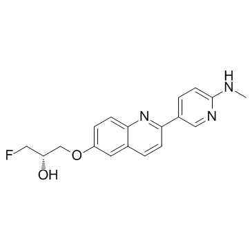
| Canonical SMILES | O[C@@H](COC1=CC=C2N=C(C3=CC=C(NC)N=C3)C=CC2=C1)CF | ||
| Formula | C18H18FN3O2 | M.Wt | 327.35 |
| Solubility | DMSO : 50 mg/mL (152.74 mM) | Storage | Store at -20°C |
| General tips | Please select the appropriate solvent to prepare the stock solution according to the
solubility of the product in different solvents; once the solution is prepared, please store it in
separate packages to avoid product failure caused by repeated freezing and thawing.Storage method
and period of the stock solution: When stored at -80°C, please use it within 6 months; when stored
at -20°C, please use it within 1 month. To increase solubility, heat the tube to 37°C and then oscillate in an ultrasonic bath for some time. |
||
| Shipping Condition | Evaluation sample solution: shipped with blue ice. All other sizes available: with RT, or with Blue Ice upon request. | ||
| Prepare stock solution | |||

|
1 mg | 5 mg | 10 mg |
| 1 mM | 3.0548 mL | 15.2742 mL | 30.5483 mL |
| 5 mM | 611 μL | 3.0548 mL | 6.1097 mL |
| 10 mM | 305.5 μL | 1.5274 mL | 3.0548 mL |
Step 1: Enter information below (Recommended: An additional animal making an allowance for loss during the experiment)
 g
μL
g
μL
Step 2: Enter the in vivo formulation (This is only the calculator, not formulation. Please contact us first if there is no in vivo formulation at the solubility Section.)
Calculation results:
Working concentration: mg/ml;
Method for preparing DMSO master liquid: mg drug pre-dissolved in μL DMSO ( Master liquid concentration mg/mL, Please contact us first if the concentration exceeds the DMSO solubility of the batch of drug. )
Method for preparing in vivo formulation: Take μL DMSO master liquid, next addμL PEG300, mix and clarify, next addμL Tween 80, mix and clarify, next add μL saline, mix and clarify.
Method for preparing in vivo formulation: Take μL DMSO master liquid, next add μL Corn oil, mix and clarify.
Note: 1. Please make sure the liquid is clear before adding the next solvent.
2. Be sure to add the solvent(s) in order. You must ensure that the solution obtained, in the previous addition, is a clear solution before proceeding to add the next solvent. Physical methods such as vortex, ultrasound or hot water bath can be used to aid dissolving.
3. All of the above co-solvents are available for purchase on the GlpBio website.
Quality Control & SDS
- View current batch:
- Purity: >98.00% Appearance: A solid
- COA (Certificate of Analysis)
- SDS (Safety Data Sheet)
- Datasheet
Average Rating: 5 (Based on Reviews and 28 reference(s) in Google Scholar.)