

3-Hydroxybutyric acid sodium |
カタログ番号GC60502
 One-Click Copy Product Info
|

3-ヒドロキシ酪酸ナトリウム (β-ヒドロキシ酪酸ナトリウム) は、I 型糖尿病で上昇する代謝産物です。
Products are for research use only. Not for human use. We do not sell to patients.
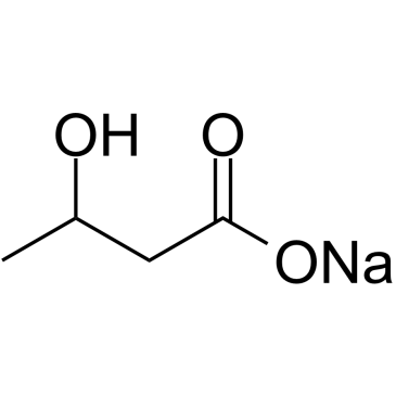
Cas No.: 150-83-4
Sample solution is provided at 25 µL, 10mM.
_1-3.$$$39978408@51.jpg');)
_1-9.$$$38538795@65.jpg');)
_1-9.$$$39112701@51.jpg');)
_1-7.$$$37316672@70.jpg');)
_366-372.$$$36198801@70.jpg');)
.$$$38565142@65.jpg');)
_1867-1883.$$$33248023@67.jpg');)
_eads6055.$$$39977546@45.jpg');)
_eadm9805.$$$40080571@45.jpg');)
_eadj3020.$$$39666821@45.jpg');)
_1-20.$$$39757301@28.jpg');)
_1-24.$$$38898113@28.jpg');)
_273-287.$$$36806353@46.jpg');)
_1-16.$$$37156877@44.jpg');)
_1-19.$$$37460805@44.jpg');)
_1291-1307.$$$34518654@46.jpg');)
3-Hydroxybutyric acid sodium is an endogenous ketone body metabolite with neuroprotective, anti-inflammatory and antioxidant effects. 3-Hydroxybutyric acid sodium enters cells as an energy substrate via monocarboxylate transporters and is converted to acetyl-CoA. 3-Hydroxybutyric acid sodium activates GPR109A and GPR41 receptors, inhibits histone deacetylases and modulates membrane lipid properties. 3-Hydroxybutyric acid sodium can be used in studies related to ketone body metabolism, cellular energy metabolism, neurological diseases such as epilepsy, cardiovascular diseases such as atherosclerosis and metabolic diseases such as methylmalonic acidemia[1-4].
In vitro, treatment of HEK293 cells with 1-32mM 3-hydroxybutyric acid sodium for 8 hours increased histone H3K9 and H3K14 acetylation and inhibited HDAC1, HDAC3 and HDAC4 activities[5]. In 5mM glucose medium, treatment of seven human breast cancer cell lines (BT20, BT474, HBL100, MCF-7, MDA-MB 231, MDA-MB 468, T47D) with 3mM 3-hydroxybutyric acid sodium for 1 to 5 days altered oxygen consumption rate and extracellular acidification rate of BT20 cells toward a more oxidative phenotype and increased extracellular acidification rate of MCF-7 and MDA-MB 468 cells[6]. Treatment of C6 cells with 1-25mM 3-hydroxybutyric acid sodium for 24 hours dose-dependently attenuated cell migration, inhibited caspase-1 activation and IL-1β maturation and reduced NLRP3 levels. Treatment of 200ng/ml LPS and 5mM ATP-induced C6 cells with 25mM 3-hydroxybutyric acid sodium for 24 hours attenuated LPS/ATP-promoted cell migration[7].
In vivo, tail vein injection of ddY mice with 3-100mg/kg/h 3-hydroxybutyric acid sodium for 30 minutes followed by exposure to hypoxic environment dose-dependently prolonged respiratory arrest latency[8]. Daily intragastric administration of apoE-/- mice with 50-200mg/kg/day 3-hydroxybutyric acid sodium for 9 weeks decreased body weight, reduced serum TNF-α, IL-6 and IL-1β levels and reduced overall aortic plaque burden, aortic root lesion area, intraplaque macrophage infiltration and lipid deposition[9]. After anterior cruciate ligament transection (ACLT) to establish an osteoarthritis model in Wistar rats, daily intraperitoneal injection of 50-200mg/kg 3-hydroxybutyric acid sodium for 8 weeks decreased serum nitric oxide, TNF-α, IL-6 and PGE2 levels, alleviated articular cartilage surface erosion and proteoglycan loss and reduced OARSI score[10].
References:
[1] Newman JC, Verdin E. β-Hydroxybutyrate: A Signaling Metabolite. Annu Rev Nutr. 2017 Aug 21;37:51-76.
[2] Soto-Mota A, Norwitz NG, Clarke K. Why a d-β-hydroxybutyrate monoester? Biochem Soc Trans. 2020 Feb 28;48(1):51-59.
[3] Wang L, Chen P, Xiao W. β-hydroxybutyrate as an Anti-Aging Metabolite. Nutrients. 2021 Sep 28;13(10):3420.
[4] Newman JC, Verdin E. β-hydroxybutyrate: much more than a metabolite. Diabetes Res Clin Pract. 2014 Nov;106(2):173-81.
[5] Shimazu T, Hirschey MD, Newman J, et al. Suppression of Oxidative Stress by β-Hydroxybutyrate, an Endogenous Histone Deacetylase Inhibitor. Science. 2013 Jan 11;339(6116):211-214.
[6] Bartmann C, Janaki Raman SR, Floter J, et al. Beta-hydroxybutyrate(3-OHB) can influence the energetic phenotype of breast cancer cells, but does not impact their proliferation and the response to chemotherapy or radiation. Cancer Metab. 2018;6:8.
[7] Shang S, Wang L, Zhang Y, et al. The Beta-Hydroxybutyrate Suppresses the Migration of Glioma Cells by Inhibition of NLRP3 Inflammasome. Cell Mol Neurobiol. 2018;38(8):1479-1489.
[8] Suzuki M, Suzuki M, Sato K, et al. Effect of β-Hydroxybutyrate, a Cerebral Function Improving Agent, on Cerebral Hypoxia, Anoxia and Ischemia in Mice and Rats. Jpn J Pharmacol. 2001;87(2):143-150.
[9] Zhang S, Li Z, Zhang Y, et al. Ketone Body 3-Hydroxybutyrate Ameliorates Atherosclerosis via Receptor Gpr109a-Mediated Calcium Influx. Adv Sci (Weinh). 2021 Jun;8(12):2003410.
[10] Zhuang H, Ren X, Zhang Y, et al. β-Hydroxybutyrate enhances chondrocyte mitophagy and reduces cartilage degeneration in osteoarthritis via the HCAR2/AMPK/PINK1/Parkin pathway. Aging Cell. 2024;23:e14294.
| Cell experiment [1]: | |
|
Cell lines |
C6 glioma cells (rat glioma cell line), LN229 cells (human glioblastoma cell line), U251 cells (human glioblastoma cell line) |
|
Preparation Method |
C6 glioma cells were maintained in Dulbecco's Modified Eagle's Medium-High glucose medium supplemented with 10% fetal bovine serum at 37°C, 5% CO2. C6/LN229/U251 cells were treated with 3-hydroxybutyric acid sodium (1-25mM) for 24 hours, then cell viability was assessed by MTT, cell migration was assessed by ECIS/wound-healing/Transwell, and NLRP3 inflammasome activation was assessed by Western blot for cleaved caspase-1 and mature IL-1β. |
|
Reaction Conditions |
1-25mM; 24h |
|
Applications |
3-hydroxybutyric acid sodium did not affect the viability of C6 glioma cells. 3-hydroxybutyric acid sodium attenuated the migration of C6, LN229 and U251 cells in a dose-dependent manner. 3-hydroxybutyric acid sodium inhibited the activation of NLRP3 inflammasome in C6 cells, reducing the levels of cleaved caspase-1 and mature IL-1β without altering pro-caspase-1 and pro-IL-1β expression. |
| Animal experiment [2]: | |
|
Animal models |
apoE-/- mice (male, 5 weeks old) |
|
Preparation Method |
Mice were fed a 1.25% high-cholesterol diet and concomitantly received daily intragastric administration of 3-hydroxybutyric acid sodium at 50-200mg/kg/day for 9 weeks; after sacrifice, serum cytokine levels, aortic plaque burden, aortic root Movat/CD68/Oil Red O staining, splenic macrophage flow cytometry and aortic protein extracts were analyzed. |
|
Dosage form |
50-200mg/kg/day; intragastric; once daily for 9 weeks |
|
Applications |
3-hydroxybutyric acid sodium decreased body weight, suppressed serum TNF-α, IL-6 and IL-1β, reduced overall aortic plaque burden, aortic root lesion area, intraplaque CD68+ macrophage infiltration and lipid deposition, decreased aortic iNOS and IL-6 protein while increasing ABCA1 protein, and lowered splenic M1 macrophage proportion. |
References: [1] Shang S, Wang L, Zhang Y, et al. The Beta-Hydroxybutyrate Suppresses the Migration of Glioma Cells by Inhibition of NLRP3 Inflammasome. Cell Mol Neurobiol. 2018 Dec;38(8):1479-1489. [2] Zhang S, Li Z, Zhang Y, et al. Ketone Body 3-Hydroxybutyrate Ameliorates Atherosclerosis via Receptor Gpr109a-Mediated Calcium Influx. Adv Sci (Weinh). 2021 May;8(9):2003410. | |
| Cas No. | 150-83-4 | SDF | |
| Canonical SMILES | CC(O)CC(O[Na])=O | ||
| Formula | C4H7NaO3 | M.Wt | 126.09 |
| 溶解度 | DMSO:10mg/ml | Storage | Store at RT |
| General tips | Please select the appropriate solvent to prepare the stock solution according to the
solubility of the product in different solvents; once the solution is prepared, please store it in
separate packages to avoid product failure caused by repeated freezing and thawing.Storage method
and period of the stock solution: When stored at -80°C, please use it within 6 months; when stored
at -20°C, please use it within 1 month. To increase solubility, heat the tube to 37°C and then oscillate in an ultrasonic bath for some time. |
||
| Shipping Condition | Evaluation sample solution: shipped with blue ice. All other sizes available: with RT, or with Blue Ice upon request. | ||
| Prepare stock solution | |||

|
1 mg | 5 mg | 10 mg |
| 1 mM | 7.9308 mL | 39.6542 mL | 79.3084 mL |
| 5 mM | 1.5862 mL | 7.9308 mL | 15.8617 mL |
| 10 mM | 793.1 μL | 3.9654 mL | 7.9308 mL |
Step 1: Enter information below (Recommended: An additional animal making an allowance for loss during the experiment)
 g
μL
g
μL
Step 2: Enter the in vivo formulation (This is only the calculator, not formulation. Please contact us first if there is no in vivo formulation at the solubility Section.)
Calculation results:
Working concentration: mg/ml;
Method for preparing DMSO master liquid: mg drug pre-dissolved in μL DMSO ( Master liquid concentration mg/mL, Please contact us first if the concentration exceeds the DMSO solubility of the batch of drug. )
Method for preparing in vivo formulation: Take μL DMSO master liquid, next addμL PEG300, mix and clarify, next addμL Tween 80, mix and clarify, next add μL saline, mix and clarify.
Method for preparing in vivo formulation: Take μL DMSO master liquid, next add μL Corn oil, mix and clarify.
Note: 1. Please make sure the liquid is clear before adding the next solvent.
2. Be sure to add the solvent(s) in order. You must ensure that the solution obtained, in the previous addition, is a clear solution before proceeding to add the next solvent. Physical methods such as vortex, ultrasound or hot water bath can be used to aid dissolving.
3. All of the above co-solvents are available for purchase on the GlpBio website.
Quality Control & SDS
- View current batch:
- Purity: >98.00% Appearance: A solid
- COA (Certificate of Analysis)
- SDS (Safety Data Sheet)
- Datasheet
Average Rating: 5 (Based on Reviews and 39 reference(s) in Google Scholar.)