

Lyso-Tracker Green |
Catalog No.GC20101
 One-Click Copy Product Info
|

Products are for research use only. Not for human use. We do not sell to patients.
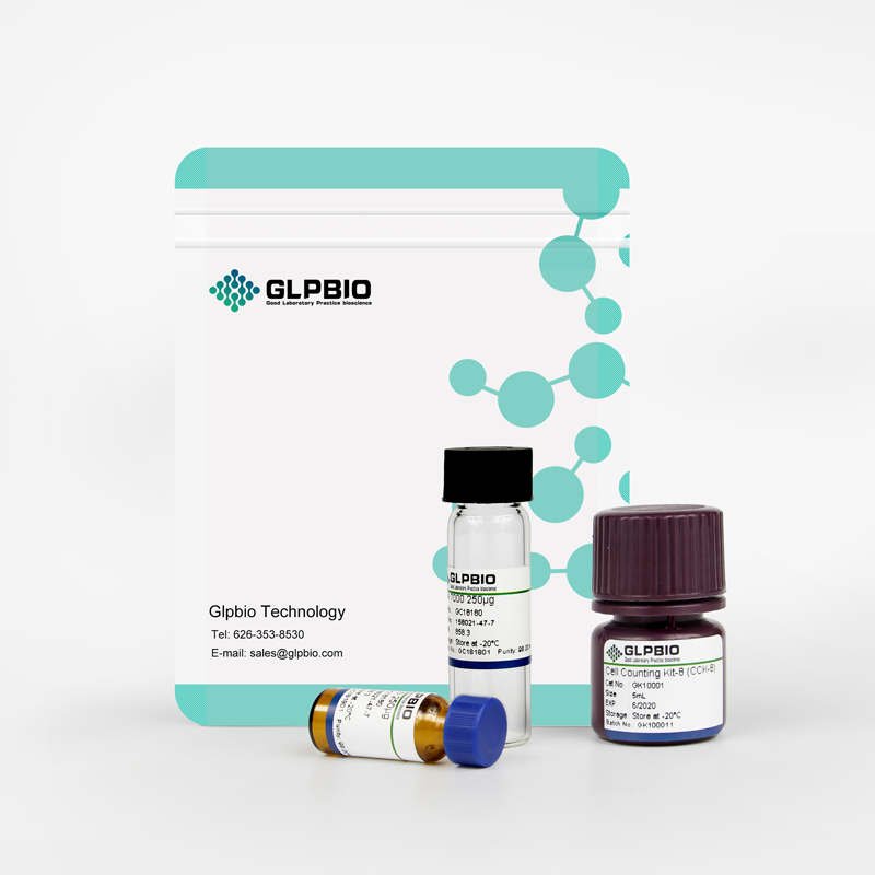
Sample solution is provided at 25 µL, 10mM.
This plan only provides a guide, please modify it to meet your specific needs.
1. Prepare Lyso-Tracker Green staining solution
(1) Take out the frozen dyeing solution, return it to room temperature, and concentrate the dyeing solution at the bottom of the tube by instant centrifugation.
(2) Preparation of working solution: Dilute the stock solution(1mM) with a suitable buffer (such as serum-free medium or PBS) to prepare a working solution with a concentration of 50-75nM.
Notes:
① Please adjust the concentration of the working fluid according to the actual situation and prepare it as needed;
② In order to reduce false positives caused by excessively high background or overload, the concentration of dyes should be as low as possible;
③ If cells are incubated in a buffer free of dye after staining, attenuation of fluorescence signals and vacuolization of cells will be observed.
2. Cell suspension staining
(1) Suspension cells: Centrifuge the suspended cells at 4°C and 1000g for 3-5 minutes, discard the supernatant, and wash twice with PBS for 5 minutes each time.
(2) Adherent cells: Wash twice with PBS, add trypsin to digest the cells, and centrifuge at 1000g for 3-5 minutes after digestion is completed.
(3) Resuspend approximately 106 cells in 0.5-1mL working solution and incubate at 37°C in the dark for 30 minutes to 2 hours. The optimal incubation time for different cells is different, please explore by yourself according to the specific experimental needs.
(4) After the incubation, centrifuge at 1000g for 5 minutes, remove the supernatant, and add PBS to wash 2-3 times, 5 minutes each time.
(5) Use pre-warmed cell culture medium to resuspend and incubate the cells.
3. Cell adhesion staining
(1) Culture adherent cells on sterile coverslips.
(2) Remove the coverslip from the culture medium, aspirate the excess culture medium, and place the coverslip in a humid environment.
(3) Add 100μL of dye working solution from one corner of the coverslip and shake gently to evenly cover all cells with the dye.
(4) Incubate at 37°C in the dark for 30 minutes to 2 hours. The optimal incubation time for different cells is different, please explore by yourself according to the specific experimental needs.
(5) After the incubation, discard the dye working solution, add PBS to wash 2-3 times, and use pre-warmed culture medium to incubate the cells.
4. Fluorescence detection: The maximum absorption/emission light of Lyso-Tracker Green is 504/511nm.
Notes:
① A kinetic study on the internalization of Lyso-Tracker revealed that the rate of uptake of this probe by living cells can be completed within a few seconds. However, the probe exhibits a lysosomal "alkalization effect", and prolonged incubation can lead to an increase in lysosomal pH. Therefore, incubating the cell at 37℃ for 1-5 minutes is a useful pH indicator;
② If the dyeing is not sufficient, it is recommended to increase the dye concentration or extend the dyeing time to allow the dye to aggregate on lysosomes;
③ Fluorescent dyes all have quenching problems. Please try to avoid light as much as possible to slow down fluorescence quenching;
For your safety and health, please wear laboratory clothes and disposable gloves when operating.
| Cas No. | SDF | ||
| Formula | C18H26BClF2N4O | M.Wt | 398.7 |
| Solubility | Storage | Store at -20℃,protect form light | |
| General tips | Please select the appropriate solvent to prepare the stock solution according to the
solubility of the product in different solvents; once the solution is prepared, please store it in
separate packages to avoid product failure caused by repeated freezing and thawing.Storage method
and period of the stock solution: When stored at -80°C, please use it within 6 months; when stored
at -20°C, please use it within 1 month. To increase solubility, heat the tube to 37°C and then oscillate in an ultrasonic bath for some time. |
||
| Shipping Condition | Evaluation sample solution: shipped with blue ice. All other sizes available: with RT, or with Blue Ice upon request. | ||
| Prepare stock solution | |||

|
1 mg | 5 mg | 10 mg |
| 1 mM | 2.5082 mL | 12.5408 mL | 25.0815 mL |
| 5 mM | 501.6 μL | 2.5082 mL | 5.0163 mL |
| 10 mM | 250.8 μL | 1.2541 mL | 2.5082 mL |
Step 1: Enter information below (Recommended: An additional animal making an allowance for loss during the experiment)
 g
μL
g
μL
Step 2: Enter the in vivo formulation (This is only the calculator, not formulation. Please contact us first if there is no in vivo formulation at the solubility Section.)
Calculation results:
Working concentration: mg/ml;
Method for preparing DMSO master liquid: mg drug pre-dissolved in μL DMSO ( Master liquid concentration mg/mL, Please contact us first if the concentration exceeds the DMSO solubility of the batch of drug. )
Method for preparing in vivo formulation: Take μL DMSO master liquid, next addμL PEG300, mix and clarify, next addμL Tween 80, mix and clarify, next add μL saline, mix and clarify.
Method for preparing in vivo formulation: Take μL DMSO master liquid, next add μL Corn oil, mix and clarify.
Note: 1. Please make sure the liquid is clear before adding the next solvent.
2. Be sure to add the solvent(s) in order. You must ensure that the solution obtained, in the previous addition, is a clear solution before proceeding to add the next solvent. Physical methods such as vortex, ultrasound or hot water bath can be used to aid dissolving.
3. All of the above co-solvents are available for purchase on the GlpBio website.
Quality Control & SDS
- View current batch:
- Appearance: A solution
- COA (Certificate of Analysis)
- SDS (Safety Data Sheet)
- Datasheet
Average Rating: 5 (Based on Reviews and 30 reference(s) in Google Scholar.)