


Necrostatin-1
A cell-permeable, potent, and selective blocker of necroptosis (EC50 = 494 nM in FADD-deficient Jurkat cells treated with TNF-α), a non apoptotic necrotic cell death pathway mediated by death-domain receptors (DRs) that offers neuroprotection in a murine model of ischemic brain injury. Necrostatin-1 is a manufactured little particle compound with the substance recipe C16H20N2O. It capabilities as an inhibitor of necroptosis, a type of modified cell passing. Necrostatin-1 explicitly focuses on the movement of RIPK1 (Receptor-Collaborating Protein Kinase 1), a critical controller of necroptosis, subsequently forestalling cell demise in different obsessive circumstances. The structural formula of necrostatin -1 is as follows:Necrostatin -1
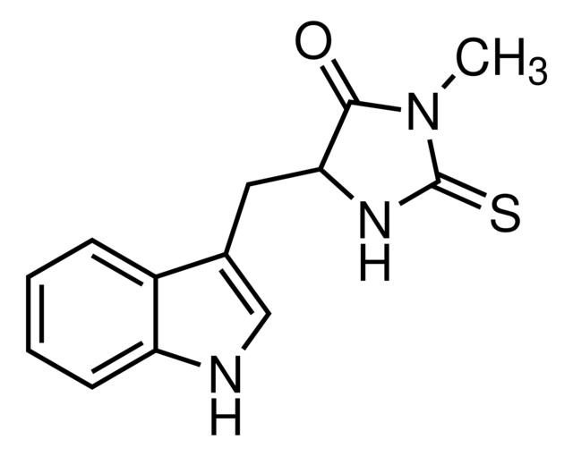
Figure1: Molecular structure of necrostatin -1
Necroptosis is an alternative mode of regulated cell death mimicking features of apoptosis and necrosis. Necroptosis requires protein RIPK3 (previously well recognized as regulator of inflammation, cell survival, and disease) and its substrate MLKL, the crucial players of this pathway. Necroptosis is induced by toll-like receptor, death receptor, interferon, and some other mediators. Shreds of evidence based on a mouse model reveals that deregulation of necroptosis has been found to be associated with pathological conditions like cancer, neurodegenerative diseases, and inflammatory diseases .Necroptosis is an alternative mode of regulated cell death mimicking features of apoptosis and necrosis. Necroptosis requires protein RIPK3 (previously well recognized as regulator of inflammation, cell survival, and disease) and its substrate MLKL, the crucial players of this pathway. Necroptosis is induced by toll-like receptor, death receptor, interferon, and some other mediators. Shreds of evidence based on a mouse model reveals that deregulation of necroptosis has been found to be associated pathological conditions like cancer, neurodegenerative diseases, and inflammatory diseases.Nec-1 halts the necroptosis signaling pathways by inhibiting RIPI signaling cascades. Furthermore, Nec-1 also affects necroptosis by targeting ROS. Nec-1 inhibits apoptosis by targeting RIP1 when the cell undergoes signaling pathway of RDA, yet it loses this anti-apoptosis effect when thepathway is RIA.
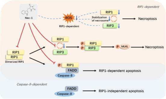
Figure 2:mechanism action of necrostatin -1 on RIP1
Necroptosis has been verified to participate in various diseases , and the protection of Nec-1 has also been authenticated in many disease models, including cardiovascular, neurological and renal diseases . The basic molecular mechanism of this protective effect is blocking necroptosis cell death by targeting RIP1. Therefore, Nec-1 administration usually results in alleviated cell death and improved cell viability. In fact, Nec-1 not only inhibits RIP1, but also reduces RIP3 expression and phosphorylation, which has been demonstrated in many disease models, including myocardial infarction (MI), mental diseases and intestinal inflammation.Ischemia reperfusion (I/R) injury is considered to be one of the major problems in flap surgery. Necroptosis is a recently discovered and caspase-3-independent programed necrosis. Necrostatin-1 (Nec-1) is a specific inhibitor of necroptosis. Reports indicate that Nec-1 provides protection in ischemic models, such as brain, kidney, and heart. The aim of this study is to investigate the influence of Nec-1 on the I/R process in rat abdominal skin flaps.Twenty male Sprague-Dawley rats, weighing 280–320 g, were randomly divided into three groups. The extended epigastric skin flap (6 cm × 9 cm) of rats was used. Three hours of complete ischemia was performed using a clamp, and the clamp was then removed to reperfusion the flap. Twenty hours after the onset of the reperfusion, the rats were assessed for flap survival and perfusion analysis. One sample (1 cm × 1 cm) was taken for H&E, TUNEL, electron microscopy, IHC stainingfor RIP-1, and ELISA analysis for caspase-3 activity.
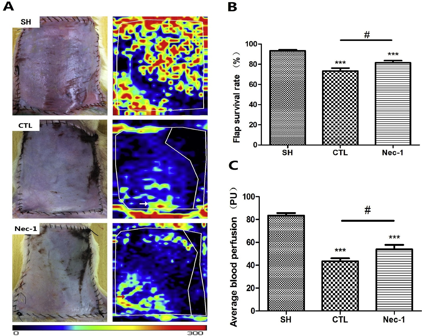
Figure 3:flap survival rate.Comparison of necrostatin 1 with other
Caspase-3 activity was clearly upregulated 24 h after reperfusion in all the three groups (Figure 7). Compared with the CTL (0.7543 ± 0.1970) and SH rats (0.1073 ± 0.0139), the rats subjected to skin flap I/R with Nec-1 treatment (0.7757 ± 0.2109) did not present with any significant change (p > 0.05) in caspase-3 activity.Caspase-3 activity in all the three groups. Compared with the SH group, the CTL and Nec-1 groups presented with a significant reduction in caspase-3 activity. However, caspase-3 activity was not significantly diminished in the Nec-1 group. Values are means ± SD; (n = 10 for each group).
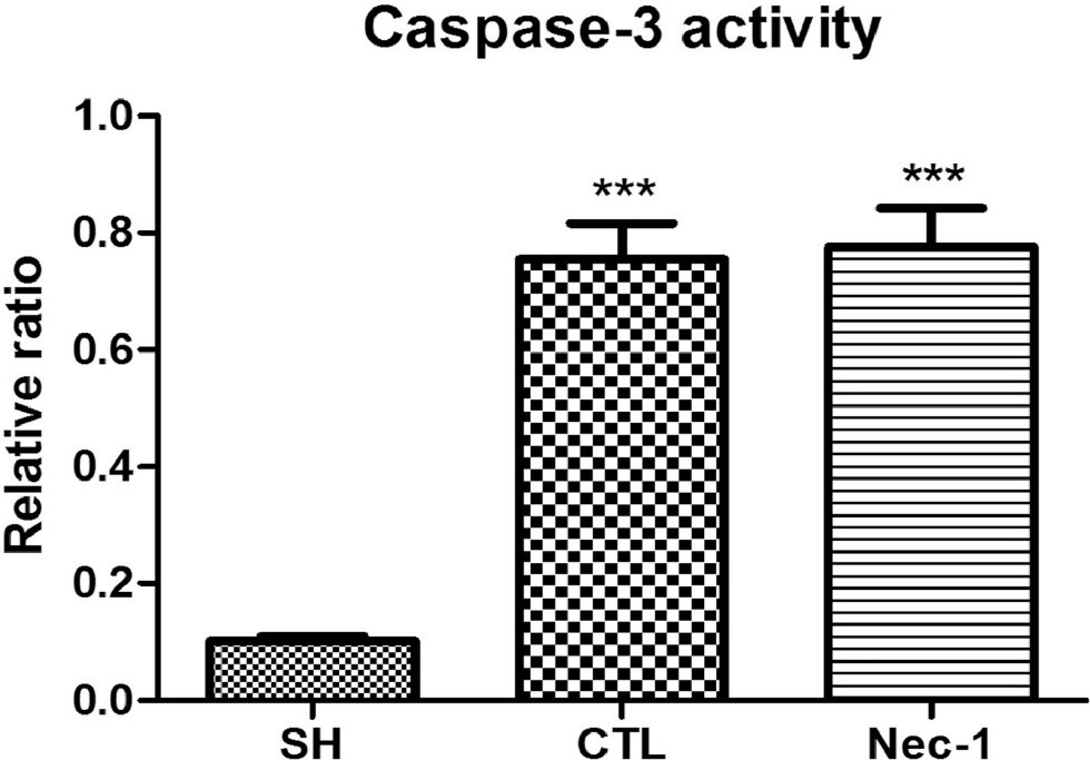
Figure 4: Caspase-3 activity of necrostatin 1
Nec-1 might protect against Al-induced cell death by reducing the phosphorylation of RIP1. Moreover, Nec-1 improved the viability of Al-treated cell to a greater extent than zVAD-fmk and 3-methyladenine, which inhibit apoptosis and autophagy respectively.Nec-1 could block necroptosis and give protection to dopaminergic neurons. They used 6-Hydroxydopamine (6-OHDA)-induced PC12 cells as a Parkinson disease model to explore the role of necroptosis in PD by examining autophagy activation.
Nec-1s does not show a paradoxical dose response in TNF-induced SIRS. (a) TNF shock-associated hypothermia. Nec-1 and Nec-1s were injected i.v. at a dose of Hi: 6 mg/kg b.w. or Lo: 0.6 mg/kg b.w. 17 min before the challenge with mTNF; i.v. Nec-1s was as protective as Nec-1 but lacked the sensitizing effect of Nec-1 at the lower dose. (b) Survival curve for the experiment described in a. Symbols of Nec-1 Hi and Nec-1s Hi coincide and are both completely protective. A cumulative result of two independent experiments is represented. Number of mice is indicated between brackets. *P<0.05, **P<0.001 and ***P<0.0001 Inhibitory effects of Nec-1 on intracellular ROS in PACs. Line graphs showing the effects of RIPK1K45A modification and Nec-1 (30 μM) on intracellular ROS levels (H2DCFDA) in the presence of (a) TLCS (500 μM) and (b) POAEE (100 μM). (c) Bar graph showing the endpoint effects of RIPK1K45A modification and Nec-1 on intracellular ROS levels at 5 h in the presence of TLCS and POAEE. Each dot represents a mouse. Changes are normalized increases in fluorescence from the baseline (F/F0) and data expressed as the mean ± SEM (n ≥ 3 mice/group; n.b. some traces have overlap and the symbols are masked). Significant differences in Nec-1 from WT controls are shown as * p < 0.05.
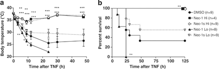
Figure 5:paradoxical dose response in TNF-induced SIRS
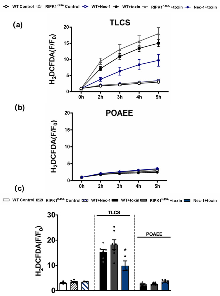
Figure 6:imhibitory response of Nec-1 on ROS in PACs
Necrostatin -1 is widely used to treat a variety of diseases due to its flexibility. It has demonstrated effectiveness against many of disorders such as Nec-1 inhibited intestinal inflammation in inflammatory bowel disease, Pretreatment with Nec-1 protects against SIRS,Nec-1 improves liver function and ameliorates pathological damage in septic rats,Nec-1 reduces cardio myocyte necrosis, improves cardiac output and prolongs cardiac allograft survival time.Necrostatin-1 has also been used in combination of diseases and strengthen its antithrepeutic properties.


Comentarios