

Calcein-AM (Calcein acetoxymethyl ester) (Synonyms: Calcein Acetoxymethyl ester, NSC 689290) |
カタログ番号GC34061
 One-Click Copy Product Info
|

Calcein-AMは、生存細胞に迅速に入る高脂溶性の活性な染料である。
Products are for research use only. Not for human use. We do not sell to patients.
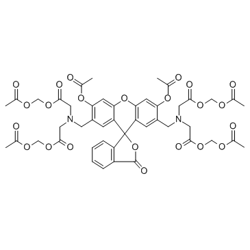
Cas No.: 148504-34-1
Sample solution is provided at 25 µL, 10mM.
- Embo Mol Med (2024):1-24.PMID:39148004
- Int J Nanomed: (2026): 611830.PMID:42615011
- Int J Biol Sci 17.11 (2021):2703.PMID:34345202
- Nat Product Bioprosp 14.1 (2024):1-13.PMID:38780674
- Iscience (2025).
- Phytomedicine 105 (2022):154330.PMID:35905566
- Molecules 28.8 (2023):3389.PMID:37110625
- Phytomedicine (2024):155463.
- J Cancer 15.14 (2024):4577-4590.
Calcein-AMは、生存細胞に迅速に入る高脂溶性の活性な染料である。細胞内のエステラーゼによってcalceinに変換され、強い緑色(530 nm)の信号を発生させ、細胞膜が完全な細胞に保持される。
in vitro実験では、Ca2+(2.5 mM)およびionophore A 23187(0.5 μM)の存在下で、ヒト赤血球(RBC)を37°Cで3時間および20時間培養した後の生存率を評価するためにcalcein-AMアッセイが使用された。CAM(Calcein-AM)の最適濃度は0.05 μMであり、0.05、0.1、0.2、および0.4 μMを試験した。0.05 μMのCAMを使用して、3人の通常のボランティアから採取されたPBMCおよび拡張されたNK細胞を染色した結果、細胞毒性に有意な減少は見られず、PBMCまたは拡張されたNK細胞のヒトNK細胞活性にもCAMの染色が有意な影響を与えなかったことが示された。in vitroでは、50μMのcalcein AMで染色された1 x 10^5リンパ球の蛍光シグナルは飽和レベルに近く、20μMのcalcein AMで染色されたリンパ球のシグナルはわずかに低かったと報告されている。Calcein-AMは、低濃度(2.5 μg/ml)でヒトリンパ腫細胞株(U-937-GTBなど)に対して細胞毒性活性を示する。in vitro実験では、混合マクロファージとTHP-1細胞(5x10^5 cells/ml)を用いて、Calcein-AM(2 µM)/propidium iodide(PI)(4.5 µM)の染色アッセイが生存細胞と死細胞の数を定量化するために使用された。さらに、OA軟骨芽細胞は24-wellプレートに2 × 10^4 cells/wellでシードされ、4時間培養された後、光のない条件下で37°Cで30分間Calcein-AM(2 μM)とPI(2 μM)で処理され、その後蛍光顕微鏡で分析された
References:
[1].Bratosin D, et al. Novel fluorescence assay using calcein-AM for the determination of human erythrocyte viability and aging. Cytometry A. 2005 Jul;66(1):78-84.
[2].Jang YY, et al. An improved flow cytometry-based natural killer cytotoxicity assay involving calcein AM staining of effector cells. Ann Clin Lab Sci. 2012 Winter;42(1):42-9.
[3].Braut-Boucher F, et al. A non-isotopic, highly sensitive, fluorimetric, cell-cell adhesion microplate assay using calcein AM-labeled lymphocytes. J Immunol Methods. 1995 Jan 13;178(1):41-51.
[4].Liminga G, et al. Cytotoxic effect of calcein acetoxymethyl ester on human tumor cell lines: drug delivery by intracellular trapping. Anticancer Drugs. 1995 Aug;6(4):578-85.
[5].Xiang N, et al. Gardnerella vaginalis induces NLRP3 inflammasome-mediated pyroptosis in macrophages and THP-1 monocytes. Exp Ther Med. 2021 Oct;22(4):1174.
[6].Zhang L, et al. MicroRNA-140-5p represses chondrocyte pyroptosis and relieves cartilage injury in osteoarthritis by inhibiting cathepsin B/Nod-like receptor protein 3. Bioengineered. 2021 Dec;12(2):9949-9964.
| 細胞実験[1]: | |
細胞株 | EPC細胞 |
準備方法 | EPC細胞を1×10^5細胞/ウェルの密度で播種し、15°Cで培養した。5 µMのカルセインAMでロードしたEPC細胞の取り込み動態を蛍光強度(FI)として測定した。 |
反応条件 | 5 µM、15°C、1〜8時間 |
アプリケーション | 取り込み動態は、1×10^5細胞/ウェルの密度で播種されたEPC細胞において、テストした7時間全体でカルセインAM標識が増加したことを示した。 |
参考文献: | |
| Cas No. | 148504-34-1 | SDF | |
| 同義語 | Calcein Acetoxymethyl ester, NSC 689290 | ||
| Chemical Name | tetrakis(acetoxymethyl) 2,2',2'',2'''-(((3',6'-diacetoxy-3-oxo-3H-spiro[isobenzofuran-1,9'-xanthene]-4',5'-diyl)bis(methylene))bis(azanetriyl))tetraacetate | ||
| Canonical SMILES | O=C1OC2(C(C=C(CN(CC(OCOC(C)=O)=O)CC(OCOC(C)=O)=O)C(OC(C)=O)=C3)=C3OC4=CC(OC(C)=O)=C(CN(CC(OCOC(C)=O)=O)CC(OCOC(C)=O)=O)C=C24)C5=C1C=CC=C5 | ||
| Formula | C46H46N2O23 | M.Wt | 994.86 |
| 溶解度 | 10 mg/mL in EtOH, MeOH, DMSO, DMF with gentle heating (37°C) for 10 minutes. | Storage | -20°C, protect from light |
| General tips | Please select the appropriate solvent to prepare the stock solution according to the
solubility of the product in different solvents; once the solution is prepared, please store it in
separate packages to avoid product failure caused by repeated freezing and thawing.Storage method
and period of the stock solution: When stored at -80°C, please use it within 6 months; when stored
at -20°C, please use it within 1 month. To increase solubility, heat the tube to 37°C and then oscillate in an ultrasonic bath for some time. |
||
| Shipping Condition | Evaluation sample solution: shipped with blue ice. All other sizes available: with RT, or with Blue Ice upon request. | ||
| Prepare stock solution | |||

|
1 mg | 5 mg | 10 mg |
| 1 mM | 1.0052 mL | 5.0258 mL | 10.0517 mL |
| 5 mM | 201 μL | 1.0052 mL | 2.0103 mL |
| 10 mM | 100.5 μL | 502.6 μL | 1.0052 mL |
Step 1: Enter information below (Recommended: An additional animal making an allowance for loss during the experiment)
 g
μL
g
μL
Step 2: Enter the in vivo formulation (This is only the calculator, not formulation. Please contact us first if there is no in vivo formulation at the solubility Section.)
Calculation results:
Working concentration: mg/ml;
Method for preparing DMSO master liquid: mg drug pre-dissolved in μL DMSO ( Master liquid concentration mg/mL, Please contact us first if the concentration exceeds the DMSO solubility of the batch of drug. )
Method for preparing in vivo formulation: Take μL DMSO master liquid, next addμL PEG300, mix and clarify, next addμL Tween 80, mix and clarify, next add μL saline, mix and clarify.
Method for preparing in vivo formulation: Take μL DMSO master liquid, next add μL Corn oil, mix and clarify.
Note: 1. Please make sure the liquid is clear before adding the next solvent.
2. Be sure to add the solvent(s) in order. You must ensure that the solution obtained, in the previous addition, is a clear solution before proceeding to add the next solvent. Physical methods such as vortex, ultrasound or hot water bath can be used to aid dissolving.
3. All of the above co-solvents are available for purchase on the GlpBio website.
Quality Control & SDS
- View current batch:
- Purity: >98.00% Appearance: A solid
- COA (Certificate of Analysis)
- SDS (Safety Data Sheet)
- Datasheet
-
Related Biological Data

Construction of breast cancer organoid in vitro for drug testing via printing homo-MA and hetero-MA bioinks:d) cell live/dead staining of the core and shell part of breast cancer organoid after exposure to cisplatin (500 umol L−1) and paclitaxel (100 nmol L−1) for 24 and 48h.
Cell viability was tested by Calcein-AM/PI Double Staining Kit. 2 mL of PBS solution containing 0.1 vol% Calcein AM(GlpBio) and 0.1 vol% PI was added into the samples and incubated for 30 min at 37 °C.
Small Methods (2024): 2400223. PMID: 38602202 IF: 12.4002 -
Related Biological Data

MSC coculture inhibits neuronal ferroptosis through mitochondrial transfer.(K) Flow cytometry analysis of LIP in Ht22 cells from culture alone, coculture with MSCs or MSC coculture combined with Cyto D treatment after Rsl3 stimulation for 24 h. Cells were incubated by Calcein-AM (FITC channel) and treated with or without Deferiprone.
After trypsinization, cells (6-well dishes) were washed twice with PBS and then incubated with 1 μM Calcein-AM(GlpBio) at 37°C for 15 min.
Redox Biology, 2023: 102871. PMID: 37699320 IF: 11.3997 -
Related Biological Data

PEF inhibits ferroptosis in C. elegans models of AD.(L) Representative images of CL4176 worms stained with Calcein-AM reagent in the presence or absence of PEF (800 μg/mL) and DFO (100 μM). (M) The bar chart shows the relative Calcein-AM intensity in CL4176 worms.
Briefly, after treatment, worms were collected and co-cultured in M9 with 0.05 μg/mL Calcein-AM(GlpBio) for 1 hour, followed by three washes in 1 mL M9.
Phytomedicine (2024): 155463. PMID: 38452694 IF: 7.8996 -
Related Biological Data

NAR reduced cellular iron concentration. (A) Chondrocytes were treated with IL-1β and FAC, with or without various concentrations of NAR for 24 hr, and then stained with a Calcein-AM fluorescent probe for 15min.
Chondrocytes were treated with IL-1β and FAC, with or without various concentrations of NAR for 24 hr, and then stained with a Calcein-AM(GlpBio) fluorescent probe for 15min.
Phytomedicine (2022): 154330. PMID: 35905566 IF: 6.6563 -
Related Biological Data

Tagitinin C induces ferroptosis in HCT116 cells.(G) Quantification of cellular LIP levels using the calcein-AM (C-AM) method. The mean fluorescence intensity (MFI) of C-AM is subtracted from the MFI of C-AM treated with DFO.
After trypsinization, the cells were washed twice with PBS followed by incubation of 2 µM calcein-acetoxymethyI ester (GLPBIO) for 30 min at 37 °C.
Int J Biol Sci 17.11 (2021): 2703-2717. PMID: 34345202 IF: 6.58
Average Rating: 5 (Based on Reviews and 1 reference(s) in Google Scholar.)