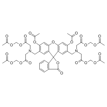
Calcein-AM (Calcein acetoxymethyl ester) |
Catalog No.: GC34061 |
Calcein-AM (Calcein acetoxymethyl ester) is cell-permeable fluorescent dye used to determine the cell viability.
Sample solution is provided at 25 µL, 10mM.
Quality Control & SDS
- View current batch:
-
Purity: >96.00%
- COA (Certificate Of Analysis)
- SDS (Safety Data Sheet)
- Datasheet
Cell experiment [1]: | |
Cell lines |
EPC cells |
Preparation Method |
Uptake kinetics of EPC cells loaded with 5 µM calcein AM seeded at 1x10⁵ cells well-1 and cultured at 15 °C. Calcein AM uptake was measured as fluorescence intensity (FI). |
Reaction Conditions |
5 µM at 15 °C, 1-8h |
Applications |
Uptake kinetics showed that for EPC cells seeded at a density of 1x10⁵ cells well-1 calcein AM labelling increased throughout the 7 h tested. |
References: [1]. Iwanowicz LR, et al. Calcein AM release-based cytotoxic cell assay for fish leucocytes. Fish Shellfish Immunol. 2004 Feb;16(2):127-37. |
Calcein-AM is a highly lipophilic vital dye that rapidly enters viable cells. It is converted by intracellular esterases to calcein that produces an intense green (530-nm) signal, and is retained by cells with intact plasma membrane.[1]
In vitro experiment it shown that calcein-AM assay used to assess human RBC viability after incubation (37°C for 3 and 20 h) in the presence of Ca2+ (2.5 mM) and ionophore A 23187 (0.5 μM).[1] 0.05 μM was the optimal concentration of CAM (Calcein-AM) for staining effector cells by testing 0.05, 0.1, 0.2, and 0.4 μM. Using 0.05 μM CAM to stain the PBMCs and expanded NK cells from three normal volunteers, the results demonstrated that there is no significant decrease in cytotoxicity and CAM staining had no significant effect on human NK cell activity in PBMCs or in expanded NK cells.[2] In vitro, 50μm calcein AM's fluorescent signal of 1 x lo5 lymphocytes was close to the saturation level, while the signal emitted by lymphocytes labeled with 20μm calcein AM was only slightly lower. [3] Calcein-AM has cytotoxic activity against human tumor cell lines (such as the human lymphoma U-937-GTB) at low concentrations (2.5 ug/ml).[4] In vitro experiment it demonstrated that in the mixed macrophages and THP-1 cells (5x10⁵ cells/ml), Calcein-AM (2 µM)/propidium Iodide (PI) (4.5 µM) staining assay Calcein-AM/PI double staining was used to quantify the number of living and dead cells as a cell death assay.[5] In addition, The cells in OA chondrocytes were seeded in 24-well plates (2 × 10⁴ cells/well), cultured for 4 h, the cells were treated with 5 μL Calcein-AM (2 μM) and 5 μL PI (2 μM) at 37°C in conditions void of light for 30 min, and then analyzed under a fluorescence microscope.[6]
References:
[1].Bratosin D, et al. Novel fluorescence assay using calcein-AM for the determination of human erythrocyte viability and aging. Cytometry A. 2005 Jul;66(1):78-84.
[2].Jang YY, et al. An improved flow cytometry-based natural killer cytotoxicity assay involving calcein AM staining of effector cells. Ann Clin Lab Sci. 2012 Winter;42(1):42-9.
[3].Braut-Boucher F, et al. A non-isotopic, highly sensitive, fluorimetric, cell-cell adhesion microplate assay using calcein AM-labeled lymphocytes. J Immunol Methods. 1995 Jan 13;178(1):41-51.
[4].Liminga G, et al. Cytotoxic effect of calcein acetoxymethyl ester on human tumor cell lines: drug delivery by intracellular trapping. Anticancer Drugs. 1995 Aug;6(4):578-85.
[5].Xiang N, et al. Gardnerella vaginalis induces NLRP3 inflammasome-mediated pyroptosis in macrophages and THP-1 monocytes. Exp Ther Med. 2021 Oct;22(4):1174.
[6].Zhang L, et al. MicroRNA-140-5p represses chondrocyte pyroptosis and relieves cartilage injury in osteoarthritis by inhibiting cathepsin B/Nod-like receptor protein 3. Bioengineered. 2021 Dec;12(2):9949-9964.
Cas No. | 148504-34-1 | SDF | |
Synonyms | Calcein Acetoxymethyl ester, NSC 689290 | ||
Chemical Name | tetrakis(acetoxymethyl) 2,2',2'',2'''-(((3',6'-diacetoxy-3-oxo-3H-spiro[isobenzofuran-1,9'-xanthene]-4',5'-diyl)bis(methylene))bis(azanetriyl))tetraacetate | ||
Canonical SMILES | O=C1OC2(C(C=C(CN(CC(OCOC(C)=O)=O)CC(OCOC(C)=O)=O)C(OC(C)=O)=C3)=C3OC4=CC(OC(C)=O)=C(CN(CC(OCOC(C)=O)=O)CC(OCOC(C)=O)=O)C=C24)C5=C1C=CC=C5 | ||
Formula | C46H46N2O23 | M.Wt | 994.86 |
Solubility | 10 mg/mL in EtOH, MeOH, DMSO, DMF with gentle heating (37°C) for 10 minutes. | Storage | -20°C, protect from light |
General tips | For obtaining a higher solubility , please warm the tube at 37 ℃ and shake it in the ultrasonic bath for a while.Stock solution can be stored below -20℃ for several months. | ||
Shipping Condition | Evaluation sample solution : ship with blue ice All other available size: ship with RT , or blue ice upon request |
Step 1: Enter information below (Recommended: An additional animal making an allowance for loss during the experiment)
Step 2: Enter the in vivo formulation (This is only the calculator, not formulation. Please contact us first if there is no in vivo formulation at the solubility Section.)
Calculation results:
Working concentration: mg/ml;
Method for preparing DMSO master liquid: mg drug pre-dissolved in μL DMSO ( Master liquid concentration mg/mL, Please contact us first if the concentration exceeds the DMSO solubility of the batch of drug. )
Method for preparing in vivo formulation: Take μL DMSO master liquid, next addμL PEG300, mix and clarify, next addμL Tween 80, mix and clarify, next add μL ddH2O, mix and clarify.
Method for preparing in vivo formulation: Take μL DMSO master liquid, next add μL Corn oil, mix and clarify.
Note: 1. Please make sure the liquid is clear before adding the next solvent.
2. Be sure to add the solvent(s) in order. You must ensure that the solution obtained, in the previous addition, is a clear solution before proceeding to add the next solvent. Physical methods such as vortex, ultrasound or hot water bath can be used to aid dissolving.
3. All of the above co-solvents are available for purchase on the GlpBio website.
Cytotoxic activity of calcein acetoxymethyl ester (Calcein/AM) on primary cultures of human haematological and solid tumours
The aim of this study was to determine the in vitro cytotoxicity of calcein acetoxymethyl ester (Calcein/AM) on primary cultures derived from solid and haematological human tumours. Calcein/AM is a fluorescent dye that localises intracellularly after esterase-dependent cellular trapping and which has shown cytotoxic activity against various established human tumour cell lines at relatively low concentrations. The semi-automated fluorometric microculture cytotoxicity assay, based on the measurement of fluorescence generated from cellular hydrolysis of fluorescein diacetate to fluorescein, in microtitre plates was used for the evaluation of Calcein/AM activity in tumour cell suspensions from patients. The cytotoxicity was measured as a survival index (SI), defined as the fluorescence as a percentage of control cultures. A total of 163 evaluable samples from various tumours were tested with continuous drug exposure. The activity of Calcein/AM was compared with representatives of six major classes of standard chemotherapeutic drugs. Calcein/AM was found to induce concentration-dependent decreases in the SI of both haematological and solid tumour cells. The ratio of solid over haematological tumour activity increased at a rate that was concentration dependent. Although it was relatively less active than cisplatin against solid tumours, Calcein/AM showed higher solid tumour activity compared to leukaemic specific agents (cytarabine and amsacrine), vincristine and doxorubicin (Dox). Among the solid tumours tested, childhood tumours, non-small cell lung cancer and sarcomas were the most sensitive to Calcein/AM. The best correlation between SI values was seen between Calcein/AM and Dox, with weaker correlations to representatives of antimetabolites, platinum compounds, topoisomerase II inhibitors, tubulin interactive agents and alkylators. Non-cytotoxic concentrations of cyclosporin A significantly potentiated calcein-induced cytotoxicity. The results show that Calcein/AM is differentially active against haematological tumours, but with substantial activity against solid tumours. The drug may represent a new class of anticancer compound with a unique means of drug delivery.
Comparison of different live/dead stainings for detection and quantification of adherent microorganisms in the initial oral biofilm
Objectives: The aim of the present study was to investigate different fluorescence-based, two-color viability assays for visualization and quantification of initial bacterial adherence and to establish reliable alternatives to the ethidium bromide staining procedure.
Materials and methods: Bacterial colonization was attained in situ on bovine enamel slabs (n = 6 subjects). Five different live/dead assays were investigated (fluorescein diacetate (FDA)/propidium iodide (PI), Syto 9/PI (BacLight?), FDA/Sytox red, Calcein acetoxymethyl (AM)/Sytox red, and carboxyfluorescein diacetate (CFDA)/Sytox red). After 120 min of oral exposure, analysis was performed with an epifluorescence microscope. Validation was carried out, using the colony-forming units for quantification and the transmission electron microscopy for visualization after staining.
Results: The average number of bacteria amounted to 2.9 ± 0.8 × 10(4) cm(-2). Quantification with Syto 9/PI and Calcein AM/Sytox red yielded an almost equal distribution of cells (Syto 9/PI 45% viable, 55% avital; Calcein AM/Sytox red 52% viable, 48% avital). The live/dead ratio of CFDA/Sytox red and FDA/Sytox red was 3:2. An aberrant dispersal was recorded with FDA/PI (viable 34%, avital 66%). The TEM analysis indicated that all staining procedures affect the structural integrity of the bacterial cells considerably.
Conclusion: The following live/dead assays are reliable techniques for differentiation of viable and avital adherent bacteria: BacLight, FDA/Sytox red, Calcein AM/Sytox red, and CFDA/Sytox red. These fluorescence-based techniques are applicable alternatives to toxic and instable conventional assays, such as the staining procedure based on ethidium bromide.
Clinical relevance: Differentiation of viable and avital adherent bacteria offers the possibility for reliable evaluation of different mouth rinses, oral medication, and disinfections.
Cytotoxic effect of calcein acetoxymethyl ester on human tumor cell lines: drug delivery by intracellular trapping
Calcein acetoxymethyl ester (calcein/AM) and some related cellular dyes with a cytoplasmic distribution were investigated with respect to cellular hydrolysis, accumulation, efflux and cytotoxicity in a panel of established human cell lines, including multidrug resistant (MDR) phenotypes. At 0.1-1 micrograms/ml, calcein/AM was highly cytotoxic against several cell lines, even after short-term exposure (30 min). Calcein/AM induced no immediate loss (3 h) of membrane integrity and the drug was more active against low compared with high density plated cells. In cell lines with the MDR phenotype and in the renal carcinoma cell line ACHN, the drug was considerably less active. Non-esterified calcein had no effect and calcein/AM was significantly more potent than other structurally related fluorescein analogs and AM esters tested. Although MDR cell lines showed a decreased cellular hydrolysis and accumulation of the dye, there was no strict relationship between cytoplasmic calcein exposure and cytotoxic activity. The rate of efflux was low in the two most sensitive cell lines, the human lymphoma U-937-GTB and its vincristine (vcr) resistant subline U-937/vcr10, while the remaining cell lines showed similar biphasic efflux patterns, including cell lines of the MDR phenotype. The results show that calcein/AM has cytotoxic activity against human tumor cell lines at low concentrations. The effect appears dependent on the intracellular trapping of the drug, although the specific cellular target remains unknown. Due to its cytotoxic efficacy and unique principle of cellular drug delivery, further investigation of calcein/AM and related compounds as potentially new anticancer agents seems warranted.
Identification of genotype-selective antitumor agents using synthetic lethal chemical screening in engineered human tumor cells
We used synthetic lethal high-throughput screening to interrogate 23,550 compounds for their ability to kill engineered tumorigenic cells but not their isogenic normal cell counterparts. We identified known and novel compounds with genotype-selective activity, including doxorubicin, daunorubicin, mitoxantrone, camptothecin, sangivamycin, echinomycin, bouvardin, NSC146109, and a novel compound that we named erastin. These compounds have increased activity in the presence of hTERT, the SV40 large and small T oncoproteins, the human papillomavirus type 16 (HPV) E6 and E7 oncoproteins, and oncogenic HRAS. We found that overexpressing hTERT and either E7 or LT increased expression of topoisomerase 2alpha and that overexpressing RAS(V12) and ST both increased expression of topoisomerase 1 and sensitized cells to a nonapoptotic cell death process initiated by erastin.
Assessment of Cell Viability with Single-, Dual-, and Multi-Staining Methods Using Image Cytometry
The ability to accurately measure cell viability is important for any cell-based assay. Traditionally, viability measurements have been performed using the trypan blue exclusion method on a hemacytometer, which allows researchers to visually distinguish viable from nonviable cells. While the trypan blue method can work for cell lines or primary cells that have been rigorously purified, in more complex samples such as PBMCs, bone marrow, whole blood, or any sample with low viability, this method can lead to errors. In recent years, advances in optics and fluorescent dyes have led to the development of automated benchtop image-based cell counters for rapid cell concentration and viability measurement. In this work, we demonstrate the use of image-based cytometry for cell viability detection using single-, dual-, or multi-stain techniques. Single-staining methods using nucleic acid stains such as EB, PI, 7-AAD, DAPI, SYTOX Green, and SYTOX Red, and enzymatic stains such as CFDA and Calcein AM, were performed. Dual-staining methods using AO/PI, CFDA/PI, Calcein AM/PI, Hoechst/PI, Hoechst/DRAQ7, and DRAQ5/DAPI that enumerate viable and nonviable cells were also performed. Finally, Hoechst/Calcein AM/PI was used for a multi-staining method. Fluorescent viability staining allows exclusion of cellular debris and nonnucleated cells from analysis, which can eliminate the need to perform purification steps during sample preparation and improve efficiency. Image cytometers increase speed and throughput, capture images for visual confirmation of results, and can greatly simplify cell count and viability measurements.
Average Rating: 5
(Based on Reviews and 1 reference(s) in Google Scholar.)GLPBIO products are for RESEARCH USE ONLY. Please make sure your review or question is research based.
Required fields are marked with *