

Cyanine5.5 amine (Synonyms: Cy 5.5 amine; Lumiprobe Cy 5.5 amine) |
カタログ番号GC59167
 One-Click Copy Product Info
|

Cyanine5.5 amineは遊離のアミノ基を含有する近赤外線蛍光色素であり、カルボキシル基、アルデヒド基、またはその他の反応性官能基を含有する生物分子と共有結合できる。
Products are for research use only. Not for human use. We do not sell to patients.
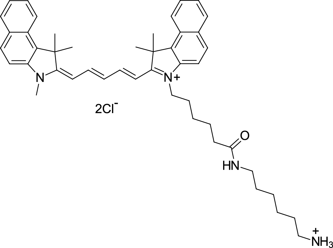
Cas No.: 2097714-44-6 (without anion), 2097714-45-7 (chloride)
Sample solution is provided at 25 µL, 10mM.
_1-3.$$$39978408@51.jpg');)
_1-9.$$$38538795@65.jpg');)
_1-9.$$$39112701@51.jpg');)
_1-7.$$$37316672@70.jpg');)
_366-372.$$$36198801@70.jpg');)
.$$$38565142@65.jpg');)
_1867-1883.$$$33248023@67.jpg');)
_eads6055.$$$39977546@45.jpg');)
_eadm9805.$$$40080571@45.jpg');)
_eadj3020.$$$39666821@45.jpg');)
_1-20.$$$39757301@28.jpg');)
_1-24.$$$38898113@28.jpg');)
_273-287.$$$36806353@46.jpg');)
_1-16.$$$37156877@44.jpg');)
_1-19.$$$37460805@44.jpg');)
_1291-1307.$$$34518654@46.jpg');)
Cyanine5.5 amineは遊離のアミノ基を含有する近赤外線蛍光色素であり、カルボキシル基、アルデヒド基、またはその他の反応性官能基を含有する生物分子と共有結合できる[1]。Cyanine5.5 amineはCyanine5.5のアミノ誘導体であり、励起波長は約648-680nmであり、発光波長は約694-710nmである[2]。Cyanine5.5 amineは高いモル吸光係数と明るい遠赤色蛍光を含有し、よって低いバックグラウンド蛍光を必要とする体内イメージングとバイオマーカーの応用に適している[3]。Cyanine5.5 amineは、pHに対して敏感ではなく、良好な光安定性と生体適合性を示す[4]。Cyanine5.5 amineは通常、生物分子の蛍光標識、ナノ粒子の調製と小動物の体内イメージング研究に使われている[5-7]。
References:
[1] Zhang E, Zhukova V, Semyonkin A, et al. Release kinetics of fluorescent dyes from PLGA nanoparticles in retinal blood vessels: In vivo monitoring and ex vivo localization. Eur J Pharm Biopharm. 2020;150:131-142.
[2] Son Phan K, Nghi Do H, Thuy Doan B, et al. The Influence of Cyanine 5.5 and Doxorubicin on Cell Cycle Arrest, Magnetic Resonance, and Near-Infrared Fluorescence Optical Imaging for Fe3O4-Encapsulated PLA-TPGS Nanoparticles. ChemMedChem. 2025;20(5):e202400586.
[3] Konig SG, Kramer R. Accessing Structurally Diverse Near-Infrared Cyanine Dyes for Folate Receptor-Targeted Cancer Cell Staining. Chemistry. 2017;23(39):9306-9312
[4] Ebert B, Riefke B, Sukowski U, Licha K. Cyanine dyes as contrast agents for near-infrared imaging in vivo: acute tolerance, pharmacokinetics, and fluorescence imaging. J Biomed Opt. 2011;16(6):066003.
[5] Li L, Xue Y, Li X, et al. Copper Nanosheet-Based Wash-Free Fluorescence Imaging of Cancer Cells. Anal Chem. 2024;96(5):2052-2058.
[6] Luo J, Fan H, Shu C, et al. Immuno-protective cationic ginsenoside Rb1 vesicles for macrophage-mediated targeted therapy of inflammatory diseases. Mater Today Bio. 2025;34:102195.
[7] Mai L, Yao A, Li J, et al. Cyanine 5.5 conjugated nanobubbles as a tumor selective contrast agent for dual ultrasound-fluorescence imaging in a mouse model. PLoS One. 2013;8(4):e61224.
網膜血管におけるCyanine5.5 amine標識のナノ粒子(Cy5.5-PLGA)のリアルタイムモニタリングのプロトコール[1]:
1.ナノ粒子懸濁液の調製
1)Cyanine5.5 amine(4mg、5.3μmol)を8mL DCMに溶解し、PLGA溶液(20mL DCMに2400mg)を添加し、N,N-diisopropylethylamine(0.8 mg、6.2μmol)とEDC(7.6mg、49μmol)を添加し、暗所、室温で48時間攪拌する。
2)脱イオン水で有機相を三回洗浄し、無水Na₂SO₄で乾燥し、酢酸エチルに溶解し、ヘキサンで沈殿させ、濾過し、減圧乾燥して、淡青色の粉末状の複合体を得る。
3)1:1の質量比で、Cy5.5標識のPLGAを未標識のPLGAと混ぜて、DCMに共に溶解し、ポリマー溶液を得る。
4)高圧水中油型エマルジョン溶媒蒸発法を使い、ポリマー溶液からナノ粒子懸濁液を調製する。
5)遠心分離またはゲル浸透クロマトグラフィーを使い、未封入の遊離染料を除去し、凍結保護剤として1%のマンニトールを添加し、凍結乾燥する。
6)新鮮に調製されたCy5.5-PLGAナノ粒子の粉末を0.5mL 1% poloxamer 188に再懸濁し、ゆっくりとボルテックスし、室温で30分間静置して、完全に分散させる。
2.体内注射とリアルタイムのイメージング
1)ラットを麻酔し、両方の目に1-2滴のNeosynephrine-POS 5%を点眼し、虹彩をリラックスさせ、Vidisic®アイゲルを塗布し、目の乾燥を防ぎ、コンタクトレンズに対しては浸潤液としても使う。
2)ラットを共焦点走査顕微鏡の下に固定し、カニューレを尾静脈に挿入し、注射器と接続する。
3)0.5mLのナノ粒子懸濁液を静脈内注射する(30-60秒、ゆっくりと注入する)。
4)網膜の蛍光を観察し、0、1、3、5、15、30、60と90分で画像を撮る。
注意事項:全ての体内イメージングのプロセスで、ラットを加熱プレートに置くこと。
3.体外網膜のイメージング
1)リアルタイムのイメージング後、上記の麻酔剤を過剰投与してラットを安楽死させ、眼球を摘出し、それを冷却されたHEPES緩衝液(135mMのNaCl、5mMのNaOH、2.5mMのKCl、7mMのMgCl₂、10mMのHEPES、10mMのグルコース、pH=7.4)に置く。
2)前眼部と硝子体を摘出し、網膜の全体を注意深く分離して、改良の培養プレートに平に広げる。
3)HEPES溶液の中で、0.1mg/mL Hoechst 33342で網膜標本全体を20分間インキュベートする。
4)網膜の標本を4%のパラホルムアルデヒドで20分間固定し、その後はHEPES溶液ですすぐ。
5)網膜の標本を顕微鏡の下で速やかに撮影する。
このプロトコールはあくまでもガイドラインであり、ご自身のニーズに合わせてご調整願います。
References:
[1] Zhang E, Zhukova V, Semyonkin A, et al. Release kinetics of fluorescent dyes from PLGA nanoparticles in retinal blood vessels: In vivo monitoring and ex vivo localization. Eur J Pharm Biopharm. 2020;150:131-142.
| Cas No. | 2097714-44-6 (without anion), 2097714-45-7 (chloride) | SDF | |
| 同義語 | Cy 5.5 amine; Lumiprobe Cy 5.5 amine | ||
| Formula | C46H58Cl2N4O | M.Wt | 753.88 |
| 溶解度 | practically insoluble in water (< 2 uM), good in polar organic solvents (DMF, DMSO, alcohols) | Storage | Store at -20°C,protect from light |
| General tips | Please select the appropriate solvent to prepare the stock solution according to the
solubility of the product in different solvents; once the solution is prepared, please store it in
separate packages to avoid product failure caused by repeated freezing and thawing.Storage method
and period of the stock solution: When stored at -80°C, please use it within 6 months; when stored
at -20°C, please use it within 1 month. To increase solubility, heat the tube to 37°C and then oscillate in an ultrasonic bath for some time. |
||
| Shipping Condition | Evaluation sample solution: shipped with blue ice. All other sizes available: with RT, or with Blue Ice upon request. | ||
| Prepare stock solution | |||

|
1 mg | 5 mg | 10 mg |
| 1 mM | 1.3265 mL | 6.6324 mL | 13.2647 mL |
| 5 mM | 265.3 μL | 1.3265 mL | 2.6529 mL |
| 10 mM | 132.6 μL | 663.2 μL | 1.3265 mL |
Step 1: Enter information below (Recommended: An additional animal making an allowance for loss during the experiment)
 g
μL
g
μL
Step 2: Enter the in vivo formulation (This is only the calculator, not formulation. Please contact us first if there is no in vivo formulation at the solubility Section.)
Calculation results:
Working concentration: mg/ml;
Method for preparing DMSO master liquid: mg drug pre-dissolved in μL DMSO ( Master liquid concentration mg/mL, Please contact us first if the concentration exceeds the DMSO solubility of the batch of drug. )
Method for preparing in vivo formulation: Take μL DMSO master liquid, next addμL PEG300, mix and clarify, next addμL Tween 80, mix and clarify, next add μL saline, mix and clarify.
Method for preparing in vivo formulation: Take μL DMSO master liquid, next add μL Corn oil, mix and clarify.
Note: 1. Please make sure the liquid is clear before adding the next solvent.
2. Be sure to add the solvent(s) in order. You must ensure that the solution obtained, in the previous addition, is a clear solution before proceeding to add the next solvent. Physical methods such as vortex, ultrasound or hot water bath can be used to aid dissolving.
3. All of the above co-solvents are available for purchase on the GlpBio website.
Quality Control & SDS
- View current batch:
- Purity: >97.00% Appearance: A solid
- COA (Certificate of Analysis)
- SDS (Safety Data Sheet)
- Datasheet
Average Rating: 5 (Based on Reviews and 10 reference(s) in Google Scholar.)