

Cyanine5.5 amine (Synonyms: Cy 5.5 amine; Lumiprobe Cy 5.5 amine) |
Catalog No.GC59167
 One-Click Copy Product Info
|

Cyanine5.5 amine은 카르복실기, 알데히드 또는 기타 반응성 기능기를 포함하는 생체 분자와 공유결합할 수 있는 자유 아미노기를 포함하는 근적외선 형광 염료이다.
Products are for research use only. Not for human use. We do not sell to patients.
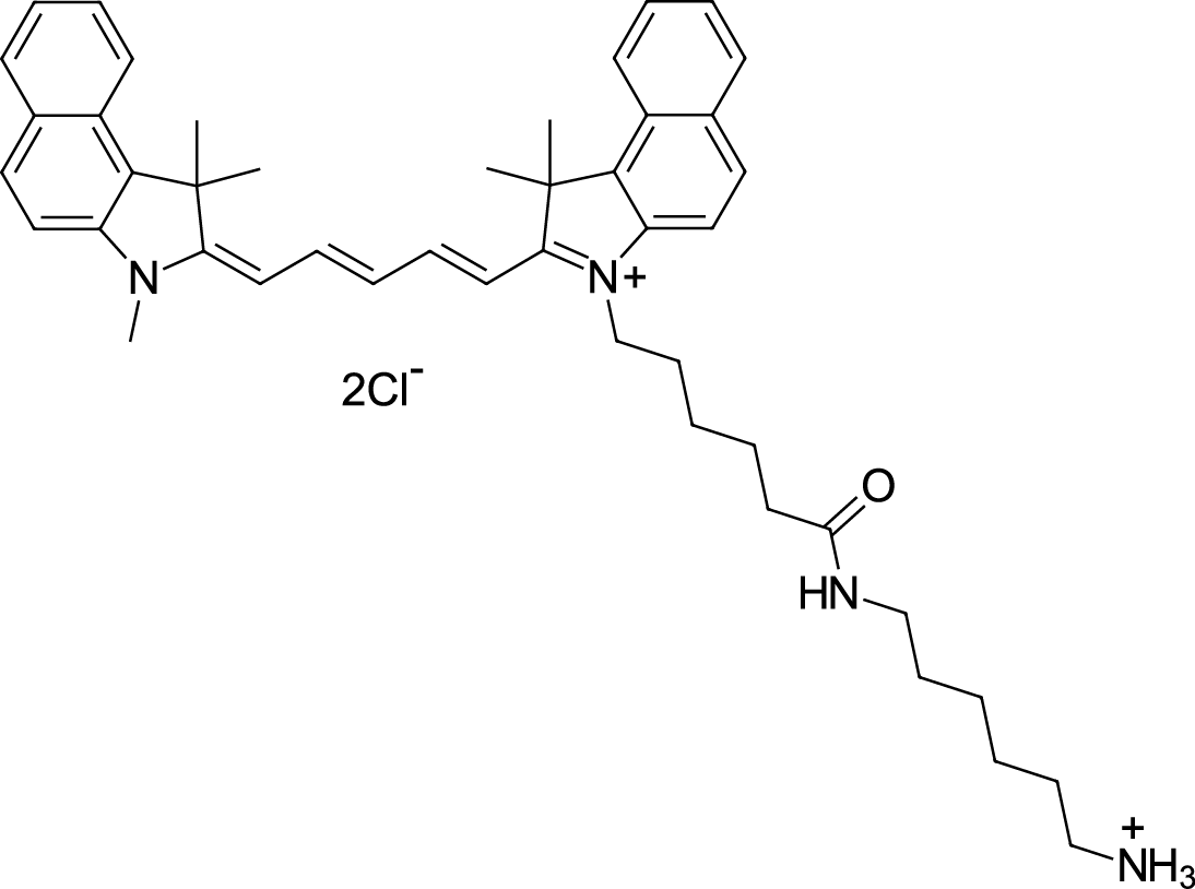
Cas No.: 2097714-44-6 (without anion), 2097714-45-7 (chloride)
Sample solution is provided at 25 µL, 10mM.
_1-3.$$$39978408@51.jpg');)
_1-9.$$$38538795@65.jpg');)
_1-9.$$$39112701@51.jpg');)
_1-7.$$$37316672@70.jpg');)
_366-372.$$$36198801@70.jpg');)
.$$$38565142@65.jpg');)
_1867-1883.$$$33248023@67.jpg');)
_eads6055.$$$39977546@45.jpg');)
_eadm9805.$$$40080571@45.jpg');)
_eadj3020.$$$39666821@45.jpg');)
_1-20.$$$39757301@28.jpg');)
_1-24.$$$38898113@28.jpg');)
_273-287.$$$36806353@46.jpg');)
_1-16.$$$37156877@44.jpg');)
_1-19.$$$37460805@44.jpg');)
_1291-1307.$$$34518654@46.jpg');)
Cyanine5.5 amine은 카르복실기, 알데히드 또는 기타 반응성 기능기를 포함하는 생체 분자와 공유결합할 수 있는 자유 아미노기를 포함하는 근적외선 형광 염료이다. Cyanine5.5 amine은 Cyanine5.5의 아미노 유도체로 여기 파장은 약 648-680nm이고 방출 파장은 약 694-710nm이다[2]. Cyanine5.5 amine은 높은 몰 소광계수와 밝은 원적색 형광을 가지고 있어 낮은 배경 형광이 필요한 생체 내 영상 및 바이오마커 응용에 적합하다[3]. Cyanine5.5 amine은 pH에 둔감하고 우수한 광안정성과 생체적합성을 나타낸다[4]. Cyanine5.5 amine은 일반적으로 생체 분자의 형광 표지, 나노입자 제조, 및 소동물 체내 영상 연구에 사용된다[5-7].
References:
[1] Zhang E, Zhukova V, Semyonkin A, et al. Release kinetics of fluorescent dyes from PLGA nanoparticles in retinal blood vessels: In vivo monitoring and ex vivo localization. Eur J Pharm Biopharm. 2020;150:131-142.
[2] Son Phan K, Nghi Do H, Thuy Doan B, et al. The Influence of Cyanine 5.5 and Doxorubicin on Cell Cycle Arrest, Magnetic Resonance, and Near-Infrared Fluorescence Optical Imaging for Fe3O4-Encapsulated PLA-TPGS Nanoparticles. ChemMedChem. 2025;20(5):e202400586.
[3] Konig SG, Kramer R. Accessing Structurally Diverse Near-Infrared Cyanine Dyes for Folate Receptor-Targeted Cancer Cell Staining. Chemistry. 2017;23(39):9306-9312
[4] Ebert B, Riefke B, Sukowski U, Licha K. Cyanine dyes as contrast agents for near-infrared imaging in vivo: acute tolerance, pharmacokinetics, and fluorescence imaging. J Biomed Opt. 2011;16(6):066003.
[5] Li L, Xue Y, Li X, et al. Copper Nanosheet-Based Wash-Free Fluorescence Imaging of Cancer Cells. Anal Chem. 2024;96(5):2052-2058.
[6] Luo J, Fan H, Shu C, et al. Immuno-protective cationic ginsenoside Rb1 vesicles for macrophage-mediated targeted therapy of inflammatory diseases. Mater Today Bio. 2025;34:102195.
[7] Mai L, Yao A, Li J, et al. Cyanine 5.5 conjugated nanobubbles as a tumor selective contrast agent for dual ultrasound-fluorescence imaging in a mouse model. PLoS One. 2013;8(4):e61224.
망막 혈관에서 Cyanine5.5 amine 표지 나노입자(Cy5.5-PLGA) 실시간 관찰을 위한 프로토콜[1]:
1.나노입자 소생된 용액 제조
1)Cyanine5.5 amine (4 mg, 5.3 μmol)을 8 mL DCM에 용해하고 PLGA 용액(2400 mg을 20 mL DCM에 용해)을 첨가하고 N,N-디이소프로필에틸아민(0.8 mg, 6.2 μmol)과 EDC(7.6 mg, 49 μmol)를 첨가해서 실온에서 48시간 암실교반합니다.
2)유기상을 탈이온수로 3회 세척하고 무수 Na₂SO₄로 건조시킨 후 에틸아세테이트에 용해하고 헥산으로 침전시킨 후 여과하고 진공 건조해서 연한 파란색 분말 형태의 접합체를 얻습니다.
3)Cy5.5 표지된 PLGA와 비표지 PLGA를 1:1 질량비로 혼합하고 함께 DCM에 용해해서 중합체 용액을 제조합니다.
4)고압 o/w 유화 용제 증발 기술을 사용해서 중합체 용액에서 나노입자 소생된 용액을 제조합니다.
5)원심분리 또는 겔 투과 크로마토그래피로 비캡슐화된 자유 염료를 제거하고1% 만니톨을 동결보호제로 첨가해서 동결건조합니다.
6)신선히 제조된 Cy5.5-PLGA 나노입자 분말을 0.5 mL 1% 폴록사머 188에 소생시키고 부드럽게 와류를 일으킨 후 실온에서 30분 동안 방치해서 완전 분산되도록 합니다.
2.체내 주사 및 실시간 이미징
1)쥐를 마취하고 양쪽 눈에 홍채를 이완시키기 위해 Neosynephrine-POS 5%를 1-2 방울 적용하고 눈이 마르는 것을 방지하기 위해 Vidisic® 안약 젤을 적용하고 접촉 렌즈의 침지 매질로도 사용하였습니다.
2)공초점 주사 현미경 하에 쥐를 고정하고 꼬리 정맥에 캐뉼라를 삽입해서 주사기를 연결합니다.
3)0.5 mL 나노입자 소생시킨 용액을 정맥내 주사합니다(30-60초에 걸쳐 서서히 투여).
4)망막 형광을 관찰하고 0, 1, 3, 5, 15, 30, 60 및 90분에 영상을 촬취합니다.
참고: 모든 생체 내 영상 과정 동안 쥐를 가열판 위에 유지합니다.
3.체외 망막 영상
1)실시간 영상 후에 상기 마취제를 과다 투여해서 쥐를 안락사시키고 안구를 적출하여 냉각된 HEPES 완충액(135 mM NaCl, 5 mM NaOH, 2.5 mM KCl, 7 mM MgCl₂, 10 mM HEPES, 10 mM 포도당, pH=7.4)에 넣습니다.
2)전방 부분과 유리체를 제거하고 전체 망막을 조심스럽게 분리해서 개량된 배양판 위에 펼칩니다.
3)HEPES 용액 중 0.1 mg/mL Hoechst 33342로 전체 장착물을 20분 동안 배양합니다.
4)평판 장착 망막을 4% 파라포름알데히드로 20분 동안 고정한 후 HEPES 용액으로 세척합니다.
5)현미경 하에서 평판 장착 망막을 즉시 영상화합니다.
본 프로토콜은 단지 가이드를 제공하고 특정 필요에 맞게 수정하시기 바랍니다.
References:
[1] Zhang E, Zhukova V, Semyonkin A, et al. Release kinetics of fluorescent dyes from PLGA nanoparticles in retinal blood vessels: In vivo monitoring and ex vivo localization. Eur J Pharm Biopharm. 2020;150:131-142.
| Cas No. | 2097714-44-6 (without anion), 2097714-45-7 (chloride) | SDF | |
| Synonyms | Cy 5.5 amine; Lumiprobe Cy 5.5 amine | ||
| Formula | C46H58Cl2N4O | M.Wt | 753.88 |
| Solubility | practically insoluble in water (< 2 uM), good in polar organic solvents (DMF, DMSO, alcohols) | Storage | Store at -20°C,protect from light |
| General tips | Please select the appropriate solvent to prepare the stock solution according to the
solubility of the product in different solvents; once the solution is prepared, please store it in
separate packages to avoid product failure caused by repeated freezing and thawing.Storage method
and period of the stock solution: When stored at -80°C, please use it within 6 months; when stored
at -20°C, please use it within 1 month. To increase solubility, heat the tube to 37°C and then oscillate in an ultrasonic bath for some time. |
||
| Shipping Condition | Evaluation sample solution: shipped with blue ice. All other sizes available: with RT, or with Blue Ice upon request. | ||
| Prepare stock solution | |||

|
1 mg | 5 mg | 10 mg |
| 1 mM | 1.3265 mL | 6.6324 mL | 13.2647 mL |
| 5 mM | 265.3 μL | 1.3265 mL | 2.6529 mL |
| 10 mM | 132.6 μL | 663.2 μL | 1.3265 mL |
Step 1: Enter information below (Recommended: An additional animal making an allowance for loss during the experiment)
 g
μL
g
μL
Step 2: Enter the in vivo formulation (This is only the calculator, not formulation. Please contact us first if there is no in vivo formulation at the solubility Section.)
Calculation results:
Working concentration: mg/ml;
Method for preparing DMSO master liquid: mg drug pre-dissolved in μL DMSO ( Master liquid concentration mg/mL, Please contact us first if the concentration exceeds the DMSO solubility of the batch of drug. )
Method for preparing in vivo formulation: Take μL DMSO master liquid, next addμL PEG300, mix and clarify, next addμL Tween 80, mix and clarify, next add μL saline, mix and clarify.
Method for preparing in vivo formulation: Take μL DMSO master liquid, next add μL Corn oil, mix and clarify.
Note: 1. Please make sure the liquid is clear before adding the next solvent.
2. Be sure to add the solvent(s) in order. You must ensure that the solution obtained, in the previous addition, is a clear solution before proceeding to add the next solvent. Physical methods such as vortex, ultrasound or hot water bath can be used to aid dissolving.
3. All of the above co-solvents are available for purchase on the GlpBio website.
Quality Control & SDS
- View current batch:
- Purity: >97.00% Appearance: A solid
- COA (Certificate of Analysis)
- SDS (Safety Data Sheet)
- Datasheet
Average Rating: 5 (Based on Reviews and 10 reference(s) in Google Scholar.)