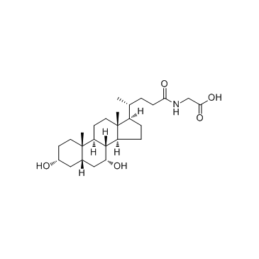
Glycochenodeoxycholic acid (Chenodeoxycholylglycine) (Synonyms: GCDCA, NSC 681056) |
| カタログ番号GC34092 |
グリコケノデオキシコール酸 (Chenodeoxycholylglycine) (Chenodeoxycholylglycine) は、ケノデオキシコール酸とグリシンから肝臓で形成される胆汁酸です。吸収のために脂肪を可溶化する洗剤として作用し、それ自体が吸収されます。グリコケノデオキシコール酸 (Chenodeoxycholylglycine) (Chenodeoxycholylglycine) は、肝細胞のアポトーシスを誘導します。
Products are for research use only. Not for human use. We do not sell to patients.

Cas No.: 640-79-9
Sample solution is provided at 25 µL, 10mM.
Glycochenodeoxycholic acid is a bile salt formed in the liver from chenodeoxycholate and glycine; used to induce hepatocyte apoptosis in research.
Chenodeoxycholate is toxic to hepatocytes, and accumulation of chenodeoxycholate in the liver during cholestasis may potentiate hepatocellular injury. At a concentration of 250μM, glycochenodeoxycholate is more toxic than either chenodeoxycholate or taurochenodeoxycholate. Glycochenodeoxycholate cytotoxicity may result from ATP depletion followed by a subsequent rise in Ca2+. The rise in Ca2+ leads to an increase in calcium-dependent degradative proteolysis and, ultimately, cell death[1]. 4 h exposure of 50 μM GCDC induces apoptosis in 42% of hepatocytes. Intracellular PKC activity decreased to 44% of controls 2 h after exposure of hepatocytes to GCDC. GCDC-induced apoptosis is associated with decreases in total cellular PKC activity, which appear to be dependent on intracellular calpain-like protease activity[2].
GCDCA induces ER-related calcium release within about ten seconds. Significant increases in activities of calpain and caspase-12 are observed after 15 h of GCDCA treatment. Bip and Chop mRNA expressions are increased with the treated GCDCA dose and incubation time. Cytochrome c release from mitochondria peaks in about 2 h of incubation[3].
[1]. Spivey JR, et al. Glycochenodeoxycholate-induced lethal hepatocellular injury in rat hepatocytes. Role of ATP depletion and cytosolic free calcium. J Clin Invest. 1993 Jul;92(1):17-24. [2]. Gonzalez B, et al. Glycochenodeoxycholic acid (GCDC) induced hepatocyte apoptosis is associated with early modulation of intracellular PKC activity. Mol Cell Biochem. 2000 Apr;207(1-2):19-27. [3]. Tsuchiya S, et al. Involvement of endoplasmic reticulum in glycochenodeoxycholic acid-induced apoptosis in rathepatocytes. Toxicol Lett. 2006 Oct 10;166(2):140-9.
Average Rating: 5 (Based on Reviews and 6 reference(s) in Google Scholar.)
GLPBIO products are for RESEARCH USE ONLY. Please make sure your review or question is research based.
Required fields are marked with *


