

ML-SI3 |
カタログ番号GC67784
 One-Click Copy Product Info
|

ML-SI3はカチオンチャネルTRPML阻害剤であり、TRPML1とTRPML2に対するIC50値はそれぞれ4.7μMと1.7μMである。
Products are for research use only. Not for human use. We do not sell to patients.
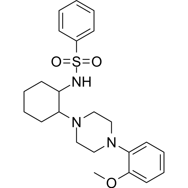
Cas No.: 891016-02-7
Sample solution is provided at 25 µL, 10mM.
_1-3.$$$39978408@51.jpg');)
_1-9.$$$38538795@65.jpg');)
_1-9.$$$39112701@51.jpg');)
_1-7.$$$37316672@70.jpg');)
_366-372.$$$36198801@70.jpg');)
.$$$38565142@65.jpg');)
_1867-1883.$$$33248023@67.jpg');)
_eads6055.$$$39977546@45.jpg');)
_eadm9805.$$$40080571@45.jpg');)
_eadj3020.$$$39666821@45.jpg');)
_1-20.$$$39757301@28.jpg');)
_1-24.$$$38898113@28.jpg');)
_273-287.$$$36806353@46.jpg');)
_1-16.$$$37156877@44.jpg');)
_1-19.$$$37460805@44.jpg');)
_1291-1307.$$$34518654@46.jpg');)
ML-SI3はカチオンチャネルTRPML阻害剤であり、TRPML1とTRPML2に対するIC50値はそれぞれ4.7μMと1.7μMである[1]。ML-SI3は、S5、S6とPH1で形成された疎水性空洞と結合し、チャネルのPI(3,5)P2依存の活性化を阻害せずに、TRPML1のチャネル活性化を遮断する[2]。ML-SI3は、ヒト胃上皮細胞株によるプロトンの分泌の刺激に広く使われている[3]。
体外で、20μMのML-SI3で24時間処理した結果、ラット海馬体ニューロン細胞のアポトーシスを著しく促進し、ミトコンドリアの機能を破壊した[4]。75µMのML-SI3で24時間処理した結果、成体した住血吸虫の表皮完全性を著しく破壊した[5]。10µMのML-SI3で24時間の処理した結果、BxPC-3細胞で、Inositol polyphosphate 4-phosphatase, type II (INPP4B)によるリソソーム関連の膜タンパク質1(LAMP1)とフィブロネクチン1(FN1)の末梢局在を拮抗した[6]。
体内で、1.5mg/kgのML-SI3を28日間、2日ごとに腹腔内注射した結果、損傷した心筋細胞のオートファジーを回復させることで、マウスにおける心筋虚血/再潅流の損傷を保護できる[7]。3週間のML-SI3の処理(16mg/kg/日;腹腔内注射)は、黒色腫のマウスモデルで、黒色腫の肺転移に対するML-SA5 (2mg/kg;腹腔内注射)の阻害作用を著しく逆転させた[8]。
References:
[1] Rühl P, Rosato A S, Urban N, et al. Estradiol analogs attenuate autophagy, cell migration and invasion by direct and selective inhibition of TRPML1, independent of estrogen receptors[J]. Scientific reports, 2021, 11(1): 8313.
[2] Schmiege P, Fine M, Li X. Atomic insights into ML-SI3 mediated human TRPML1 inhibition[J]. Structure, 2021, 29(11): 1295-1302. e3.
[3] Mueller A U, Andersen G, Richter P, et al. Activation of the TRPML1 Ion Channel Induces Proton Secretion in the Human Gastric Parietal Cell Line HGT-1[J]. International Journal of Molecular Sciences, 2024, 25(16): 8829.
[4] Peng T, Xie Y, Zhao S, et al. TRPML1 ameliorates seizures-related neuronal injury by regulating autophagy and lysosomal biogenesis via Ca2+/TFEB signaling pathway[J]. Biochimica et Biophysica Acta (BBA)-Molecular Basis of Disease, 2024, 1870(8): 167477.
[5] Bais S, Norwillo A, Ruthel G, et al. Schistosome TRPML channels play a role in neuromuscular activity and tegumental integrity[J]. Biochimie, 2022, 194: 108-117.
[6] Saffi G T, Kleine N, Salmena L. The lipid phosphatase INPP4B controls pancreatic cancer cell migration and invasion by regulating fibronectin exocytosis[J]. Journal of Biological Chemistry, 2025: 110716.
[7] Xing Y, Sui Z, Liu Y, et al. Blunting TRPML1 channels protects myocardial ischemia/reperfusion injury by restoring impaired cardiomyocyte autophagy[J]. Basic research in cardiology, 2022, 117(1): 20.
[8] Xing Y, Wei X, Liu Y, et al. Autophagy inhibition mediated by MCOLN1/TRPML1 suppresses cancer metastasis via regulating a ROS-driven TP53/p53 pathway[J]. Autophagy, 2022, 18(8): 1932-1954.
| 細胞実験[1]: | |
細胞株 | ラット海馬体ニューロン細胞 |
準備方法 | 10%のウシ胎児血清(FBS)、1%のペニシリン-ストレプトマイシン溶液、1%のグルタミン酸と88%の神経基礎培地で構成された培地でラット海馬体ニューロン細胞を培養した。適切な密度で細胞を培養プレートに播種した。4-6時間処理培養した後、2%のB27、1%のペニシリン-ストレプトマイシン溶液、1%のグルタミン酸と96%の神経基礎培地で構成された維持培地で成長培地を交換した。維持培地を2-3日ごとに交換した。培養の10日目に、培養されたニューロンをランダムに五つのグループに分けた:(1)CON群(Mg2+を含有する環境で培養)、(2)無Mg2+群(Mg2+なしの環境に暴露)、(3)DMSO群(DMSOで24時間事前処理し、その後は無Mg2+の環境に暴露)、(4)ML-SA1群(20μMのML-SA1で24事前処理し、その後は無Mg2+の環境に暴露)、そして(5)ML-SI3群(20μMのML-SI3で24時間事前処理し、その後は無Mg2+の環境に暴露)。神経細胞の生存率を分析した。 |
反応条件 | 20μM;24時間 |
アプリケーション | ML-SI3の処理は、神経細胞の生存率を抑制し、アポトーシスを悪化させた。 |
| 動物実験 [2]: | |
動物モデル | BALB/cヌードマウス |
準備方法 | 200µlの最終体積に0.5×10⁶個のA-375-luc細胞を再懸濁し、続いてそれを約6週齢のBALB/cヌードマウスの尾にゆっくり注射した。マウスを三つのグループに分ける。PBS、ML-SA5 (2mg/kg)またはML-SA5 (2mg/kg)+ML-SI3 (16mg/kg)を3週間、毎日腹腔内注射した。その後、イメージングの10分前に、0.2mlのD-fluorescein (30mg/ml)をマウスの腹腔に注射した。黒色腫の転移を同定するために、生物発光を毎週モニタリングする。 |
投与形態 | 16mg/kg/日、3週間;腹腔内注射 |
アプリケーション | ML-SI3の処理は、マウスで、黒色腫の肺転移に対するML-SA5の阻害作用を著しく逆転させた。 |
References: | |
| Cas No. | 891016-02-7 | SDF | |
| Formula | C23H31N3O3S | M.Wt | 429.58 |
| 溶解度 | DMSO : 50 mg/mL (116.39 mM; Need ultrasonic) | Storage | Store at -20°C |
| General tips | Please select the appropriate solvent to prepare the stock solution according to the
solubility of the product in different solvents; once the solution is prepared, please store it in
separate packages to avoid product failure caused by repeated freezing and thawing.Storage method
and period of the stock solution: When stored at -80°C, please use it within 6 months; when stored
at -20°C, please use it within 1 month. To increase solubility, heat the tube to 37°C and then oscillate in an ultrasonic bath for some time. |
||
| Shipping Condition | Evaluation sample solution: shipped with blue ice. All other sizes available: with RT, or with Blue Ice upon request. | ||
| Prepare stock solution | |||

|
1 mg | 5 mg | 10 mg |
| 1 mM | 2.3279 mL | 11.6393 mL | 23.2786 mL |
| 5 mM | 465.6 μL | 2.3279 mL | 4.6557 mL |
| 10 mM | 232.8 μL | 1.1639 mL | 2.3279 mL |
Step 1: Enter information below (Recommended: An additional animal making an allowance for loss during the experiment)
 g
μL
g
μL
Step 2: Enter the in vivo formulation (This is only the calculator, not formulation. Please contact us first if there is no in vivo formulation at the solubility Section.)
Calculation results:
Working concentration: mg/ml;
Method for preparing DMSO master liquid: mg drug pre-dissolved in μL DMSO ( Master liquid concentration mg/mL, Please contact us first if the concentration exceeds the DMSO solubility of the batch of drug. )
Method for preparing in vivo formulation: Take μL DMSO master liquid, next addμL PEG300, mix and clarify, next addμL Tween 80, mix and clarify, next add μL saline, mix and clarify.
Method for preparing in vivo formulation: Take μL DMSO master liquid, next add μL Corn oil, mix and clarify.
Note: 1. Please make sure the liquid is clear before adding the next solvent.
2. Be sure to add the solvent(s) in order. You must ensure that the solution obtained, in the previous addition, is a clear solution before proceeding to add the next solvent. Physical methods such as vortex, ultrasound or hot water bath can be used to aid dissolving.
3. All of the above co-solvents are available for purchase on the GlpBio website.
Quality Control & SDS
- View current batch:
- Purity: >99.00% Appearance: A solid
- COA (Certificate of Analysis)
- SDS (Safety Data Sheet)
- Datasheet
Average Rating: 5 (Based on Reviews and 30 reference(s) in Google Scholar.)