

ML-SI3 |
Katalog-Nr.GC67784
 One-Click Copy Product Info
|

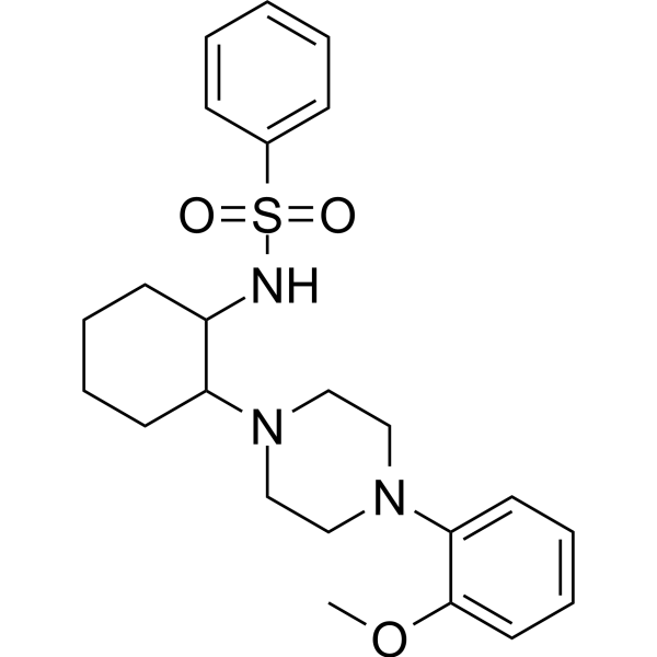
ML-SI3 is cation channel TRPML inhibitor, with IC50 values of 4.7μM and 1.7μM for TRPML1 and TRPML2, respectively.
Products are for research use only. Not for human use. We do not sell to patients.
Cas No.: 891016-02-7
Sample solution is provided at 25 µL, 10mM.
_1-3.$$$39978408@51.jpg');)
_1-9.$$$38538795@65.jpg');)
_1-9.$$$39112701@51.jpg');)
_1-7.$$$37316672@70.jpg');)
_366-372.$$$36198801@70.jpg');)
.$$$38565142@65.jpg');)
_1867-1883.$$$33248023@67.jpg');)
_eads6055.$$$39977546@45.jpg');)
_eadm9805.$$$40080571@45.jpg');)
_eadj3020.$$$39666821@45.jpg');)
_1-20.$$$39757301@28.jpg');)
_1-24.$$$38898113@28.jpg');)
_273-287.$$$36806353@46.jpg');)
_1-16.$$$37156877@44.jpg');)
_1-19.$$$37460805@44.jpg');)
_1291-1307.$$$34518654@46.jpg');)
ML-SI3 is cation channel TRPML inhibitor, with IC50 values of 4.7μM and 1.7μM for TRPML1 and TRPML2, respectively [1]. ML-SI3 binds to the hydrophobic cavity created by S5, S6, and PH1, blocking channel activation of TRPML1 without inhibiting PI(3,5)P2-dependent activation of the channel [2]. ML-SI3 has been widely used to stimulate the secretion of protons by human gastric epithelial cell lines[3].
In vitro, ML-SI3 treatment at 20μM for 24 hours significantly promoted the apoptosis of rat hippocampal neuronal cells and disrupted mitochondrial function[4]. The 75µM ML-SI3 treatment for 24 hours significantly disrupted the epidermal integrity of adult schistosomes[5]. The 24-hour treatment with 10µM ML-SI3 antagonized the peripheral localization of lysosome-associated membrane protein 1 (LAMP1) and fibronectin 1 (FN1) induced by Inositol polyphosphate 4-phosphatase, type II (INPP4B) in BxPC-3 cells[6].
In vivo, ML-SI3 treatment via intraperitoneal injection at a dose of 1.5mg/kg every 2 days for 28 days can protect the myocardial ischemia/reperfusion injury in mice by restoring the autophagy of damaged myocardial cells[7]. ML-SI3 treatment (16mg/kg/day; i.p.) for 3 weeks significantly reversed the inhibitory effect of ML-SA5 (2mg/kg; i.p.) on the lung metastasis of melanoma in mouse model of melanoma[8].
References:
[1] Rühl P, Rosato A S, Urban N, et al. Estradiol analogs attenuate autophagy, cell migration and invasion by direct and selective inhibition of TRPML1, independent of estrogen receptors[J]. Scientific reports, 2021, 11(1): 8313.
[2] Schmiege P, Fine M, Li X. Atomic insights into ML-SI3 mediated human TRPML1 inhibition[J]. Structure, 2021, 29(11): 1295-1302. e3.
[3] Mueller A U, Andersen G, Richter P, et al. Activation of the TRPML1 Ion Channel Induces Proton Secretion in the Human Gastric Parietal Cell Line HGT-1[J]. International Journal of Molecular Sciences, 2024, 25(16): 8829.
[4] Peng T, Xie Y, Zhao S, et al. TRPML1 ameliorates seizures-related neuronal injury by regulating autophagy and lysosomal biogenesis via Ca2+/TFEB signaling pathway[J]. Biochimica et Biophysica Acta (BBA)-Molecular Basis of Disease, 2024, 1870(8): 167477.
[5] Bais S, Norwillo A, Ruthel G, et al. Schistosome TRPML channels play a role in neuromuscular activity and tegumental integrity[J]. Biochimie, 2022, 194: 108-117.
[6] Saffi G T, Kleine N, Salmena L. The lipid phosphatase INPP4B controls pancreatic cancer cell migration and invasion by regulating fibronectin exocytosis[J]. Journal of Biological Chemistry, 2025: 110716.
[7] Xing Y, Sui Z, Liu Y, et al. Blunting TRPML1 channels protects myocardial ischemia/reperfusion injury by restoring impaired cardiomyocyte autophagy[J]. Basic research in cardiology, 2022, 117(1): 20.
[8] Xing Y, Wei X, Liu Y, et al. Autophagy inhibition mediated by MCOLN1/TRPML1 suppresses cancer metastasis via regulating a ROS-driven TP53/p53 pathway[J]. Autophagy, 2022, 18(8): 1932-1954.
| Cell experiment [1]: | |
Cell lines | Rat hippocampal neuronal cells |
Preparation Method | The rat hippocampal neuronal cells were cultured in a growth medium, which consisted of 10% fetal bovine serum (FBS), 1% penicillin-streptomycin solution, 1% glutamine, and 88% neural basal medium. The cells were seeded at an appropriate density on the culture plates. After the initial culture for 4-6 hours, the growth medium was replaced with a maintenance medium, which consisted of 2% B27, 1% penicillin-streptomycin solution, 1% glutamine, and 96% neural basal medium. The maintenance medium was changed every 2-3 days. On the 10th day of culture, the cultured neurons were randomly divided into five groups: (1) CON group (cultured in an environment containing Mg2+), (2) Mg2+-free group (exposed to an environment without Mg2+), (3) DMSO group (pre-treated with DMSO for 24 hours before exposure to the Mg2+-free environment), (4) ML-SA1 group (pre-treated with 20μM ML-SA1 for 24 hours before exposure to the Mg2+-free environment), and (5) ML-SI3 group (pre-treated with 20μM ML-SI3 for 24 hours before exposure to the Mg2+-free environment). The survival of the nerve cells was analyzed. |
Reaction Conditions | 20μM; 24h |
Applications | ML-SI3 treatment suppressed viability of nerve cells and aggravated apoptosis. |
| Animal experiment [2]: | |
Animal models | BALB/c nude mouse |
Preparation Method | Resuspend 0.5×10⁶ A-375-luc cells to a final volume of 200µl, and then slowly inject them into the tail of approximately 6-week-old BALB/c nude mice. Divide the mice into three groups. Administer intraperitoneal injections of PBS, ML-SA5 (2mg/kg), or ML-SA5 (2mg/kg)+ML-SI3 (16mg/kg) every day for three weeks. Then, 10 minutes before imaging, inject 0.2ml of D-fluorescein (30mg/ml) into the mice's abdominal cavity. Monitor bioluminescence weekly to identify melanoma metastasis. |
Dosage form | 16mg/kg/day for 3 weeks; i.p. |
Applications | ML-SI3 treatment significantly reversed the inhibitory effect of ML-SA5 on the lung metastasis of melanoma in mice. |
References: | |
| Cas No. | 891016-02-7 | SDF | |
| Formula | C23H31N3O3S | M.Wt | 429.58 |
| Löslichkeit | DMSO : 50 mg/mL (116.39 mM; Need ultrasonic) | Storage | Store at -20°C |
| General tips | Please select the appropriate solvent to prepare the stock solution according to the
solubility of the product in different solvents; once the solution is prepared, please store it in
separate packages to avoid product failure caused by repeated freezing and thawing.Storage method
and period of the stock solution: When stored at -80°C, please use it within 6 months; when stored
at -20°C, please use it within 1 month. To increase solubility, heat the tube to 37°C and then oscillate in an ultrasonic bath for some time. |
||
| Shipping Condition | Evaluation sample solution: shipped with blue ice. All other sizes available: with RT, or with Blue Ice upon request. | ||
| Prepare stock solution | |||

|
1 mg | 5 mg | 10 mg |
| 1 mM | 2.3279 mL | 11.6393 mL | 23.2786 mL |
| 5 mM | 465.6 μL | 2.3279 mL | 4.6557 mL |
| 10 mM | 232.8 μL | 1.1639 mL | 2.3279 mL |
Step 1: Enter information below (Recommended: An additional animal making an allowance for loss during the experiment)
 g
μL
g
μL
Step 2: Enter the in vivo formulation (This is only the calculator, not formulation. Please contact us first if there is no in vivo formulation at the solubility Section.)
Calculation results:
Working concentration: mg/ml;
Method for preparing DMSO master liquid: mg drug pre-dissolved in μL DMSO ( Master liquid concentration mg/mL, Please contact us first if the concentration exceeds the DMSO solubility of the batch of drug. )
Method for preparing in vivo formulation: Take μL DMSO master liquid, next addμL PEG300, mix and clarify, next addμL Tween 80, mix and clarify, next add μL saline, mix and clarify.
Method for preparing in vivo formulation: Take μL DMSO master liquid, next add μL Corn oil, mix and clarify.
Note: 1. Please make sure the liquid is clear before adding the next solvent.
2. Be sure to add the solvent(s) in order. You must ensure that the solution obtained, in the previous addition, is a clear solution before proceeding to add the next solvent. Physical methods such as vortex, ultrasound or hot water bath can be used to aid dissolving.
3. All of the above co-solvents are available for purchase on the GlpBio website.
Quality Control & SDS
- View current batch:
- Purity: >99.00% Appearance: A solid
- COA (Certificate of Analysis)
- SDS (Safety Data Sheet)
- Datasheet
Average Rating: 5 (Based on Reviews and 30 reference(s) in Google Scholar.)